Tag Archives: biomimetics

Predicting how a memristor functions

An April 3, 2017 news item on Nanowerk announces a new memristor development (Note: A link has been removed),

Researchers from the CNRS [Centre national de la recherche scientifique; France] , Thales, and the Universities of Bordeaux, Paris-Sud, and Evry have created an artificial synapse capable of learning autonomously. They were also able to model the device, which is essential for developing more complex circuits. The research was published in Nature Communications (“Learning through ferroelectric domain dynamics in solid-state synapses”)

An April 3, 2017 CNRS press release, which originated the news item, provides a nice introduction to the memristor concept before providing a few more details about this latest work (Note: A link has been removed),

One of the goals of biomimetics is to take inspiration from the functioning of the brain [also known as neuromorphic engineering or neuromorphic computing] in order to design increasingly intelligent machines. This principle is already at work in information technology, in the form of the algorithms used for completing certain tasks, such as image recognition; this, for instance, is what Facebook uses to identify photos. However, the procedure consumes a lot of energy. Vincent Garcia (Unité mixte de physique CNRS/Thales) and his colleagues have just taken a step forward in this area by creating directly on a chip an artificial synapse that is capable of learning. They have also developed a physical model that explains this learning capacity. This discovery opens the way to creating a network of synapses and hence intelligent systems requiring less time and energy.

Our brain’s learning process is linked to our synapses, which serve as connections between our neurons. The more the synapse is stimulated, the more the connection is reinforced and learning improved. Researchers took inspiration from this mechanism to design an artificial synapse, called a memristor. This electronic nanocomponent consists of a thin ferroelectric layer sandwiched between two electrodes, and whose resistance can be tuned using voltage pulses similar to those in neurons. If the resistance is low the synaptic connection will be strong, and if the resistance is high the connection will be weak. This capacity to adapt its resistance enables the synapse to learn.

Although research focusing on these artificial synapses is central to the concerns of many laboratories, the functioning of these devices remained largely unknown. The researchers have succeeded, for the first time, in developing a physical model able to predict how they function. This understanding of the process will make it possible to create more complex systems, such as a series of artificial neurons interconnected by these memristors.

As part of the ULPEC H2020 European project, this discovery will be used for real-time shape recognition using an innovative camera1 : the pixels remain inactive, except when they see a change in the angle of vision. The data processing procedure will require less energy, and will take less time to detect the selected objects. The research involved teams from the CNRS/Thales physics joint research unit, the Laboratoire de l’intégration du matériau au système (CNRS/Université de Bordeaux/Bordeaux INP), the University of Arkansas (US), the Centre de nanosciences et nanotechnologies (CNRS/Université Paris-Sud), the Université d’Evry, and Thales.


Image synapse


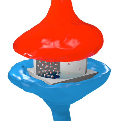
© Sören Boyn / CNRS/Thales physics joint research unit.

Artist’s impression of the electronic synapse: the particles represent electrons circulating through oxide, by analogy with neurotransmitters in biological synapses. The flow of electrons depends on the oxide’s ferroelectric domain structure, which is controlled by electric voltage pulses.


Here’s a link to and a citation for the paper,

Learning through ferroelectric domain dynamics in solid-state synapses by Sören Boyn, Julie Grollier, Gwendal Lecerf, Bin Xu, Nicolas Locatelli, Stéphane Fusil, Stéphanie Girod, Cécile Carrétéro, Karin Garcia, Stéphane Xavier, Jean Tomas, Laurent Bellaiche, Manuel Bibes, Agnès Barthélémy, Sylvain Saïghi, & Vincent Garcia. Nature Communications 8, Article number: 14736 (2017) doi:10.1038/ncomms14736 Published online: 03 April 2017

This paper is open access.

Thales or Thales Group is a French company, from its Wikipedia entry (Note: Links have been removed),

Thales Group (French: [talɛs]) is a French multinational company that designs and builds electrical systems and provides services for the aerospace, defence, transportation and security markets. Its headquarters are in La Défense[2] (the business district of Paris), and its stock is listed on the Euronext Paris.

The company changed its name to Thales (from the Greek philosopher Thales,[3] pronounced [talɛs] reflecting its pronunciation in French) from Thomson-CSF in December 2000 shortly after the £1.3 billion acquisition of Racal Electronics plc, a UK defence electronics group. It is partially state-owned by the French government,[4] and has operations in more than 56 countries. It has 64,000 employees and generated €14.9 billion in revenues in 2016. The Group is ranked as the 475th largest company in the world by Fortune 500 Global.[5] It is also the 10th largest defence contractor in the world[6] and 55% of its total sales are military sales.[4]

The ULPEC (Ultra-Low Power Event-Based Camera) H2020 [Horizon 2020 funded) European project can be found here,

The long term goal of ULPEC is to develop advanced vision applications with ultra-low power requirements and ultra-low latency. The output of the ULPEC project is a demonstrator connecting a neuromorphic event-based camera to a high speed ultra-low power consumption asynchronous visual data processing system (Spiking Neural Network with memristive synapses). Although ULPEC device aims to reach TRL 4, it is a highly application-oriented project: prospective use cases will b…

Finally, for anyone curious about Thales, the philosopher (from his Wikipedia entry), Note: Links have been removed,

Thales of Miletus (/ˈθeɪliːz/; Greek: Θαλῆς (ὁ Μῑλήσιος), Thalēs; c. 624 – c. 546 BC) was a pre-Socratic Greek/Phoenician philosopher, mathematician and astronomer from Miletus in Asia Minor (present-day Milet in Turkey). He was one of the Seven Sages of Greece. Many, most notably Aristotle, regard him as the first philosopher in the Greek tradition,[1][2] and he is otherwise historically recognized as the first individual in Western civilization known to have entertained and engaged in scientific philosophy.[3][4]

The inside scoop on beetle exoskeletons

In the past I’ve covered work on the Namib beetle and its bumps which allow it to access condensation from the air in one of the hottest places on earth and work on jewel beetles and how their structural colo(u)r is derived. Now, there’s research into a beetle’s body armor from the University of Nebraska-Lincoln according to a Feb. 22, 2017 news item on ScienceDaily,

Beetles wear a body armor that should weigh them down — think medieval knights and turtles. In fact, those hard shells protecting delicate wings are surprisingly light, allowing even flight.

Better understanding the structure and properties of beetle exoskeletons could help scientists engineer lighter, stronger materials. Such materials could, for example, reduce gas-guzzling drag in vehicles and airplanes and reduce the weight of armor, lightening the load for the 21st-century knight.

But revealing exoskeleton architecture at the nanoscale has proven difficult. Nebraska’s Ruiguo Yang, assistant professor of mechanical and materials engineering, and his colleagues found a way to analyze the fibrous nanostructure. …

A Feb. 22, 2017 University of Nebraska-Lincoln news release by Gillian Klucas (also on EurekAlert), which originated the news item, describes skeletons and the work in more detail,

The lightweight exoskeleton is composed of chitin fibers just around 20 nanometers in diameter (a human hair measures approximately 75,000 nanometers in diameter) and packed and piled into layers that twist in a spiral, like a spiral staircase. The small diameter and helical twisting, known as Bouligand, make the structure difficult to analyze.

Yang and his team developed a method of slicing down the spiral to reveal a surface of cross-sections of fibers at different orientations. From that viewpoint, the researchers were able to analyze the fibers’ mechanical properties with the aid of an atomic force microscope. This type of microscope applies a tiny force to a test sample, deforms the sample and monitors the sample’s response. Combining the experimental procedure and theoretical analysis, the researchers were able to reveal the nanoscale architecture of the exoskeleton and the material properties of the nanofibers.

Yang holds a piece of the atomic force microscope used to measure the beetle's surface. A small wire can barely be seen in the middle of the piece. Unseen is a two-nano-size probe attached to the wire, which does the actual measuring.

Craig Chandler | University Communication

Yang holds a piece of the atomic force microscope used to measure the beetle’s surface. A small wire can barely be seen in the middle of the piece. Unseen is a two-nano-size probe attached to the wire, which does the actual measuring.

They made their discoveries in the common figeater beetle, Cotinis mutabilis, a metallic green native of the western United States. But the technique can be used on other beetles and hard-shelled creatures and might also extend to artificial materials with fibrous structures, Yang said.

Comparing beetles with differing demands on their exoskeletons, such as defending against predators or environmental damage, could lead to evolutionary insights as well as a better understanding of the relationship between structural features and their properties.

Yang’s co-authors are Alireza Zaheri and Horacio Espinosa of Northwestern University; Wei Gao of the University of Texas at San Antonio; and Cheryl Hayashi of the University of California, Riverside.

Here’s a link to and a citation for the paper,

Exoskeletons: AFM Identification of Beetle Exocuticle: Bouligand Structure and Nanofiber Anisotropic Elastic Properties by Ruiguo Yang, Alireza Zaheri,Wei Gao, Charely Hayashi, Horacio D. Espinosa. Adv. Funct. Mater. vol. 27 (6) 2017 DOI: 10.1002/adfm.201770031 First published: 8 February 2017

This paper is behind a paywall.

Brown recluse spider, one of the world’s most venomous spiders, shows off unique spinning technique

Caption: American Brown Recluse Spider is pictured. Credit: Oxford University

According to scientists from Oxford University this deadly spider could teach us a thing or two about strength. From a Feb. 15, 2017 news item on ScienceDaily,

Brown recluse spiders use a unique micro looping technique to make their threads stronger than that of any other spider, a newly published UK-US collaboration has discovered.

One of the most feared and venomous arachnids in the world, the American brown recluse spider has long been known for its signature necro-toxic venom, as well as its unusual silk. Now, new research offers an explanation for how the spider is able to make its silk uncommonly strong.

Researchers suggest that if applied to synthetic materials, the technique could inspire scientific developments and improve impact absorbing structures used in space travel.

The study, published in the journal Material Horizons, was produced by scientists from Oxford University’s Department of Zoology, together with a team from the Applied Science Department at Virginia’s College of William & Mary. Their surveillance of the brown recluse spider’s spinning behaviour shows how, and to what extent, the spider manages to strengthen the silk it makes.

A Feb. 15, 2017 University of Oxford press release, which originated the news item,  provides more detail about the research,

From observing the arachnid, the team discovered that unlike other spiders, who produce round ribbons of thread, recluse silk is thin and flat. This structural difference is key to the thread’s strength, providing the flexibility needed to prevent premature breakage and withstand the knots created during spinning which give each strand additional strength.

Professor Hannes Schniepp from William & Mary explains: “The theory of knots adding strength is well proven. But adding loops to synthetic filaments always seems to lead to premature fibre failure. Observation of the recluse spider provided the breakthrough solution; unlike all spiders its silk is not round, but a thin, nano-scale flat ribbon. The ribbon shape adds the flexibility needed to prevent premature failure, so that all the microloops can provide additional strength to the strand.”

By using computer simulations to apply this technique to synthetic fibres, the team were able to test and prove that adding even a single loop significantly enhances the strength of the material.

William & Mary PhD student Sean Koebley adds: “We were able to prove that adding even a single loop significantly enhances the toughness of a simple synthetic sticky tape. Our observations open the door to new fibre technology inspired by the brown recluse.”

Speaking on how the recluse’s technique could be applied more broadly in the future, Professor Fritz Vollrath, of the Department of Zoology at Oxford University, expands: “Computer simulations demonstrate that fibres with many loops would be much, much tougher than those without loops. This right away suggests possible applications. For example carbon filaments could be looped to make them less brittle, and thus allow their use in novel impact absorbing structures. One example would be spider-like webs of carbon-filaments floating in outer space, to capture the drifting space debris that endangers astronaut lives’ and satellite integrity.”

Here’s a link to and a citation for the paper,

Toughness-enhancing metastructure in the recluse spider’s looped ribbon silk by
S. R. Koebley, F. Vollrath, and H. C. Schniepp. Mater. Horiz., 2017, Advance Article DOI: 10.1039/C6MH00473C First published online 15 Feb 2017

This paper is open access although you may need to register with the Royal Society of Chemistry’s publishing site to get access.

Steering a synthetic nanorobot using light

This news comes from the University of Hong Kong. A Nov. 8, 2016 news item on Nanowerk throws some light on the matter (Note: A link has been removed),

A team of researchers led by Dr Jinyao Tang of the Department of Chemistry, the University of Hong Kong, has developed the world’s first light-seeking synthetic Nano robot. With size comparable to a blood cell, those tiny robots have the potential to be injected into patients’ bodies, helping surgeons to remove tumors and enabling more precise engineering of targeted medications. The findings have been published in October [2016] earlier in leading scientific journal Nature Nanotechnology (“Programmable artificial phototactic microswimmer”).

An Oct. 24, 2016 University of Hong Kong press release (also on EurekAlert), which originated the news item, expands on the theme,

It has been a dream in science fiction for decades that tiny robots can fundamentally change our daily life. The famous science fiction  movie “Fantastic  Voyage” is a very good example, with a group of scientists driving their miniaturized nano-submarine inside human body to repair a damaged brain. In the film “Terminator  2,” billions of nanorobots were assembled into the amazing shapeshifting body: the T-1000. In the real world, it is quite challenging to make and design a sophisticated nanorobot with advanced functions.

The Nobel Prize in Chemistry 2016 was awarded to three scientists for “the design and synthesis of molecular machines.” They developed a set of mechanical components at molecular scale which may be  assembled into  more complicated nanomachines  to  manipulate single  molecule such as DNA or proteins in the future. The development of tiny nanoscale machines for biomedical applications has been a major trend of scientific research in recent years. Any breakthroughs will potentially open the door to new knowledge and treatments of diseases and development of new drugs.

One difficulty in nanorobot design is to make these nanostructures sense and respond to the environment. Given each nanorobot is only a few micrometer in size which is ~50 times smaller than the diameter of a human hair, it  is very difficult  to  squeeze  normal electronic sensors and circuits into  nanorobots with reasonable price. Currently, the only method to remotely control nanorobots is to  incorporate tiny magnetic inside the nanorobot and guide the motion via external magnetic field.

The  nanorobot developed by Dr Tang’s team use light as the propelling  force, and is the first research team globally to explore the light-guided nanorobots and demonstrated its feasibility and effectiveness. In their paper published in Nature  Nanotechnology, Dr Tang’s team  demonstrated  the  unprecedented ability of these light-controlled nanorobots as they are “dancing”  or even spell a word under light control. With a novel  nanotree structure, the nanorobots can respond to the light shining on it like  moths  being drawn to flames. Dr Tang described the motions as if “they can “see” the light and drive itself towards it”.

The team gained inspiration from natural green algae
for the nanorobot design. In nature, some green algae have evolved  with  the  ability  of  sensing  light  around  it.  Even just a single cell, these green  algae can sense the intensity of light and swim  towards the light source for photosynthesis. Dr  Jinyao  Tang’s team successfully developed the nanorobots after over three years’ efforts. With a novel nanotree structure, they are composed of two  common and low-price semiconductor materials: silicon  and titanium oxide. During  the  synthesis, silicon  and titanium oxide are shaped into nanowire and then further arranged into a tiny nanotree heterostructure.

Dr Tang said: “Although the current nanorobot cannot be used for disease treatment yet, we are working on the next generation nanorobotic system which is more efficient and biocompatible.”

“Light is a more effective option to communicate between microscopic world and macroscopic world. We can conceive that more complicated instructions can be sent to nanorobots which provide scientists with a new tool to further develop more functions into nanorobot and get us one step closer to daily life applications,” he added.

Here’s a link to and a citation for the paper,

Programmable artificial phototactic microswimmer by Baohu Dai, Jizhuang Wang, Ze Xiong, Xiaojun Zhan, Wei Dai, Chien-Cheng Li, Shien-Ping Feng, & Jinyao Tang.  Nature Nanotechnology (2016)  doi:10.1038/nnano.2016.187 Published online 17 October 2016

So, this ‘bot’ seems to be a microbot or microrobot with some nanoscale features. In any event, the paper is behind a paywall.

Slip sliding away—making surfaces bacteria can’t grasp onto

Here’s another biomimicry story with a connection to Harvard University. From a Nov. 1, 2016 Beth Israel Deaconess Medical Center (Harvard Medical School Teaching Hospital) news release (also on EurekAlert),

Implanted medical devices like catheters, surgical mesh and dialysis systems are ideal surfaces on which bacteria can colonize and form hard-to-kill sheets called biofilms. Known as biofouling, this contamination of devices is responsible for more than half of the 1.7 million hospital-acquired infections in the United States each year.

In a report published in Biomaterials today, a team of scientists at Beth Israel Deaconess Medical Center (BIDMC), the Wyss Institute for Biologically Inspired Engineering and the John A. Paulson School of Engineering and Applied Sciences (SEAS) at Harvard University has demonstrated that an innovative, ultra-low adhesive coating prevented bacteria from attaching to surfaces treated with it, reducing bacterial adhesion by more than 98 percent in laboratory tests.

“Device related infections remain a significant problem in medicine, burdening society with millions of dollars in health care costs,” said Elliot Chaikof, MD, PhD, chair of the Roberta and Stephen R. Weiner Department of Surgery and Surgeon-in-Chief at BIDMC and an associate faculty member at the Wyss Institute. “Antibiotics alone will not solve this problem. We need to use new approaches to minimize the risk of infection, and this strategy is a very important step in that direction.”

The self-healing slippery surface coatings – known as ‘slippery liquid-infused porous surfaces’ (SLIPS) – were developed by Joanna Aizenberg, PhD, a Wyss Institute core faculty member, Professor of Chemistry and Chemical Biology and the Amy Smith Berylson Professor of Materials Science at SEAS at Harvard University. Inspired by the carnivorous Nepenthes pitcher plant that uses the slippery surface of its leaves to trap insects, Aizenberg engineered surface coatings that work to repel a variety of substances across a broad range of temperature, pressure and other environmental conditions. They are stable when exposed to UV light, and are low-cost and simple to manufacture. The current study is the first to demonstrate that SLIPS not only limit the ability of bacteria to adhere to surfaces, but also impede infection in an animal model.

SLIPS has been mentioned here before, most recently in a March 2, 2016 posting and before that in an Oct. 14, 2014 posting which appears to be precursor work for this latest research.

Getting back to the Nov. 1, 2016 news release, here’s more about plans for SLIPS and about recent trials,

“We are developing SLIPS recipes for a variety of medical applications by working with different medical-grade materials, ensuring the stability of the coating, and carefully pairing the non-fouling properties of the SLIPS materials to specific contaminates, environments and performance requirements,” said Aizenberg. “Here we have extended our repertoire and applied the SLIPS concept very convincingly to medical-grade lubricants, demonstrating its enormous potential in implanted devices prone to bacterial fouling and infection.”

In a series of trials, the researchers tested three SLIPS lubricants for their anti-adhesive qualities. First, they incubated disks of SLIPS-coated medical material ePTFE – a microporous form of Teflon – in a broth of Staphylococcus aureus (S. aureus), a generally harmless bacterium found in the nose and on skin, but one of the most common causes of hospital-acquired infections. After 48 hours, the three variations of SLIPS-treated disks demonstrated 98.3, 99.1 and 99.7 percent reductions in bacterial adhesion.

To test the material’s stability, the scientists performed the same experiment after soaking the SLIPS-coated samples for up to 21 days in a solution meant to simulate conditions inside a living mammal. After exposing these disks to S. aureus for 48 hours, the researchers found similar, nearly 100 percent reductions in bacterial adhesion.

Widely used clinically, medical mesh is particularly susceptible to bacterial infection. In another set of experiments to test the material’s biocompatibility, Chaikof and colleagues implanted small squares of SLIPS-treated mesh into murine models, injecting the site with S. aureus 24 hours later. Three days later, when the researchers removed the implanted mesh, they found little to no infection, compared with an infection rate of more than 90 percent among controls.

“Today, patients who receive implants often require antibiotics to keep the risk of bacterial infection at bay,” the authors wrote. “SLIPS coatings one day could obviate the widespread use of antibiotics and minimize the development of antibiotic resistant micro-organisms.”

“SLIPs have many promising medical applications that are in a very early stage of evaluation,” said Chaikof. “Clearly, there’s more work to be done before its introduction into the clinic, but this is one of a few studies that reinforces the exciting opportunities presented by this strategy to improve device performance and clinical outcomes.”

Here’s a link to and a citation for the paper,

An immobilized liquid interface prevents device associated bacterial infection in vivo by Jiaxuan Chen, Caitlin Howell, Carolyn A. Haller, Madhukar S. Patel, Perla Ayala, Katherine A. Moravec, Erbin Dai, Liying Liu, Irini Sotiri, Michael Aizenberg, Joanna Aizenberg, Elliot L. Chaikof. Biomaterials Volume 113, January 2017, Pages 80–92  http://dx.doi.org/10.1016/j.biomaterials.2016.09.028

This paper is behind a paywall.

Ocean-inspired coatings for organic electronics

An Oct. 19, 2016 news item on phys.org describes the advantages a new coating offers and the specific source of inspiration,

In a development beneficial for both industry and environment, UC Santa Barbara [University of California at Santa Barbara] researchers have created a high-quality coating for organic electronics that promises to decrease processing time as well as energy requirements.

“It’s faster, and it’s nontoxic,” said Kollbe Ahn, a research faculty member at UCSB’s Marine Science Institute and corresponding author of a paper published in Nano Letters.

In the manufacture of polymer (also known as “organic”) electronics—the technology behind flexible displays and solar cells—the material used to direct and move current is of supreme importance. Since defects reduce efficiency and functionality, special attention must be paid to quality, even down to the molecular level.

Often that can mean long processing times, or relatively inefficient processes. It can also mean the use of toxic substances. Alternatively, manufacturers can choose to speed up the process, which could cost energy or quality.

Fortunately, as it turns out, efficiency, performance and sustainability don’t always have to be traded against each other in the manufacture of these electronics. Looking no further than the campus beach, the UCSB researchers have found inspiration in the mollusks that live there. Mussels, which have perfected the art of clinging to virtually any surface in the intertidal zone, serve as the model for a molecularly smooth, self-assembled monolayer for high-mobility polymer field-effect transistors—in essence, a surface coating that can be used in the manufacture and processing of the conductive polymer that maintains its efficiency.

An Oct. 18, 2016 UCSB news release by Sonia Fernandez, which originated the news item, provides greater technical detail,

More specifically, according to Ahn, it was the mussel’s adhesion mechanism that stirred the researchers’ interest. “We’re inspired by the proteins at the interface between the plaque and substrate,” he said.

Before mussels attach themselves to the surfaces of rocks, pilings or other structures found in the inhospitable intertidal zone, they secrete proteins through the ventral grove of their feet, in an incremental fashion. In a step that enhances bonding performance, a thin priming layer of protein molecules is first generated as a bridge between the substrate and other adhesive proteins in the plaques that tip the byssus threads of their feet to overcome the barrier of water and other impurities.

That type of zwitterionic molecule — with both positive and negative charges — inspired by the mussel’s native proteins (polyampholytes), can self-assemble and form a sub-nano thin layer in water at ambient temperature in a few seconds. The defect-free monolayer provides a platform for conductive polymers in the appropriate direction on various dielectric surfaces.

Current methods to treat silicon surfaces (the most common dielectric surface), for the production of organic field-effect transistors, requires a batch processing method that is relatively impractical, said Ahn. Although heat can hasten this step, it involves the use of energy and increases the risk of defects.

With this bio-inspired coating mechanism, a continuous roll-to-roll dip coating method of producing organic electronic devices is possible, according to the researchers. It also avoids the use of toxic chemicals and their disposal, by replacing them with water.

“The environmental significance of this work is that these new bio-inspired primers allow for nanofabrication on silicone dioxide surfaces in the absence of organic solvents, high reaction temperatures and toxic reagents,” said co-author Roscoe Lindstadt, a graduate student researcher in UCSB chemistry professor Bruce Lipshutz’s lab. “In order for practitioners to switch to newer, more environmentally benign protocols, they need to be competitive with existing ones, and thankfully device performance is improved by using this ‘greener’ method.”

Here’s a link to and a citation for the research paper,

Molecularly Smooth Self-Assembled Monolayer for High-Mobility Organic Field-Effect Transistors by Saurabh Das, Byoung Hoon Lee, Roscoe T. H. Linstadt, Keila Cunha, Youli Li, Yair Kaufman, Zachary A. Levine, Bruce H. Lipshutz, Roberto D. Lins, Joan-Emma Shea, Alan J. Heeger, and B. Kollbe Ahn. Nano Lett., 2016, 16 (10), pp 6709–6715
DOI: 10.1021/acs.nanolett.6b03860 Publication Date (Web): September 27, 2016

Copyright © 2016 American Chemical Society

This paper is behind a paywall but the scientists have made an illustration available,

An artist's concept of a zwitterionic molecule of the type secreted by mussels to prime surfaces for adhesion Photo Credit: Peter Allen

An artist’s concept of a zwitterionic molecule of the type secreted by mussels to prime surfaces for adhesion Photo Credit: Peter Allen

Cicada wings for anti-reflective surfaces

This bioinspired piece of research comes courtesy of China. From an Oct. 11, 2016 news item on Nanowerk,

A team of Shanghai Jiao Tong University researchers has used the shape of cicada wings as a template to create antireflective structures fabricated with one of the most intriguing semiconductor materials, titanium dioxide (TiO2). The antireflective structures they produced are capable of suppressing visible light — 450 to 750 nanometers — at different angles of incidence.

An Oct. 11,2016 American Institute of Physics news release, which originated the news item, explains why the researchers focused on cicada wings and how their observations led to a new anti-reflective material,

Why cicada wings? The surfaces of the insect’s wings are composed of highly ordered, tiny vertical “nano-nipple” arrays, according to the researchers. As they report this week in Applied Physics Letters, from AIP Publishing, the resulting biomorphic TiO2 surface they created with antireflective structures shows a significant decrease in reflectivity.

“This can be attributed to an optimally graded refractive index profile between air and the TiO2 via antireflective structures on the surface,” explained Wang Zhang, associate professor at State Key Laboratory of Metal Matrix Composites, Shanghai Jiao Tong University in China.

Small spaces between the ordered nano-antireflective structures “can be thought of as a light-transfer path that let incident light rays into the interior surface of the biomorphic TiO2 — allowing the incident light rays to completely enter the structure,” Zhang continued. “The multiple reflective and scattering effects of the antireflective structures prevented the incident light from returning to the outside atmosphere.”

Significantly, the team’s work relies on “a simple and low-cost sol-gel (wet chemical) method to fabricate biomorphic TiO2 with precise subwavelength antireflective surfaces,” Zhang pointed out. “The TiO2 was a purely anatase phase (a mineral form of TiO2), which has unique antireflective surfaces. This led to an optimally graded refractive index and, ultimately, to angle-dependent antireflective properties within the visible light range.”

In terms of applications, the team’s biomorphic TiO2 antireflective structures “show great potential for photovoltaic devices such as solar cells,” Zhang said. “We expect our work to inspire and motivate engineers to develop antireflective surfaces with unique structures for various practical applications.”

Even after high calcination at 500 C, the antireflective structures retain their morphology and high-performance antireflection properties. These qualities should enable the coatings to withstand harsh environments and make them suitable for long-term applications.

In the future, the team plans “to reduce the optical losses in solar cells by using materials with a higher refractive index such as tantalum pentoxide or any other semiconductor materials,” Zhang said.

I. Photograph and scanning electron microscope characterizations of a black cicada wing (Cryptympana atrata Fabricius). II. Synthesis process of biomorphic TiO2 with ordered nano-nipple array structures. III. Counter map angle-dependent antireflection of biomorphic TiO2 and non-templated TiO2, respectively. CREDIT: Shanghai Jiao Tong University

I. Photograph and scanning electron microscope characterizations of a black cicada wing (Cryptympana atrata Fabricius).
II. Synthesis process of biomorphic TiO2 with ordered nano-nipple array structures.
III. Counter map angle-dependent antireflection of biomorphic TiO2 and non-templated TiO2, respectively.
CREDIT: Shanghai Jiao Tong University

Here’s a link to and a citation for the paper,

Angle dependent antireflection property of TiO2 inspired by cicada wings by Imran Zada, Wang Zhang, Yao Li, Peng Sun, Nianjin Cai, Jiajun Gu, Qinglei Liu, Huilan Su, and Di Zhang.  Appl. Phys. Lett. 109, 153701 (2016); http://dx.doi.org/10.1063/1.4962903

This paper appears to be open access.

Noniridescent photonics inspired by tarantulas

Last year, I was quite taken with a structural colour story centering on tarantulas which was featured in my Dec. 7, 2015 posting.

Cobalt Blue Tarantula [downloaded from http://www.tarantulaguide.com/tarantula-pictures/cobalt-blue-tarantula-4/]

Cobalt Blue Tarantula [downloaded from http://www.tarantulaguide.com/tarantula-pictures/cobalt-blue-tarantula-4/]

On Oct. 17, 2016 I was delighted to receive an email with the latest work from the same team who this time around crowdfunded resources to complete their research. Before moving on to the paper, here’s more from the team’s crowdfunder on Experiment was titled “The Development of Non-iridescent Structurally Colored Material Inspired by Tarantula Hairs,”

Many vibrant colors in nature are produced by nanostructures rather than pigments. But their application is limited by iridescence – changing hue and brightness with viewing angles. This project aims to mimic the nanostructures that tarantulas use to produce bright, non-iridescent blue colors to inspire next-generation, energy efficient, wide-angle color displays. Moreover, one day non-iridescent structural colorants may replace costly and toxic pigments and dyes.

What is the context of this research?

We recently discovered that some tarantulas produce vivid blue colors using unique nanostructures not found in other blue organisms like birds and Morpho butterflies. We described a number of different nanostructures that help explain how blue color evolved at least eight times within tarantulas. These colors are also remarkably non-iridescent so that they stay bright blue even at wide viewing angles, unlike the “flashy” structural colors seen in many birds and butterflies. We hypothesize that although the hue is produced by multilayer nanostructure, it is the hierarchical morphology of the hairs controls iridescence. We would like to validate our results from preliminary optical simulations by making nano-3D printed physical prototypes with and without key features of the tarantula hairs.

What is the significance of this project?

While iridescence can make a flashy signal to a mating bird or butterfly, it isn’t so useful in optical technology. This limits the application of structural colors in human contexts, even though they can be more vibrant and resist fading better than traditional pigment-based colors. For example, despite being energy efficient and viewable in direct sunlight, this butterfly-inspired color display, that utilizes principles of structural colors, has never made it into the mainstream because iridescence limits its viewing angle. We believe this limitation could be overcome using tarantula-inspired nanostructures that could be mass-produced in an economically viable way through top-down approaches. Those nanostructures may even be used to replace pigments and dyes someday!

What are the goals of the project?

We have designed five models that vary in complexity, incorporating successively more details of real tarantula hairs. We would like to fabricate those five designs by 3D nano-printing, so that we can test our hypothesis experimentally and determine which features produce blue and which remove iridescence. We’ll start making those designs as soon as we reach our goal and the project is fully funded. Once these designs are made, we will compare the angle-dependency of the colors produced by each design through angle-resolved reflectance spectrometry. We’ll also compare them visually through photography by taking series of shots from different angles similar to Fig. S4. Through those steps, we’ll be able to identify how each feature of the complex nanostructure contributes to color.

Budget
Ultra-high resolution (nano-scale) 3D printing
$6,000
To fund nano 3D printing completely
$1,700

This project has been designed using Biomimicry Thinking, and is a follow-up to our published, well-received tarantula research. In order to test our hypothesis, we are planning to use Photonic Professional GT by nanoscribe to fabricate tarantula hair-inspired prototypes by 3D printing nanostructures within millimeter sized swatches. To be able to 3D print nanostructures across these relatively large-sized swatches is critical to the success of our project. Currently, there’s no widely-accessible technology out there that meets our needs other than Photonic Professional GT. However, the estimated cost just for 3D printing those nanostructures alone is $20,000. So far, we have successfully raised and allocated $13,000 of research funds through conventional means, but we are still $7,000 short. Initial trial of our most complex prototype was a success. Therefore, we’re here, seeking your help. Please help us make this nano fabrication happen, and make this project a success! Thank you!

The researchers managed to raise $7, 708.00 in total, making this paper possible,

Tarantula-Inspired Noniridescent Photonics with Long-Range Order by Bor-Kai Hsiung, Radwanul Hasan Siddique, Lijia Jiang, Ying Liu, Yongfeng Lu, Matthew D. Shawkey, and Todd A. Blackledge. Advanced Materials DOI: 10.1002/adom.201600599 Version of Record online: 11 OCT 2016

© 2016 WILEY-VCH Verlag GmbH & Co. KGaA, Weinheim

This paper is behind a paywall but I did manage to get my hands on a copy. So here are a few highlights from the paper,

Pigment-based colorants are used for applications ranging from textiles to packaging to cosmetics.[1] However, structural-based alternatives can be more vibrant, durable, and eco-friendly relative to pigmentary colors.[2] Moreover, optical nanostructures are highly tunable, they can achieve a full color gamut by slight alterations to spacing.[3] However, light interference and/or diffraction from most photonic structures results in iridescence,[4] which limits their broader applications. Iridescent colors that change hue when viewed from different directions are useful for niche markets, such as security and anticounterfeiting, {emphasis mine} [5] but are not desirable for most applications, such as paints, coatings, electronic displays, and apparels. Hence, fabricating a photonic structure that minimizes iridescence is a key step to unlocking the potential applications of structural colors.

Noniridescent structural colors in nature are produced by coherent scattering of light by quasi-ordered, amorphous photonic structures (i.e., photonic glass),[6–10] or photonic polycrystals [9,11–14] that possess only short-range order. Iridescence is thought to be a fundamental component of photonic structures with long-range order, such as multilayers.[4] However, the complexity of short-range order photonic structures prohibits their design and fabrication using top-down approaches while bottom-up synthesis using colloidal suspension[15,16] or self-assembly[17–20] lack the tight controls over the spatial and temporal scales needed for industrial mass production. Photonic structures with long-range order are easier to model mathematically. Hence, long-range order photonic structures are intrinsically suitable for top-down fabrication, where precise feature placement and scalability can be guaranteed.

Recently, we found blue color produced by multilayer interference on specialized hairs from two species of blue tarantulas (Poecilotheria metallica (Figure 1a,b) and Lampropelma violaceopes) that was largely angle independent.[21] We hypothesize that the iridescent effects of the multilayer are reduced by hierarchical structuring of the hairs. Specifically, the hairs have: (1) high degrees of rotational symmetry, (2) hierarchy—with subcylindrical multilayers surrounding a larger, overarching multilayer cylinder, and (3) nanoscale surface grooves. Because all of these structures co-occur on the tarantulas, it is impossible to decouple them simply by observing nature. Here, we use optical simulation and nano-3D rapid prototyping to demonstrate that introducing design features seen in these tarantulas onto a multilayer photonic structure nearly eliminates iridescence. As far as we are aware, this is the first known example of a noniridescent structural color produced by a photonic structure with both short and long-range order. This opens up an array of new possibilities for photonic structure design and fabrication to produce noniridescent structural colors and is a key first step to achieving economically viable solutions for mass production of noniridescent structural color.  … (p. 1 PDF)

There is a Canadian security and anti-counterfeiting company (Nanotech Security Corp.), inspired by the Morpho butterfly and its iridescent blue, which got its start in Bozena Kaminska’s laboratory at Simon Fraser University (Vancouver, Canada).

Getting back to the paper, after a few twists and turns, they conclude with this,

This approach of producing noniridescent structural colors using photonic structures with long-range order (i.e., modified multilayer) has, to our knowledge, not been explored previously. Our findings reaffirm the value of using nature and the biomimetic process as a tool for innovation and our approach also may help to overcome the current inability of colloidal self-assembly to achieve pure noniridescent structural red due to single-particle scattering and/or multiple scattering.[25] As a result, our research provides a new and easy way for designing structural colorants with customizable hues (see Figure S6, Supporting Information, as one of the potential examples) and iridescent effects to satisfy the needs of different applications. While nano-3D printing of these nanostructures is not viable for mass production, it does identify the key features that are necessary for top-down fabrication. With promising nanofabrication techniques, such as preform drawing[26]—a generally scalable methodology that has been demonstrated for fabricating particles with complex internal architectures and continuously tunable diameters down to nanometer scale[27] – it is possible to mass produce these “designer structural colorants” in an economically viable manner. Our discovery of how to produce noniridescent structural colors using long-range order may therefore lead to a more sustainable future that does not rely upon toxic and wasteful synthetic pigments and dyes. (p. 5)

I’m glad to have gotten caught up with the work. Thank you, Bor-Kai Hsiung.

What’s a science historian doing in the field of synthetic biology?

Dominic Berry’s essay on why he, a science historian, is involved in a synthetic biology project takes some interesting twists and turns, from a Sept. 2, 2016 news item on phys.org,

What are synthetic biologists doing to plants, and what are plants doing to synthetic biology? This question frames a series of laboratory observations that I am pursuing across the UK as part of the Engineering Life project, which is dedicated to exploring what it might mean to engineer biology. I contribute to the project through a focus on plant scientists and my training in the history and philosophy of science. For plant scientists the engineering of biology can take many forms not all of which are captured by the category ‘synthetic biology’. Scientists that aim to create modified organisms are more inclined to refer to themselves as the latter, while other plant scientists will emphasise an integration of biological work with methods or techniques from engineering without adopting the identity of synthetic biologist. Accordingly, different legacies in the biosciences (from molecular biology to biomimetics) can be drawn upon depending on the features of the project at hand. These category and naming problems are all part of a larger set of questions that social and natural scientists continue to explore together. For the purposes of this post the distinctions between synthetic biology and the broader engineering of biology do not matter greatly, so I will simply refer to synthetic biology throughout.

Berry’s piece was originally posted Sept. 1, 2016 by Stephen Burgess on the PLOS (Public Library of Science) Synbio (Synthetic Biology blog). In this next bit Berry notes briefly why science historians and scientists might find interaction and collaboration fruitful (Note: Links have been removed),

It might seem strange that a historian is focused so closely on the present. However, I am not alone, and one recent author has picked out projects that suggest it is becoming a trend. This is only of interest for readers of the PLOS Synbio blog because it flags up that there are historians of science available for collaboration (hello!), and plenty of historical scholarship to draw upon to see your work in a new light, or rediscover forgotten research programs, or reconsider current practices, precisely as a recent Nature editorial emphasised for all sciences.

The May 17, 2016 Nature editorial ‘Second Thoughts’, mentioned in Berry’s piece, opens provocatively and continues in that vein (Note: A link has been removed),

The thought experiment has a noble place in research, but some thoughts are deemed more noble than others. Darwin and Einstein could let their minds wander and imagine the consequences of certain actions or natural laws. But scientists and historians who try to estimate what might have happened if, say, Darwin had fallen off the Beagle and drowned, are often accused of playing parlour games.

What if Darwin had toppled overboard before he joined the evolutionary dots? That discussion seems useful, because it raises interesting questions about the state of knowledge, then and now, and how it is communicated and portrayed. In his 2013 book Darwin Deleted — in which the young Charles is, indeed, lost in a storm — the historian Peter Bowler argued that the theory of evolution would have emerged just so, but with the pieces perhaps placed in a different order, and therefore less antagonistic to religious society.

In this week’s World View, another historian offers an alternative pathway for science: what if the ideas of Gregor Mendel on the inheritance of traits had been challenged more robustly and more successfully by a rival interpretation by the scientist W. F. R. Weldon? Gregory Radick argues that a twentieth-century genetics driven more by Weldon’s emphasis on environmental context would have weakened the dominance of the current misleading impression that nature always trumps nurture.

Here is Berry on the importance of questions,

The historian can ask: What traditions and legacies are these practitioners either building on or reacting against? How do these ideas cohere (or remain incoherent) for individuals and laboratories? Is a new way of understanding and investigating biology being created, and if so, where can we find evidence of it? Have biologists become increasingly concerned with controlling biological phenomena rather than understanding them? How does the desire to integrate engineering with biology sit within the long history of the establishment of biological science over the course of the 19th and 20th centuries?

Berry is an academic and his piece reflects an academic writing style with its complicated sentence structures and muted conclusions. If you have the patience, it is a good read on a topic that isn’t discussed all that often.

Robots built from living tissue

Biohybrid robots, as they are known, are built from living tissue but not in a Frankenstein kind of way as Victoria Webster PhD candidate at Case Western Reserve University (US) explains in her Aug. 9, 2016 essay on The Conversation (also on phys.org as an Aug. 10, 2016 news item; Note: Links have been removed),

Researchers are increasingly looking for solutions to make robots softer or more compliant – less like rigid machines, more like animals. With traditional actuators – such as motors – this can mean using air muscles or adding springs in parallel with motors. …

But there’s a growing area of research that’s taking a different approach. By combining robotics with tissue engineering, we’re starting to build robots powered by living muscle tissue or cells. These devices can be stimulated electrically or with light to make the cells contract to bend their skeletons, causing the robot to swim or crawl. The resulting biobots can move around and are soft like animals. They’re safer around people and typically less harmful to the environment they work in than a traditional robot might be. And since, like animals, they need nutrients to power their muscles, not batteries, biohybrid robots tend to be lighter too.

Webster explains how these biobots are built,

Researchers fabricate biobots by growing living cells, usually from heart or skeletal muscle of rats or chickens, on scaffolds that are nontoxic to the cells. If the substrate is a polymer, the device created is a biohybrid robot – a hybrid between natural and human-made materials.

If you just place cells on a molded skeleton without any guidance, they wind up in random orientations. That means when researchers apply electricity to make them move, the cells’ contraction forces will be applied in all directions, making the device inefficient at best.

So to better harness the cells’ power, researchers turn to micropatterning. We stamp or print microscale lines on the skeleton made of substances that the cells prefer to attach to. These lines guide the cells so that as they grow, they align along the printed pattern. With the cells all lined up, researchers can direct how their contraction force is applied to the substrate. So rather than just a mess of firing cells, they can all work in unison to move a leg or fin of the device.

Researchers sometimes mimic animals when creating their biobots (Note: Links have been removed),

Others have taken their cues from nature, creating biologically inspired biohybrids. For example, a group led by researchers at California Institute of Technology developed a biohybrid robot inspired by jellyfish. This device, which they call a medusoid, has arms arranged in a circle. Each arm is micropatterned with protein lines so that cells grow in patterns similar to the muscles in a living jellyfish. When the cells contract, the arms bend inwards, propelling the biohybrid robot forward in nutrient-rich liquid.

More recently, researchers have demonstrated how to steer their biohybrid creations. A group at Harvard used genetically modified heart cells to make a biologically inspired manta ray-shaped robot swim. The heart cells were altered to contract in response to specific frequencies of light – one side of the ray had cells that would respond to one frequency, the other side’s cells responded to another.

Amazing, eh? And, this is quite a recent video; it was published on YouTube on July 7, 2016.

Webster goes on to describe work designed to make these robots hardier and more durable so they can leave the laboratory,

… Here at Case Western Reserve University, we’ve recently begun to investigate … by turning to the hardy marine sea slug Aplysia californica. Since A. californica lives in the intertidal region, it can experience big changes in temperature and environmental salinity over the course of a day. When the tide goes out, the sea slugs can get trapped in tide pools. As the sun beats down, water can evaporate and the temperature will rise. Conversely in the event of rain, the saltiness of the surrounding water can decrease. When the tide eventually comes in, the sea slugs are freed from the tidal pools. Sea slugs have evolved very hardy cells to endure this changeable habitat.

We’ve been able to use Aplysia tissue to actuate a biohybrid robot, suggesting that we can manufacture tougher biobots using these resilient tissues. The devices are large enough to carry a small payload – approximately 1.5 inches long and one inch wide.

Webster has written a fascinating piece and, if you have time, I encourage you to read it in its entirety.