[downloaded from http://www.e-architect.co.uk/israel/tel-aviv-university-center-for-nanoscience-and-nanotechnology-competition]
The image above from e-architect shows part of the reason why l’Atelier d’Architecture Michel Rémon was announced as the winner of the international architectural competition for Tel Aviv University’s Nanoscience and Nanotechnology Center. A May 4, 2016 news item on Dexigner provides some explanation,
…
“The final choice of the Nano building reflects the synergy between the technical needs defined by the research teams and our desire to provide an open and welcoming research environment,” commented Joseph Klafter, President of Tel Aviv University. “I have no doubt that the new building will help inspire outstanding research and global collaborations.”
The project for Tel Aviv University presents a matrix of vertical lines creating a “skin” covering the three-storey building. The structure will enable natural light control and balance out the interior-exterior ratio. Visually, the building will not feature windows or doors. [emphasis mine] Among the energy efficiency solutions suggested by the company is special glass to optimize sun energy, natural ventilation, solar panels to cool the building and a rainwater collection system. …
In place of a main door or entry, it seems, according to the image, this building will have an opening. I wonder what they mean ‘special glass’. Are the walls underneath those white strips supposed to be glass? That would explain the lack of obvious windows but how do you cool a ‘transparent’ building and deal with the glare during summer and deal with heat loss in the winter? Presumably the ‘special’ glass will address those issues.
Unfortunately, there isn’t much information available. L’Atelier d’Architecture Michel Rémon doesn’t have an announcement about this latest success on the company website. As for Tel Aviv University’s Center for Nanoscience and Nanotechnology, their website also doesn’t have an announcement.
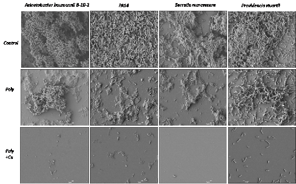
This anti-biofilm acts as an anti-adhesive and is another approach to dealing with unwanted bacteria on medical implants and on marine equipment. From an April 25, 2016 news item about the Israeli research on ScienceDaily,
Researchers at Ben-Gurion University of the Negev (BGU) have developed an innovative anti-biofilm coating, which has significant anti-adhesive potential for a variety of medical and industrial applications.
According to the research published in Advanced Materials Interfaces, anti-adhesive patches that are developed from naturally occurring biomaterials can prevent destructive bacterial biofilm from forming on metal surfaces when they are immersed in water and other damp environments.
“Our solution addresses a pervasive need to design environmentally friendly materials to impede dangerous surface bacteria growth,” the BGU researchers from the Avram and Stella Goldstein-Goren Department of Biotechnology Engineering explain. “This holds tremendous potential for averting biofilm formed by surface-anchored bacteria and could have a tremendous impact.”
Above: SEM micrographs of A. baumannii, P. aeruginosa (PA14), S. marcescens and P.stuartii biofilm architectures. The untreated control surface shows intricate bacteria densely embedded in the matrix. Biofilms were grown statically on the different surfaces.
The anti-adhesive could be used on medical implants, devices and surgical equipment where bacteria can contribute to chronic diseases, resist antibiotic treatment and thereby compromise the body’s defense system. The prevention of aquatic biofouling on ships and bridges is one of the industrial applications.
Both the University of Georgia (US) and the American Associates Ben-Gurion University of the Negev (Israel) have issued press releases about a joint research project resulting in the world’s smallest diode.
Researchers at the University of Georgia and at Ben-Gurion University in Israel have demonstrated for the first time that nanoscale electronic components can be made from single DNA molecules. Their study, published in the journal Nature Chemistry, represents a promising advance in the search for a replacement for the silicon chip.
The finding may eventually lead to smaller, more powerful and more advanced electronic devices, according to the study’s lead author, Bingqian Xu.
“For 50 years, we have been able to place more and more computing power onto smaller and smaller chips, but we are now pushing the physical limits of silicon,” said Xu, an associate professor in the UGA College of Engineering and an adjunct professor in chemistry and physics. “If silicon-based chips become much smaller, their performance will become unstable and unpredictable.”
To find a solution to this challenge, Xu turned to DNA. He says DNA’s predictability, diversity and programmability make it a leading candidate for the design of functional electronic devices using single molecules.
In the Nature Chemistry paper, Xu and collaborators at Ben-Gurion University of the Negev describe using a single molecule of DNA to create the world’s smallest diode. A diode is a component vital to electronic devices that allows current to flow in one direction but prevents its flow in the other direction.
Xu and a team of graduate research assistants at UGA isolated a specifically designed single duplex DNA of 11 base pairs and connected it to an electronic circuit only a few nanometers in size. After the measured current showed no special behavior, the team site-specifically intercalated a small molecule named coralyne into the DNA. They found the current flowing through the DNA was 15 times stronger for negative voltages than for positive voltages, a necessary feature of a diode.
“This finding is quite counterintuitive because the molecular structure is still seemingly symmetrical after coralyne intercalation,” Xu said.
A theoretical model developed by Yanantan Dubi of Ben-Gurion University indicated the diode-like behavior of DNA originates from the bias voltage-induced breaking of spatial symmetry inside the DNA molecule after the coralyne is inserted.
“Our discovery can lead to progress in the design and construction of nanoscale electronic elements that are at least 1,000 times smaller than current components,” Xu said.
The research team plans to continue its work, with the goal of constructing additional molecular devices and enhancing the performance of the molecular diode.
The world’s smallest diode, the size of a single molecule, has been developed collaboratively by U.S. and Israeli researchers from the University of Georgia and Ben-Gurion University of the Negev (BGU).
…
“Creating and characterizing the world’s smallest diode is a significant milestone in the development of molecular electronic devices,” explains Dr. Yoni Dubi, a researcher in the BGU Department of Chemistry and Ilse Katz Institute for Nanoscale Science and Technology. “It gives us new insights into the electronic transport mechanism.”
Continuous demand for more computing power is pushing the limitations of present day methods. This need is driving researchers to look for molecules with interesting properties and find ways to establish reliable contacts between molecular components and bulk materials in an electrode, in order to mimic conventional electronic elements at the molecular scale.
An example for such an element is the nanoscale diode (or molecular rectifier), which operates like a valve to facilitate electronic current flow in one direction. A collection of these nanoscale diodes, or molecules, has properties that resemble traditional electronic components such as a wire, transistor or rectifier. The emerging field of single molecule electronics may provide a way to overcome Moore’s Law– the observation that over the history of computing hardware the number of transistors in a dense integrated circuit has doubled approximately every two years – beyond the limits of conventional silicon integrated circuits.
Prof. Bingqian Xu’s group at the College of Engineering at the University of Georgia took a single DNA molecule constructed from 11 base pairs and connected it to an electronic circuit only a few nanometers in size. When they measured the current through the molecule, it did not show any special behavior. However, when layers of a molecule called “coralyne,” were inserted (or intercalated) between layers of DNA, the behavior of the circuit changed drastically. The current jumped to 15 times larger negative vs. positive voltages–a necessary feature for a nano diode. “In summary, we have constructed a molecular rectifier by intercalating specific, small molecules into designed DNA strands,” explains Prof. Xu.
Dr. Dubi and his student, Elinor Zerah-Harush, constructed a theoretical model of the DNA molecule inside the electric circuit to better understand the results of the experiment. “The model allowed us to identify the source of the diode-like feature, which originates from breaking spatial symmetry inside the DNA molecule after coralyne is inserted.”
There’s an April 4, 2016 posting on the Nanoclast blog (on the IEEE [Institute of Electrical and Electronics Engineers] website) which provides a brief overview and a link to a previous essay, Whatever Happened to the Molecular Computer?
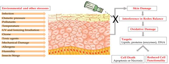
A Feb. 29, 2016 news item on ScienceDaily announces a new development concerning free radicals that may be helpful with skin diseases and pathologies,
Researchers at The Hebrew University of Jerusalem have developed a nanotechnology-based delivery system containing a protective cellular pathway inducer that activates the body’s natural defense against free radicals efficiently, a development that could control a variety of skin pathologies and disorders.
The human skin is constantly exposed to various pollutants, UV rays, radiation and other stressors that exist in our day-to-day environment. When they filter into the body they can create Reactive Oxygen Species (ROS) – oxygen molecules known as Free Radicals, which are able to damage and destroy cells, including lipids, proteins and DNA.
In the skin – the largest organ of the body – an excess of ROS can lead to various skin conditions, including inflammatory diseases, pigmenting disorders, wrinkles and some types of skin cancer, and can also affect internal organs. This damage is known as Oxidative Stress.
The body is naturally equipped with defense mechanisms to counter oxidative stress. It has anti-oxidants and, more importantly, anti-oxidant enzymes that attack the ROS before they cause damage.
In a review article published in the journal Cosmetics, a PhD student from The Hebrew University of Jerusalem, working in collaboration with researchers at the Technion – Israel Institute of Technology, suggested an innovative way to invigorate the body to produce antioxidant enzymes, while maintaining skin cell redox balance – a gentle equilibrium between Reactive Oxygen Species and their detoxification.
“The approach of using the body’s own defense system is very effective. We showed that activation of the body’s defense system with the aid of a unique delivery system is feasible, and may leverage dermal cure,” said Hebrew University researcher Maya Ben-Yehuda Greenwald.
Ben-Yehuda Greenwald showed that applying nano-size droplets of microemulsion liquids containing a cellular protective pathway inducer into the skin activates the natural skin defense systems.
“Currently, there are many scientific studies supporting the activation of the body’s defense mechanisms. However, none of these studies has demonstrated the use of a nanotechnology-based delivery system to do so,” Ben-Yehuda Greenwald said.
Production of antioxidant enzymes in the body is signaled in the DNA by activation of Nrf2 – a powerful protein that exists in every cell in our body. This Nrf2 cellular-protective signaling pathway is a major intersection of many other signaling pathways affecting each other and determining cell functionality and fate. Nrf2 is capable of coordinating the cellular response to internal as well as external stressors by tight regulation of phase-II protective enzymes, such as the antioxidant enzymes.
Ben-Yehuda Greenwald has also discovered a new family of compounds capable of activating the Nrf2 pathway. Moreover, by incorporating them into the unique delivery system she has developed, she managed to efficiently stimulate the activation of the Nrf2 pathway and mimic the activity of the body’s’ natural way of coping with a variety of stress conditions.
“The formula we have created could be used in topical medication for treating skin conditions. Our formula could be used both as preventive means and for treatment of various skin conditions, such as infections, over-exposure to UV irradiation, inflammatory conditions, and also internal disease,” she said.
While the researchers focused on the skin, the formulation could prove to be effective in enhancing the body’s natural protection against the damaging effects of ROS in other parts of the body, such as inflammation in cardiovascular diseases, heart attack, cancer, multiple sclerosis and Alzheimer’s.
Here’s an image provided by Ben-Yehuda Greenwald illustrating the team’s work,
Caption: These are the consequences of skin exposure to stressors. Credit: Maya Ben-Yehuda Greenwald
If you want to learn how something works, one strategy is to take it apart and put it back together again [also known as reverse engineering]. For 10 years, a global initiative called the Blue Brain Project–hosted at the Ecole Polytechnique Federale de Lausanne (EPFL)–has been attempting to do this digitally with a section of juvenile rat brain. The project presents a first draft of this reconstruction, which contains over 31,000 neurons, 55 layers of cells, and 207 different neuron subtypes, on October 8 [2015] in Cell.
Heroic efforts are currently being made to define all the different types of neurons in the brain, to measure their electrical firing properties, and to map out the circuits that connect them to one another. These painstaking efforts are giving us a glimpse into the building blocks and logic of brain wiring. However, getting a full, high-resolution picture of all the features and activity of the neurons within a brain region and the circuit-level behaviors of these neurons is a major challenge.
Henry Markram and colleagues have taken an engineering approach to this question by digitally reconstructing a slice of the neocortex, an area of the brain that has benefitted from extensive characterization. Using this wealth of data, they built a virtual brain slice representing the different neuron types present in this region and the key features controlling their firing and, most notably, modeling their connectivity, including nearly 40 million synapses and 2,000 connections between each brain cell type.
“The reconstruction required an enormous number of experiments,” says Markram, of the EPFL. “It paves the way for predicting the location, numbers, and even the amount of ion currents flowing through all 40 million synapses.”
Once the reconstruction was complete, the investigators used powerful supercomputers to simulate the behavior of neurons under different conditions. Remarkably, the researchers found that, by slightly adjusting just one parameter, the level of calcium ions, they could produce broader patterns of circuit-level activity that could not be predicted based on features of the individual neurons. For instance, slow synchronous waves of neuronal activity, which have been observed in the brain during sleep, were triggered in their simulations, suggesting that neural circuits may be able to switch into different “states” that could underlie important behaviors.
“An analogy would be a computer processor that can reconfigure to focus on certain tasks,” Markram says. “The experiments suggest the existence of a spectrum of states, so this raises new types of questions, such as ‘what if you’re stuck in the wrong state?'” For instance, Markram suggests that the findings may open up new avenues for explaining how initiating the fight-or-flight response through the adrenocorticotropic hormone yields tunnel vision and aggression.
The Blue Brain Project researchers plan to continue exploring the state-dependent computational theory while improving the model they’ve built. All of the results to date are now freely available to the scientific community at https://bbp.epfl.ch/nmc-portal.
Published by the renowned journal Cell, the paper is the result of a massive effort by 82 scientists and engineers at EPFL and at institutions in Israel, Spain, Hungary, USA, China, Sweden, and the UK. It represents the culmination of 20 years of biological experimentation that generated the core dataset, and 10 years of computational science work that developed the algorithms and built the software ecosystem required to digitally reconstruct and simulate the tissue.
The Hebrew University of Jerusalem’s Prof. Idan Segev, a senior author of the research paper, said: “With the Blue Brain Project, we are creating a digital reconstruction of the brain and using supercomputer simulations of its electrical behavior to reveal a variety of brain states. This allows us to examine brain phenomena within a purely digital environment and conduct experiments previously only possible using biological tissue. The insights we gather from these experiments will help us to understand normal and abnormal brain states, and in the future may have the potential to help us develop new avenues for treating brain disorders.”
Segev, a member of the Hebrew University’s Edmond and Lily Safra Center for Brain Sciences and director of the university’s Department of Neurobiology, sees the paper as building on the pioneering work of the Spanish anatomist Ramon y Cajal from more than 100 years ago: “Ramon y Cajal began drawing every type of neuron in the brain by hand. He even drew in arrows to describe how he thought the information was flowing from one neuron to the next. Today, we are doing what Cajal would be doing with the tools of the day: building a digital representation of the neurons and synapses, and simulating the flow of information between neurons on supercomputers. Furthermore, the digitization of the tissue is open to the community and allows the data and the models to be preserved and reused for future generations.”
While a long way from digitizing the whole brain, the study demonstrates that it is feasible to digitally reconstruct and simulate brain tissue, and most importantly, to reveal novel insights into the brain’s functioning. Simulating the emergent electrical behavior of this virtual tissue on supercomputers reproduced a range of previous observations made in experiments on the brain, validating its biological accuracy and providing new insights into the functioning of the neocortex. This is a first step and a significant contribution to Europe’s Human Brain Project, which Henry Markram founded, and where EPFL is the coordinating partner.
Cell has made a video abstract available (it can be found with the Hebrew University of Jerusalem press release)
Here’s a link to and a citation for the paper,
Reconstruction and Simulation of Neocortical Microcircuitry by Henry Markram, Eilif Muller, Srikanth Ramaswamy, Michael W. Reimann, Marwan Abdellah, Carlos Aguado Sanchez, Anastasia Ailamaki, Lidia Alonso-Nanclares, Nicolas Antille, Selim Arsever, Guy Antoine Atenekeng Kahou, Thomas K. Berger, Ahmet Bilgili, Nenad Buncic, Athanassia Chalimourda, Giuseppe Chindemi, Jean-Denis Courcol, Fabien Delalondre, Vincent Delattre, Shaul Druckmann, Raphael Dumusc, James Dynes, Stefan Eilemann, Eyal Gal, Michael Emiel Gevaert, Jean-Pierre Ghobril, Albert Gidon, Joe W. Graham, Anirudh Gupta, Valentin Haenel, Etay Hay, Thomas Heinis, Juan B. Hernando, Michael Hines, Lida Kanari, Daniel Keller, John Kenyon, Georges Khazen, Yihwa Kim, James G. King, Zoltan Kisvarday, Pramod Kumbhar, Sébastien Lasserre, Jean-Vincent Le Bé, Bruno R.C. Magalhães, Angel Merchán-Pérez, Julie Meystre, Benjamin Roy Morrice, Jeffrey Muller, Alberto Muñoz-Céspedes, Shruti Muralidhar, Keerthan Muthurasa, Daniel Nachbaur, Taylor H. Newton, Max Nolte, Aleksandr Ovcharenko, Juan Palacios, Luis Pastor, Rodrigo Perin, Rajnish Ranjan, Imad Riachi, José-Rodrigo Rodríguez, Juan Luis Riquelme, Christian Rössert, Konstantinos Sfyrakis, Ying Shi, Julian C. Shillcock, Gilad Silberberg, Ricardo Silva, Farhan Tauheed, Martin Telefont, Maria Toledo-Rodriguez, Thomas Tränkler, Werner Van Geit, Jafet Villafranca Díaz, Richard Walker, Yun Wang, Stefano M. Zaninetta, Javier DeFelipe, Sean L. Hill, Idan Segev, Felix Schürmann. Cell, Volume 163, Issue 2, p456–492, 8 October 2015 DOI: http://dx.doi.org/10.1016/j.cell.2015.09.029
This paper appears to be open access.
My most substantive description of the Blue Brain Project , previous to this, was in a Jan. 29, 2013 posting featuring the European Union’s (EU) Human Brain project and involvement from countries that are not members.
* I edited a redundant lede (That’s a virtual slice of a rat brain.), moved the second sentence to the lede while adding this: *about this virtual brain slice* on Oct. 16, 2015 at 0955 hours PST.
According to an August 25, 2015 news item on Nanotechnology Now, a security and defence conference (NanoSD 2015) will be held in September 2015 in Spain,
Nano for Security & Defense International Conference (NanoSD2015) will be held in Madrid, Spain (September 22-25, 2015). The conference will provide an opportunity to discuss general issues and important impacts of nanotechnology in the development of security and defense. A broad range of defense and security technologies and applications, such as nanostructures, nanosensors, nano energy sources, and nanoelectronics which are influencing these days will be discussed.
The NanoSD 2015 website notes this on its homepage,
After a first edition organised in Avila [Spain], NanoSD 2015 will again provide an opportunity to discuss general issues and important impacts of nanotechnology in the development of security and defense. …
It is evident that nanotechnology can bring many innovations into the defense world such as new innovate products, materials and power sources. Therefore, NanoSD 2015 will present current developments, research findings and relevant information on nanotechnology that will impact the security and defense.
Do not miss presentations from well known institutions
Lawrence Livermore National Laboratory (USA) | Ministry of Economy, Industry and Digital (France) | European Defence Agency (Belgium) | Metamaterial Technologies Inc. (Canada) | Graphenea (Spain) | Consiglio Nazionale delle Ricerche (Italy) | Gemalto SA (France) | ICFO (Spain) | The University of Texas at Dallas (USA) | International Commercialisation Alliance of Israel | Grupo Antolin (Spain), among others
Do not miss the opportunity to meet the key players of the Security & Defense industry. Prices starting from 350€ and 495€ for students and seniors respectively.
My most recent piece on nanotechnology and security is an Aug. 19, 2014 posting about a then upcoming NATO (North Atlantic Treaty Organization) workshop on aiding chemical and biological defenses. It took place in Sept. 2014 in Turkey.
What impact does a droplet make on a solid surface? It’s not the first question that comes to my mind but scientists have been studying it for over a century. From an Aug. 5, 2015 news item on Nanowerk (Note: A link has been removed),
Studies of the impact a droplet makes on solid surfaces hark back more than a century. And until now, it was generally believed that a droplet’s impact on a solid surface could always be separated into two phases: spreading and retracting. But it’s much more complex than that, as a team of researchers from City University of Hong Kong, Ariel University in Israel, and Dalian University of Technology in China report in the journal Applied Physics Letters, from AIP Publishing (“Controlling drop bouncing using surfaces with gradient features”).
“During the spreading phase, the droplet undergoes an inertia-dominant acceleration and spreads into a ‘pancake’ shape,” explained Zuankai Wang, an associate professor within the Department of Mechanical and Biomedical Engineering at the City University of Hong Kong. “And during the retraction phase, the drop minimizes its surface energy and pulls back inward.”
Remarkably, on gold standard superhydrophobic–a.k.a. repellant–surfaces such as lotus leaves, droplets jump off at the end of the retraction stage due to the minimal energy dissipation during the impact process. This is attributed to the presence of an air cushion within the rough surface.
There exists, however, a classical limit in terms of the contact time between droplets and the gold standard superhydrophobic materials inspired by lotus leaves.
As the team previously reported in the journal Nature Physics, it’s possible to shape the droplet to bounce from the surface in a pancake shape directly at the end of the spreading stage without going through the receding process. As a result, the droplet can be shed away much faster.
“Interestingly, the contact time is constant under a wide range of impact velocities,” said Wang. “In other words: the contact time reduction is very efficient and robust, so the novel surface behaves like an elastic spring. But the real magic lies within the surface texture itself.”
To prevent the air cushion from collapsing or water from penetrating into the surface, conventional wisdom suggests the use of nanoscale posts with small inter-post spacings. “The smaller the inter-post spacings, the greater the impact velocity the small inter-post can withstand,” he elaborated. “By contrast, designing a surface with macrostructures–tapered sub-millimeter post arrays with a wide spacing–means that a droplet will shed from it much faster than any previously engineered materials.”
What the New Results Show
Despite exciting progress, rationally controlling the contact time and quantitatively predicting the critical Weber number–a number used in fluid mechanics to describe the ratio between deforming inertial forces and stabilizing cohesive forces for liquids flowing through a fluid medium–for the occurrence of pancake bouncing remained elusive.
So the team experimentally demonstrated that the drop bouncing is intricately influenced by the surface morphology. “Under the same center-to-center post spacing, surfaces with a larger apex angle can give rise to more pancake bouncing, which is characterized by a significant contact time reduction, smaller critical Weber number, and a wider Weber number range,” according to co-authors Gene Whyman and Edward Bormashenko, both professors at Ariel University.
Wang and colleagues went on to develop simple harmonic spring models to theoretically reveal the dependence of timescales associated with the impinging drop and the critical Weber number for pancake bouncing on the surface morphology. “The insights gained from this work will allow us to rationally design various surfaces for many practical applications,” he added.
The team’s novel surfaces feature a shortened contact time that prevents or slows ice formation. “Ice formation and its subsequent buildup hinder the operation of modern infrastructures–including aircraft, offshore oil platforms, air conditioning systems, wind turbines, power lines, and telecommunications equipment,” Wang said.
At supercooled temperatures, which involves lowering the temperature of a liquid or gas below its freezing point without it solidifying, the longer a droplet remains in contact with a surface before bouncing off the greater the chances are of it freezing in place. “Our new surface structure can be used to help prevent aircraft wings and engines from icing,” he said.
This is highly desirable, because a very light coating of snow or ice–light enough to be barely visible–is known to reduce the performance of airplanes and even cause crashes. One such disaster occurred in 2009, and called attention to the dangers of in-flight icing after it caused Air France Flight 447 flying from Rio de Janeiro to Paris to crash into the Atlantic Ocean.
Beyond anti-icing for aircraft, “turbine blades in power stations and wind farms can also benefit from an anti-icing surface by gaining a boost in efficiency,” he added.
As you can imagine, this type of nature-inspired surface shows potential for a tremendous range of other applications as well–everything from water and oil separation to disease transmission prevention.
The next step for the team? To “develop bioinspired ‘active’ materials that are adaptive to their environments and capable of self-healing,” said Wang.
Finally, here’s an illustration of the pancake bounce,
Droplet hitting tapered posts shows “pancake” bouncing characterized by lifting off the surface of the end of spreading without retraction. Credit- Z.Wang/HKU
There is also a pancake bounce video which you can view here on EurekAlert.
Scientists are studying the disappearing act of this ocean-dwelling copepod. Credit: Kaj Maney, www.liquidguru.com Courtesy: American Chemical Society
Now, you’ve seen a sea sapphire. Here’s more about them and the interest they hold for experts in photonics, from a July 15, 2015 news item on ScienceDaily,
Sapphirina, or sea sapphire, has been called “the most beautiful animal you’ve never seen,” and it could be one of the most magical. Some of the tiny, little-known copepods appear to flash in and out of brilliantly colored blue, violet or red existence. Now scientists are figuring out the trick to their hues and their invisibility. The findings appear in the Journal of the American Chemical Society and could inspire the next generation of optical technologies.
Copepods are tiny aquatic crustaceans that live in both fresh and salt water. Some males of the ocean-dwelling Sapphirina genus display striking, iridescent colors that scientists think play a role in communication and mate recognition. The shimmering animals’ colors result when light bounces off of the thin, hexagonal crystal plates that cover their backs. These plates also help them vanish, if only fleetingly. Scientists didn’t know specifically what factors contributed to creating different shades. Scientists at the Weizmann Institute [Israel] and the Interuniversity Institute for Marine Sciences in Eilat [Israel] wanted to investigate the matter.
The researchers measured the light reflectance — which determines color — of live Sapphirina males and the spacing between crystal layers. They found that changes of reflectance depended on the thickness of the spacing. And for at least one particular species, when light hits an animal at a 45-degree angle, reflectance shifts out of the visible light range and into the ultraviolet, and it practically disappears. Their results could help inform the design of artificial photonic crystal structures, which have many potential uses in reflective coatings, optical mirrors and optical displays.
To sum this up, the colour and the invisibility properties are due to thin, hexagonal crystal plates and the spacing of these plates, in other words, structural colour, which is usually achieved at the nanoscale.
Here’s a link to and a citation for the paper,
Structural Basis for the Brilliant Colors of the Sapphirinid Copepods by Dvir Gur, Ben Leshem, Maria Pierantoni, Viviana Farstey, Dan Oron, Steve Weiner, and Lia Addadi. J. Am. Chem. Soc., 2015, 137 (26), pp 8408–8411 DOI: 10.1021/jacs.5b05289 Publication Date (Web): June 22, 2015
For anyone who’s interested, Lynn Kimlicka has a nice explanation of structural colour in a July 22, 2015 posting on the Something About Science blog where she discusses some recent research iridescence in bird feathers and synthetic melanin. She also shares a picture of her budgie and its iridescent feathers. The ‘melanin’ research was mentioned here in a May 19, 2015 posting where I also provide a link to a great 2013 piece on structural throughout the animal and plant kingdoms by Cristina Luiggi for The Scientist.
Understanding how nanostructures can affect optical properties could be leading to new ways of managing light. A July 23, 2015 news item on ScienceDaily describes a project at the University of Delaware dedicated to “changing the color of light,”
Researchers at the University of Delaware have received a $1 million grant from the W.M. Keck Foundation to explore a new idea that could improve solar cells, medical imaging and even cancer treatments. Simply put, they want to change the color of light.
“A ray of light contains millions and millions of individual units of light called photons,” says project leader Matthew Doty. “The energy of each photon is directly related to the color of the light — a photon of red light has less energy than a photon of blue light. You can’t simply turn a red photon into a blue one, but you can combine the energy from two or more red photons to make one blue photon.”
This process, called “photon upconversion,” isn’t new, Doty says. However, the UD team’s approach to it is.
They want to design a new kind of semiconductor nanostructure that will act like a ratchet. It will absorb two red photons, one after the other, to push an electron into an excited state when it can emit a single high-energy (blue) photon.
These nanostructures will be so teeny they can only be viewed when magnified a million times under a high-powered electron microscope.
“Think of the electrons in this structure as if they were at a water park,” Doty says. “The first red photon has only enough energy to push an electron half-way up the ladder of the water slide. The second red photon pushes it the rest of the way up. Then the electron goes down the slide, releasing all of that energy in a single process, with the emission of the blue photon. The trick is to make sure the electron doesn’t slip down the ladder before the second photon arrives. The semiconductor ratchet structure is how we trap the electron in the middle of the ladder until the second photon arrives to push it the rest of the way up.”
The UD team will develop new semiconductor structures containing multiple layers of different materials, such as aluminum arsenide and gallium bismuth arsenide, each only a few nanometers thick. This “tailored landscape” will control the flow of electrons into states with varying potential energy, turning once-wasted photons into useful energy.
The UD team has shown theoretically that their semiconductors could reach an upconversion efficiency of 86 percent, which would be a vast improvement over the 36 percent efficiency demonstrated by today’s best materials. What’s more, Doty says, the amount of light absorbed and energy emitted by the structures could be customized for a variety of applications, from lightbulbs to laser-guided surgery.
How do you even begin to make structures so tiny they can only be seen with an electron microscope? In one technique the UD team will use, called molecular beam epitaxy, nanostructures will be built by depositing layers of atoms one at a time. Each structure will be tested to see how well it absorbs and emits light, and the results will be used to tailor the structure to improve performance.
The researchers also will develop a milk-like solution filled with millions of identical individual nanoparticles, each one containing multiple layers of different materials. The multiple layers of this structure, like multiple candy shells in an M&M, will implement the photon ratchet idea. Through such work, the team envisions a future upconversion “paint” that could be easily applied to solar cells, windows and other commercial products.
Improving medical tests and treatments
While the initial focus of the three-year project will be on improving solar energy harvesting, the team also will explore biomedical applications.
A number of diagnostic tests and medical treatments, ranging from CT [computed tomography] and PET [positron emission tomography] scans to chemotherapy, rely on the release of fluorescent dyes and pharmaceutical drugs. Ideally, such payloads are delivered both at specific disease sites and at specific times, but this is hard to control in practice.
The UD team aims to develop an upconversion nanoparticle that can be triggered by light to release its payload. The goal is to achieve the controlled release of drug therapies even deep within diseased human tissue while reducing the peripheral damage to normal tissue by minimizing the laser power required.
“This is high-risk, high-reward research,” Doty says. “High-risk because we don’t yet have proof-of-concept data. High-reward because it has such a huge potential impact in renewable energy to medicine. It’s amazing to think that this same technology could be used to harvest more solar energy and to treat cancer. We’re excited to get started!”
The June 27, 2015 news item on Nanotechnology Now includes two pieces of business news (I am more interested in the second),
Knight Therapeutics Inc. (TSX:GUD) (“Knight” or the “Company”), a leading Canadian specialty pharmaceutical company, announced today that it has (1) extended a secured loan of US$15 million to Pro Bono Bio PLC (“Pro Bono Bio”), the world’s leading healthcare nanotechnology company, and (2) entered into an exclusive distribution agreement with Pro Bono Bio to commercialize its wide range of nanotechnology products, medical devices and drug delivery technologies in select territories.
The secured loan of US$15 million, which matures on June 25, 2018, will bear interest at 12% per annum plus other additional consideration. The interest rate will decrease to 10% if Pro Bono Bio meets certain equity-fundraising targets. The loan is secured by a charge over the assets of Pro Bono Bio and its affiliates which includes but is not limited to Flexiseq™, an innovative topical pain product that has sales of more than 3 million units since its U.K. launch last year.
As part of the license agreement, Knight obtained the exclusive Quebec and Israeli distribution rights to Pro Bono Bio’s innovative Flexiseq™ range of pain relief products and its promising SEQuaderma™ derma-cosmetic range of products, both of which are expected to launch in Quebec within the next 12 months. In addition, Knight obtained the exclusive Canadian and Israeli rights to two earlier stage product groups: blood factor products for the treatment of Hemophiliacs, and diagnostic devices designed for the automated detection of peripheral arterial disease. [emphasis mine]
John Mayo, Chairman and CEO of Pro Bono Bio, said, “We worked night and day to find a good distribution and strategic partner to help our North American team launch our existing products and drive growth. We welcome the good Knight on our quest to deliver to Canadian and American consumers’ best-in-class, drug-free nanotechnology products that are safe, effective and of the highest quality: truly the holy grail!”
“When you donate to charity, you always receive back more than you give. I hope this truism also holds true for this Pro Bono world!” said Jonathan Ross Goodman, President and CEO of Knight. “We look forward to the late 2015 launch of Flexiseq™ and SEQuaderma™ in La Belle Province.”
The news release also provides a description of the drugs and the companies, along with a disclaimer,
About Flexiseq™
Flexiseq™ is a topically applied drug-free gel which is clinically proven to safely relieve the pain and improve the joint stiffness associated with osteoarthritis (OA). Flexiseq™ is unique – it lubricates your joints to address joint damage. Pain is relieved and joint function improved because it lubricates away the friction and associated wear and tear on a user’s joints.
About SEQuaderma™
SEQuaderma™ Dermatology Products are a unique range of active dermatology solutions specifically designed to address the symptoms and, in some cases, the causes of the targeted conditions, leading to reduced recurrence. SEQuaderma™ Dermatology Products are suitable for long term use and can be used on their own or in between drug treatments to reduce exposure to adverse events; they will not compromise any other medication and are suitable for those with multiple conditions.
About Pro Bono Bio PLC
Pro Bono Bio PLC is the world’s leading healthcare nanotechnology company offering health and lifestyle products, headquartered in London with presence in Europe, Africa and Asia and due to launch in North America. [emphasis mine]
About Knight Therapeutics Inc.
Knight Therapeutics Inc., headquartered in Montreal, Canada, is a specialty pharmaceutical company focused on acquiring or in-licensing innovative pharmaceutical products for the Canadian and select international markets. Knight’s shares trade on TSX under the symbol GUD. For more information about Knight Therapeutics Inc., please visit the Company’s web site at www.gud-knight.com or www.sedar.com.
Forward-Looking Statement [disclaimer]
This document contains forward-looking statements for the Company and its subsidiaries. These forward looking statements, by their nature, necessarily involve risks and uncertainties that could cause actual results to differ materially from those contemplated by the forward-looking statements. The Company considers the assumptions on which these forward-looking statements are based to be reasonable at the time they were prepared, but cautions the reader that these assumptions regarding future events, many of which are beyond the control of the Company and its subsidiaries, may ultimately prove to be incorrect. Factors and risks, which could cause actual results to differ materially from current expectations are discussed in the Company’s Annual Report and in the Company’s Annual Information Form for the year ended December 31, 2014. The Company disclaims any intention or obligation to update or revise any forward-looking statements whether as a result of new information or future events, except as required by law.
Pro Bono Bio, an international pharmaceutical company, develops and commercializes new medicines in the Russian Federation. Its products include FLEXISEQ, a pain relieving gel containing absorbing nanostructures (Sequessomes) for the treatment of pain associated with osteoarthritis; EXOSEQ, which delivers Sequessomes to the upper dermal layers of the skin for the treatment of inflammatory conditions, such as eczema and seborrhoeic dermatitis; and ROSSOSEQ, which distributes Sequessome vesicles into lower dermal tissues in the skin to treat psoriasis and atopic eczema conditions. The company also develops blood products, CV diagnostics, anti-infectives, and biological drugs. Pro Bono Bio was …
Detailed Description
Moscow,
Russia
Founded in 2011
www.probonobio.com
Key Executives for Pro Bono Bio
Mr. John Mayo
Chief Executive Officer
Mr. Michael Earl
Chief Operating Officer
Compensation as of Fiscal Year 2014.
Pro Bono Bio Key Developments
Pro Bono Bio Appoints Jason Flowerday as CEO of North American Operations
Jun 26 15
Pro Bono Bio launched its North American operations with headquarters based in Toronto, Canada and secured USD 15 million in funding to accelerate the global launches of FLEXISEQ and SEQUADERMA as well as help fund its ambitious research and development programs that continue to place Pro Bono Bio at the forefront of nanotechnology healthcare development. Pro Bono Bio has recently appointed a North American CEO, Jason Flowerday, to build-out the North American operations and set its strategy for entering both the Canadian and US markets over the next three quarters.
Pro Bono Bio Launches its North American Operations
Jun 26 15
These are interesting developments for both Montréal (Québec) and Toronto (Ontario). As for whether or not Pro Bono Bio is Russian or British, I imagine the legal entity which is the company is Russian while the operations (headquarters as previously noted) are based in the UK.
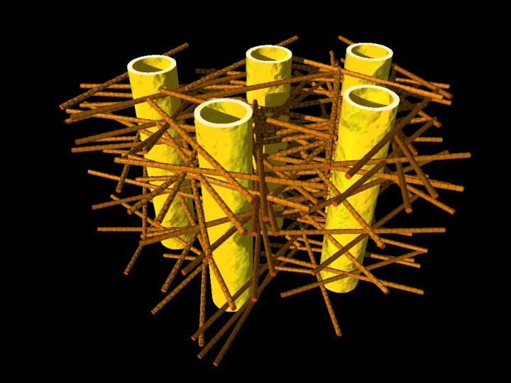
Caption: Illustration shows complex biostructure of dentin: the dental tubuli (yellow hollow cylinders, diameters appr. 1 micrometer) are surrounded by layers of mineralized collagen fibers (brown rods). The tiny mineral nanoparticles are embedded in the mesh of collagen fibers and not visible here. Credit: JB Forien @Charité
Human teeth have to serve for a lifetime, despite being subjected to huge forces. But the high failure resistance of dentin in teeth is not fully understood. An interdisciplinary team led by scientists of Charite Universitaetsmedizin Berlin has now analyzed the complex structure of dentin. At the synchrotron sources BESSY II at HZB, Berlin, Germany, and the European Synchrotron Radiation Facility ESRF, Grenoble, France, they could reveal that the mineral particles are precompressed.
The internal stress works against crack propagation and increases resistance of the biostructure.
Engineers use internal stresses to strengthen materials for specific technical purposes. Now it seems that evolution has long ‘known’ about this trick, and has put it to use in our natural teeth. Unlike bones, which are made partly of living cells, human teeth are not able to repair damage. Their bulk is made of dentin, a bonelike material consisting of mineral nanoparticles. These mineral nanoparticles are embedded in collagen protein fibres, with which they are tightly connected. In every tooth, such fibers can be found, and they lie in layers, making teeth tough and damage resistant. Still, it was not well understood, how crack propagation in teeth can be stopped.
The press release goes on to describe the new research and the teams which investigated the role of the mineral nanoparticles with regard to compression and cracking,
Now researchers from Charite Julius-Wolff-Institute, Berlin have been working with partners from Materials Engineering Department of Technische Universitaets Berlin, MPI of Colloids and Interfaces, Potsdam and Technion – Israel Institute of Technology, Haifa, to examine these biostructures more closely. They performed Micro-beam in-situ stress experiments in the mySpot BESSY facility of HZB, Berlin, Germany and analyzed the local orientation of the mineral nanoparticles using the nano-imaging facility of the European Synchrotron Radiation Facility (ESRF) in Grenoble, France.
When the tiny collagen fibers shrink, the attached mineral particles become increasingly compressed, the science team found out. “Our group was able to use changes in humidity to demonstrate how stress appears in the mineral in the collagen fibers, Dr. Paul Zaslansky from Julius Wolff-Institute of Charite Berlin explains. “The compressed state helps to prevents cracks from developing and we found that compression takes place in such a way that cracks cannot easily reach the tooth inner parts, which could damage the sensitive pulp. In this manner, compression stress helps to prevent cracks from rushing through the tooth.
The scientists also examined what happens if the tight mineral-protein link is destroyed by heating: In that case, dentin in teeth becomes much weaker. We therefore believe that the balance of stresses between the particles and the protein is important for the extended survival of teeth in the mouth, Charite scientist Jean-Baptiste Forien says. Their results may explain why artificial tooth replacements usually do not work as well as healthy teeth do: they are simply too passive, lacking the mechanisms found in the natural tooth structures, and consequently fillings cannot sustain the stresses in the mouth as well as teeth do. “Our results might inspire the development of tougher ceramic structures for tooth repair or replacement, Zaslansky hopes.
Experiments took place as part of the DFG project “Biomimetic Materials Research: Functionality by Hierarchical Structuring of Materials (SPP1420).
![[downloaded from http://www.e-architect.co.uk/israel/tel-aviv-university-center-for-nanoscience-and-nanotechnology-competition]](http://www.frogheart.ca/wp-content/uploads/2016/05/Tel-Aviv-University-Nano-Center.jpg)