Tag Archives: brain

Graphene and neurons in a UK-Italy-Spain collaboration

There’s been a lot of talk about using graphene-based implants in the brain due to the material’s flexibility along with its other properties. A step forward has been taking according to a Jan. 29, 2016 news item on phys.org,

Researchers have successfully demonstrated how it is possible to interface graphene – a two-dimensional form of carbon – with neurons, or nerve cells, while maintaining the integrity of these vital cells. The work may be used to build graphene-based electrodes that can safely be implanted in the brain, offering promise for the restoration of sensory functions for amputee or paralysed patients, or for individuals with motor disorders such as epilepsy or Parkinson’s disease.

A Jan. 29, 2016 Cambridge University press release (also on EurekAlert), which originated the news item, provides more detail,

Previously, other groups had shown that it is possible to use treated graphene to interact with neurons. However the signal to noise ratio from this interface was very low. By developing methods of working with untreated graphene, the researchers retained the material’s electrical conductivity, making it a significantly better electrode.

“For the first time we interfaced graphene to neurons directly,” said Professor Laura Ballerini of the University of Trieste in Italy. “We then tested the ability of neurons to generate electrical signals known to represent brain activities, and found that the neurons retained their neuronal signalling properties unaltered. This is the first functional study of neuronal synaptic activity using uncoated graphene based materials.”

Our understanding of the brain has increased to such a degree that by interfacing directly between the brain and the outside world we can now harness and control some of its functions. For instance, by measuring the brain’s electrical impulses, sensory functions can be recovered. This can be used to control robotic arms for amputee patients or any number of basic processes for paralysed patients – from speech to movement of objects in the world around them. Alternatively, by interfering with these electrical impulses, motor disorders (such as epilepsy or Parkinson’s) can start to be controlled.

Scientists have made this possible by developing electrodes that can be placed deep within the brain. These electrodes connect directly to neurons and transmit their electrical signals away from the body, allowing their meaning to be decoded.

However, the interface between neurons and electrodes has often been problematic: not only do the electrodes need to be highly sensitive to electrical impulses, but they need to be stable in the body without altering the tissue they measure.

Too often the modern electrodes used for this interface (based on tungsten or silicon) suffer from partial or complete loss of signal over time. This is often caused by the formation of scar tissue from the electrode insertion, which prevents the electrode from moving with the natural movements of the brain due to its rigid nature.

Graphene has been shown to be a promising material to solve these problems, because of its excellent conductivity, flexibility, biocompatibility and stability within the body.

Based on experiments conducted in rat brain cell cultures, the researchers found that untreated graphene electrodes interfaced well with neurons. By studying the neurons with electron microscopy and immunofluorescence the researchers found that they remained healthy, transmitting normal electric impulses and, importantly, none of the adverse reactions which lead to the damaging scar tissue were seen.

According to the researchers, this is the first step towards using pristine graphene-based materials as an electrode for a neuro-interface. In future, the researchers will investigate how different forms of graphene, from multiple layers to monolayers, are able to affect neurons, and whether tuning the material properties of graphene might alter the synapses and neuronal excitability in new and unique ways. “Hopefully this will pave the way for better deep brain implants to both harness and control the brain, with higher sensitivity and fewer unwanted side effects,” said Ballerini.

“We are currently involved in frontline research in graphene technology towards biomedical applications,” said Professor Maurizio Prato from the University of Trieste. “In this scenario, the development and translation in neurology of graphene-based high-performance biodevices requires the exploration of the interactions between graphene nano- and micro-sheets with the sophisticated signalling machinery of nerve cells. Our work is only a first step in that direction.”

“These initial results show how we are just at the tip of the iceberg when it comes to the potential of graphene and related materials in bio-applications and medicine,” said Professor Andrea Ferrari, Director of the Cambridge Graphene Centre. “The expertise developed at the Cambridge Graphene Centre allows us to produce large quantities of pristine material in solution, and this study proves the compatibility of our process with neuro-interfaces.”

The research was funded by the Graphene Flagship [emphasis mine],  a European initiative which promotes a collaborative approach to research with an aim of helping to translate graphene out of the academic laboratory, through local industry and into society.

Here’s a link to and a citation for the paper,

Graphene-Based Interfaces Do Not Alter Target Nerve Cells by Alessandra Fabbro, Denis Scaini, Verónica León, Ester Vázquez, Giada Cellot, Giulia Privitera, Lucia Lombardi, Felice Torrisi, Flavia Tomarchio, Francesco Bonaccorso, Susanna Bosi, Andrea C. Ferrari, Laura Ballerini, and Maurizio Prato. ACS Nano, 2016, 10 (1), pp 615–623 DOI: 10.1021/acsnano.5b05647 Publication Date (Web): December 23, 2015

Copyright © 2015 American Chemical Society

This paper is behind a paywall.

There are a couple things I found a bit odd about this project. First, all of the funding is from the Graphene Flagship initiative. I was expecting to see at least some funding from the European Union’s other mega-sized science initiative, the Human Brain Project. Second, there was no mention of Spain nor were there any quotes from the Spanish researchers. For the record, the Spanish institutions represented were: University of Castilla-La Mancha, Carbon Nanobiotechnology Laboratory, and the Basque Foundation for Science.
Nanotechnology and cybersecurity risks

Gregory Carpenter has written a gripping (albeit somewhat exaggerated) piece for Signal, a publication of the  Armed Forces Communications and Electronics Association (AFCEA) about cybersecurity issues and  nanomedicine endeavours. From Carpenter’s Jan. 1, 2016 article titled, When Lifesaving Technology Can Kill; The Cyber Edge,

The exciting advent of nanotechnology that has inspired disruptive and lifesaving medical advances is plagued by cybersecurity issues that could result in the deaths of people that these very same breakthroughs seek to heal. Unfortunately, nanorobotic technology has suffered from the same security oversights that afflict most other research and development programs.

Nanorobots, or small machines [or nanobots[, are vulnerable to exploitation just like other devices.

At the moment, the issue of cybersecurity exploitation is secondary to making nanobots, or nanorobots, dependably functional. As far as I’m aware, there is no such nanobot. Even nanoparticles meant to function as packages for drug delivery have not been perfected (see one of the controversies with nanomedicine drug delivery described in my Nov. 26, 2015 posting).

That said, Carpenter’s point about cybersecurity is well taken since security features are often overlooked in new technology. For example, automated banking machines (ABMs) had woefully poor (inadequate, almost nonexistent) security when they were first introduced.

Carpenter outlines some of the problems that could occur, assuming some of the latest research could be reliably  brought to market,

The U.S. military has joined the fray of nanorobotic experimentation, embarking on revolutionary research that could lead to a range of discoveries, from unraveling the secrets of how brains function to figuring out how to permanently purge bad memories. Academia is making amazing advances as well. Harnessing progress by Harvard scientists to move nanorobots within humans, researchers at the University of Montreal, Polytechnique Montreal and Centre Hospitalier Universitaire Sainte-Justine are using mobile nanoparticles inside the human brain to open the blood-brain barrier, which protects the brain from toxins found in the circulatory system.

A different type of technology presents a risk similar to the nanoparticles scenario. A DARPA-funded program known as Restoring Active Memory (RAM) addresses post-traumatic stress disorder, attempting to overcome memory deficits by developing neuroprosthetics that bridge gaps in an injured brain. In short, scientists can wipe out a traumatic memory, and they hope to insert a new one—one the person has never actually experienced. Someone could relish the memory of a stroll along the French Riviera rather than a terrible firefight, even if he or she has never visited Europe.

As an individual receives a disruptive memory, a cyber criminal could manage to hack the controls. Breaches of the brain could become a reality, putting humans at risk of becoming zombie hosts [emphasis mine] for future virus deployments. …

At this point, the ‘zombie’ scenario Carpenter suggests seems a bit over-the-top but it does hearken to the roots of the zombie myth where the undead aren’t mindlessly searching for brains but are humans whose wills have been overcome. Mike Mariani in an Oct. 28, 2015 article for The Atlantic has presented a thought-provoking history of zombies,

… the zombie myth is far older and more rooted in history than the blinkered arc of American pop culture suggests. It first appeared in Haiti in the 17th and 18th centuries, when the country was known as Saint-Domingue and ruled by France, which hauled in African slaves to work on sugar plantations. Slavery in Saint-Domingue under the French was extremely brutal: Half of the slaves brought in from Africa were worked to death within a few years, which only led to the capture and import of more. In the hundreds of years since, the zombie myth has been widely appropriated by American pop culture in a way that whitewashes its origins—and turns the undead into a platform for escapist fantasy.

The original brains-eating fiend was a slave not to the flesh of others but to his own. The zombie archetype, as it appeared in Haiti and mirrored the inhumanity that existed there from 1625 to around 1800, was a projection of the African slaves’ relentless misery and subjugation. Haitian slaves believed that dying would release them back to lan guinée, literally Guinea, or Africa in general, a kind of afterlife where they could be free. Though suicide was common among slaves, those who took their own lives wouldn’t be allowed to return to lan guinée. Instead, they’d be condemned to skulk the Hispaniola plantations for eternity, an undead slave at once denied their own bodies and yet trapped inside them—a soulless zombie.

I recommend reading Mariani’s article although I do have one nit to pick. I can’t find a reference to brain-eating zombies until George Romero’s introduction of the concept in his movies. This Zombie Wikipedia entry seems to be in agreement with my understanding (if I’m wrong, please do let me know and, if possible, provide a link to the corrective text).

Getting back to Carpenter and cybersecurity with regard to nanomedicine, while his scenarios may seem a trifle extreme it’s precisely the kind of thinking you need when attempting to anticipate problems. I do wish he’d made clear that the technology still has a ways to go.

Titanium dioxide nanoparticles and the brain

This research into titanium dioxide nanoparticles and possible effects on your brain should they pass the blood-brain barrier comes from the University of Nebraska-Lincoln (US) according to a Dec. 15, 2015 news item on Nanowerk (Note: A link has been removed),

Even moderate concentrations of a nanoparticle used to whiten certain foods, milk and toothpaste could potentially compromise the brain’s most numerous cells, according to a new study from the University of Nebraska-Lincoln (Nanoscale, “Mitochondrial dysfunction and loss of glutamate uptake in primary astrocytes exposed to titanium dioxide nanoparticles”).

A Dec. 14, 2015 University of Nebraska-Lincoln news release, which originated the news item, provides more detail (Note: Links have been removed),

The researchers examined how three types of titanium dioxide nanoparticles [rutile, anatase, and commercially available P25 TiO2 nanoparticles], the world’s second-most abundant nanomaterial, affected the functioning of astrocyte cells. Astrocytes help regulate the exchange of signal-carrying neurotransmitters in the brain while also supplying energy to the neurons that process those signals, among many other functions.

The team exposed rat-derived astrocyte cells to nanoparticle concentrations well below the extreme levels that have been shown to kill brain cells but are rarely encountered by humans. At the study’s highest concentration of 100 parts per million, or PPM, two of the nanoparticle types still killed nearly two-thirds of the astrocytes within a day. That mortality rate fell to between half and one-third of cells at 50 PPM, settling to about one-quarter at 25 PPM.

Yet the researchers found evidence that even surviving cells are severely impaired by exposure to titanium dioxide nanoparticles. Astrocytes normally take in and process a neurotransmitter called glutamate that plays wide-ranging roles in cognition, memory and learning, along with the formation, migration and maintenance of other cells.

When allowed to accumulate outside cells, however, glutamate becomes a potent toxin that kills neurons and may increase the risk of neurodegenerative diseases such as Alzheimer’s and Parkinson’s. The study reported that one of the nanoparticle types reduced the astrocytes’ uptake of glutamate by 31 percent at concentrations of just 25 PPM. Another type decreased that uptake by 45 percent at 50 PPM.

The team further discovered that the nanoparticles upset the intricate balance of protein dynamics occurring within astrocytes’ mitochondria, the cellular organelles that help regulate energy production and contribute to signaling among cells. Titanium dioxide exposure also led to other signs of mitochondrial distress, breaking apart a significant proportion of the mitochondrial network at 100 PPM.

“These events are oftentimes predecessors of cell death,” said Oleh Khalimonchuk, a UNL assistant professor of biochemistry who co-authored the study. “Usually, people are looking at those ultimate consequences, but what happens before matters just as much. Those little damages add up over time. Ultimately, they’re going to cause a major problem.”

Khalimonchuk and fellow author Srivatsan Kidambi, assistant professor of chemical and biomolecular engineering, cautioned that more research is needed to determine whether titanium dioxide nanoparticles can avoid digestion and cross the blood-brain barrier that blocks the passage of many substances. [emphasis mine]

However, the researchers cited previous studies that have discovered these nanoparticles in the brain tissue of animals with similar blood-brain barriers. [emphasis mine] The concentrations of nanoparticles found in those specimens served as a reference point for the levels examined in the new study.

“There’s evidence building up now that some of these particles can actually cross the (blood-brain) barrier,” Khalimonchuk said. “Few molecules seem to be able to do so, but it turns out that there are certain sites in the brain where you can get this exposure.”

Kidambi said the team hopes the study will help facilitate further research on the presence of nanoparticles in consumer and industrial products.

“We’re hoping that this study will get some discussion going, because these nanoparticles have not been regulated,” said Kidambi, who also holds a courtesy appointment with the University of Nebraska Medical Center. “If you think about anything white – milk, chewing gum, toothpaste, powdered sugar – all these have nanoparticles in them.

“We’ve found that some nanoparticles are safe and some are not, so we are not saying that all of them are bad. Our reasoning is that … we need to have a classification of ‘safe’ versus ‘not safe,’ along with concentration thresholds (for each type). It’s about figuring out how the different forms affect the biology of cells.

I notice the researchers are being careful about alarming anyone unduly while emphasizing the importance of this research. For anyone curious enough to read the paper, here’s a link to and a citation for it,

Mitochondrial dysfunction and loss of glutamate uptake in primary astrocytes exposed to titanium dioxide nanoparticles by Christina L. Wilson, Vaishaali Natarajan, Stephen L. Hayward, Oleh Khalimonchuk and   Srivatsan Kidambi. Nanoscale, 2015,7, 18477-18488 DOI: 10.1039/C5NR03646A First published online 31 Jul 2015

This is paper is open access although you may need to register on the site.

Final comment, I note this was published online way back in July 2015. Either the paper version of the journal was just published and that’s what’s being promoted or the media people thought they’d try to get some attention for this work by reissuing the publicity. Good on them! It’s hard work getting people to notice things when there is so much information floating around.

New tool for mapping neuronal connections in the brain

This work comes from the US Naval Research Laboratory according to a Nov. 17, 2015 news item on Nanowerk (Note: A link has been removed),

Research biologists, chemists and theoreticians at the U.S. Naval Research Laboratory (NRL), are on pace to develop the next generation of functional materials that could enable the mapping of the complex neural connections in the brain (“Electric Field Modulation of Semiconductor Quantum Dot Photoluminescence: Insights Into the Design of Robust Voltage-Sensitive Cellular Imaging Probes”). The ultimate goal is to better understand how the billions of neurons in the brain communicate with one another during normal brain function, or dysfunction, as result of injury or disease.

“There is tremendous interest in mapping all the neuron connections in the human brain,” said Dr. James Delehanty, research biologist, Center for Biomolecular Science and Engineering. “To do that we need new tools or materials that allow us to see how large groups of neurons communicate with one another while, at the same time, being able to focus in on a single neuron’s activity. Our most recent work potentially opens the integration of voltage-sensitive nanomaterials into live cells and tissues in a variety of configurations to achieve real-time imaging capabilities not currently possible.”

A Nov. 17, 2015 US Naval Research Laboratory (NRL) news release on EurekAlert, which originated the news item, provides more details,

The basis of neuron communication is the time-dependent modulation of the strength of the electric field that is maintained across the cell’s plasma membrane. This is called an action potential. Among the nanomaterials under consideration for application in neuronal action potential imaging are quantum dots (QDs) — crystalline semiconductor nanomaterials possessing a number of advantageous photophysical attributes.

“QDs are very bright and photostable so you can look at them for long times and they allow for tissue imaging configurations that are not compatible with current materials, for example, organic dyes,” Delehanty added. “Equally important, we’ve shown here that QD brightness tracks, with very high fidelity, the time-resolved electric field strength changes that occur when a neuron undergoes an action potential. Their nanoscale size make them ideal nanoscale voltage sensing materials for interfacing with neurons and other electrically active cells for voltage sensing.”

QDs are small, bright, photo-stable materials that possess nanosecond fluorescence lifetimes. They can be localized within or on cellular plasma membranes and have low cytotoxicity when interfaced with experimental brain systems. Additionally, QDs possess two-photon action cross-section orders of magnitude larger than organic dyes or fluorescent proteins. Two-photon imaging is the preferred imaging modality for imaging deep (millimeters) into the brain and other tissues of the body.

In their most recent work, the NRL researchers showed that an electric field typical of those found in neuronal membranes results in suppression of the QD photoluminescence (PL) and, for the first time, that QD PL is able to track the action potential profile of a firing neuron with millisecond time resolution. This effect is shown to be connected with electric-field-driven QD ionization and consequent QD PL quenching, in contradiction with conventional wisdom that suppression of the QD PL is attributable to the quantum confined Stark effect — the shifting and splitting of spectral lines of atoms and molecules due to presence of an external electric field.

“The inherent superior photostability properties of QDs coupled with their voltage sensitivity could prove advantageous to long-term imaging capabilities that are not currently attainable using traditional organic voltage sensitive dyes,” Delehanty said. “We anticipate that continued research will facilitate the rational design and synthesis of voltage-sensitive QD probes that can be integrated in a variety of imaging configurations for the robust functional imaging and sensing of electrically active cells.”

Here’s a link to and a citation for the paper,

Electric Field Modulation of Semiconductor Quantum Dot Photoluminescence: Insights Into the Design of Robust Voltage-Sensitive Cellular Imaging Probes by Clare E. Rowland, Kimihiro Susumu, Michael H. Stewart, Eunkeu Oh, Antti J. Mäkinen, Thomas J. O’Shaughnessy, Gary Kushto, Mason A. Wolak, Jeffrey S. Erickson, Alexander L. Efros, Alan L. Huston, and James B. Delehanty. Nano Lett., 2015, 15 (10), pp 6848–6854 DOI: 10.1021/acs.nanolett.5b02725 Publication Date (Web): September 28, 2015

Copyright © 2015 American Chemical Society

This paper is behind a paywall.

Blue Brain Project builds a digital piece of brain

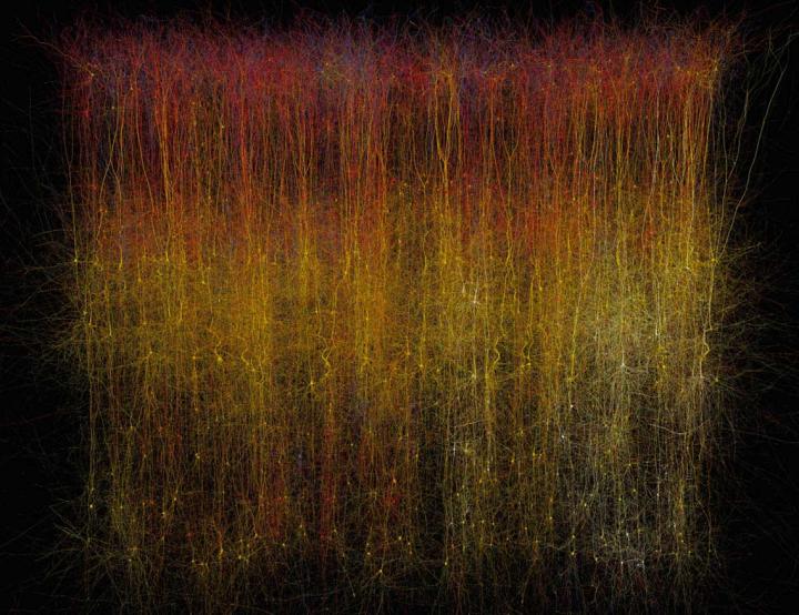
Caption: This is a photo of a virtual brain slice. Credit: Makram et al./Cell 2015

Caption: This is a photo of a virtual brain slice. Credit: Makram et al./Cell 2015

Here’s more *about this virtual brain slice* from an Oct. 8, 2015 Cell (magazine) news release on EurekAlert,

If you want to learn how something works, one strategy is to take it apart and put it back together again [also known as reverse engineering]. For 10 years, a global initiative called the Blue Brain Project–hosted at the Ecole Polytechnique Federale de Lausanne (EPFL)–has been attempting to do this digitally with a section of juvenile rat brain. The project presents a first draft of this reconstruction, which contains over 31,000 neurons, 55 layers of cells, and 207 different neuron subtypes, on October 8 [2015] in Cell.

Heroic efforts are currently being made to define all the different types of neurons in the brain, to measure their electrical firing properties, and to map out the circuits that connect them to one another. These painstaking efforts are giving us a glimpse into the building blocks and logic of brain wiring. However, getting a full, high-resolution picture of all the features and activity of the neurons within a brain region and the circuit-level behaviors of these neurons is a major challenge.

Henry Markram and colleagues have taken an engineering approach to this question by digitally reconstructing a slice of the neocortex, an area of the brain that has benefitted from extensive characterization. Using this wealth of data, they built a virtual brain slice representing the different neuron types present in this region and the key features controlling their firing and, most notably, modeling their connectivity, including nearly 40 million synapses and 2,000 connections between each brain cell type.

“The reconstruction required an enormous number of experiments,” says Markram, of the EPFL. “It paves the way for predicting the location, numbers, and even the amount of ion currents flowing through all 40 million synapses.”

Once the reconstruction was complete, the investigators used powerful supercomputers to simulate the behavior of neurons under different conditions. Remarkably, the researchers found that, by slightly adjusting just one parameter, the level of calcium ions, they could produce broader patterns of circuit-level activity that could not be predicted based on features of the individual neurons. For instance, slow synchronous waves of neuronal activity, which have been observed in the brain during sleep, were triggered in their simulations, suggesting that neural circuits may be able to switch into different “states” that could underlie important behaviors.

“An analogy would be a computer processor that can reconfigure to focus on certain tasks,” Markram says. “The experiments suggest the existence of a spectrum of states, so this raises new types of questions, such as ‘what if you’re stuck in the wrong state?'” For instance, Markram suggests that the findings may open up new avenues for explaining how initiating the fight-or-flight response through the adrenocorticotropic hormone yields tunnel vision and aggression.

The Blue Brain Project researchers plan to continue exploring the state-dependent computational theory while improving the model they’ve built. All of the results to date are now freely available to the scientific community at https://bbp.epfl.ch/nmc-portal.

An Oct. 8, 2015 Hebrew University of Jerusalem press release on the Canadian Friends of the Hebrew University of Jerusalem website provides more detail,

Published by the renowned journal Cell, the paper is the result of a massive effort by 82 scientists and engineers at EPFL and at institutions in Israel, Spain, Hungary, USA, China, Sweden, and the UK. It represents the culmination of 20 years of biological experimentation that generated the core dataset, and 10 years of computational science work that developed the algorithms and built the software ecosystem required to digitally reconstruct and simulate the tissue.

The Hebrew University of Jerusalem’s Prof. Idan Segev, a senior author of the research paper, said: “With the Blue Brain Project, we are creating a digital reconstruction of the brain and using supercomputer simulations of its electrical behavior to reveal a variety of brain states. This allows us to examine brain phenomena within a purely digital environment and conduct experiments previously only possible using biological tissue. The insights we gather from these experiments will help us to understand normal and abnormal brain states, and in the future may have the potential to help us develop new avenues for treating brain disorders.”

Segev, a member of the Hebrew University’s Edmond and Lily Safra Center for Brain Sciences and director of the university’s Department of Neurobiology, sees the paper as building on the pioneering work of the Spanish anatomist Ramon y Cajal from more than 100 years ago: “Ramon y Cajal began drawing every type of neuron in the brain by hand. He even drew in arrows to describe how he thought the information was flowing from one neuron to the next. Today, we are doing what Cajal would be doing with the tools of the day: building a digital representation of the neurons and synapses, and simulating the flow of information between neurons on supercomputers. Furthermore, the digitization of the tissue is open to the community and allows the data and the models to be preserved and reused for future generations.”

While a long way from digitizing the whole brain, the study demonstrates that it is feasible to digitally reconstruct and simulate brain tissue, and most importantly, to reveal novel insights into the brain’s functioning. Simulating the emergent electrical behavior of this virtual tissue on supercomputers reproduced a range of previous observations made in experiments on the brain, validating its biological accuracy and providing new insights into the functioning of the neocortex. This is a first step and a significant contribution to Europe’s Human Brain Project, which Henry Markram founded, and where EPFL is the coordinating partner.

Cell has made a video abstract available (it can be found with the Hebrew University of Jerusalem press release)

Here’s a link to and a citation for the paper,

Reconstruction and Simulation of Neocortical Microcircuitry by Henry Markram, Eilif Muller, Srikanth Ramaswamy, Michael W. Reimann, Marwan Abdellah, Carlos Aguado Sanchez, Anastasia Ailamaki, Lidia Alonso-Nanclares, Nicolas Antille, Selim Arsever, Guy Antoine Atenekeng Kahou, Thomas K. Berger, Ahmet Bilgili, Nenad Buncic, Athanassia Chalimourda, Giuseppe Chindemi, Jean-Denis Courcol, Fabien Delalondre, Vincent Delattre, Shaul Druckmann, Raphael Dumusc, James Dynes, Stefan Eilemann, Eyal Gal, Michael Emiel Gevaert, Jean-Pierre Ghobril, Albert Gidon, Joe W. Graham, Anirudh Gupta, Valentin Haenel, Etay Hay, Thomas Heinis, Juan B. Hernando, Michael Hines, Lida Kanari, Daniel Keller, John Kenyon, Georges Khazen, Yihwa Kim, James G. King, Zoltan Kisvarday, Pramod Kumbhar, Sébastien Lasserre, Jean-Vincent Le Bé, Bruno R.C. Magalhães, Angel Merchán-Pérez, Julie Meystre, Benjamin Roy Morrice, Jeffrey Muller, Alberto Muñoz-Céspedes, Shruti Muralidhar, Keerthan Muthurasa, Daniel Nachbaur, Taylor H. Newton, Max Nolte, Aleksandr Ovcharenko, Juan Palacios, Luis Pastor, Rodrigo Perin, Rajnish Ranjan, Imad Riachi, José-Rodrigo Rodríguez, Juan Luis Riquelme, Christian Rössert, Konstantinos Sfyrakis, Ying Shi, Julian C. Shillcock, Gilad Silberberg, Ricardo Silva, Farhan Tauheed, Martin Telefont, Maria Toledo-Rodriguez, Thomas Tränkler, Werner Van Geit, Jafet Villafranca Díaz, Richard Walker, Yun Wang, Stefano M. Zaninetta, Javier DeFelipe, Sean L. Hill, Idan Segev, Felix Schürmann. Cell, Volume 163, Issue 2, p456–492, 8 October 2015 DOI: http://dx.doi.org/10.1016/j.cell.2015.09.029

This paper appears to be open access.

My most substantive description of the Blue Brain Project , previous to this, was in a Jan. 29, 2013 posting featuring the European Union’s (EU) Human Brain project and involvement from countries that are not members.

* I edited a redundant lede (That’s a virtual slice of a rat brain.), moved the second sentence to the lede while adding this:  *about this virtual brain slice* on Oct. 16, 2015 at 0955 hours PST.

Nanoscale imaging of a mouse brain

Researchers have developed a new brain imaging tool they would like to use as a founding element for a national brain observatory. From a July 30, 2015 news item on Azonano,

A new imaging tool developed by Boston scientists could do for the brain what the telescope did for space exploration.

In the first demonstration of how the technology works, published July 30 in the journal Cell, the researchers look inside the brain of an adult mouse at a scale previously unachievable, generating images at a nanoscale resolution. The inventors’ long-term goal is to make the resource available to the scientific community in the form of a national brain observatory.

A July 30, 2015 Cell Press news release on EurekAlert, which originated the news item, expands on the theme,

“I’m a strong believer in bottom up-science, which is a way of saying that I would prefer to generate a hypothesis from the data and test it,” says senior study author Jeff Lichtman, of Harvard University. “For people who are imagers, being able to see all of these details is wonderful and we’re getting an opportunity to peer into something that has remained somewhat intractable for so long. It’s about time we did this, and it is what people should be doing about things we don’t understand.”

The researchers have begun the process of mining their imaging data by looking first at an area of the brain that receives sensory information from mouse whiskers, which help the animals orient themselves and are even more sensitive than human fingertips. The scientists used a program called VAST, developed by co-author Daniel Berger of Harvard and the Massachusetts Institute of Technology, to assign different colors and piece apart each individual “object” (e.g., neuron, glial cell, blood vessel cell, etc.).

“The complexity of the brain is much more than what we had ever imagined,” says study first author Narayanan “Bobby” Kasthuri, of the Boston University School of Medicine. “We had this clean idea of how there’s a really nice order to how neurons connect with each other, but if you actually look at the material it’s not like that. The connections are so messy that it’s hard to imagine a plan to it, but we checked and there’s clearly a pattern that cannot be explained by randomness.”

The researchers see great potential in the tool’s ability to answer questions about what a neurological disorder actually looks like in the brain, as well as what makes the human brain different from other animals and different between individuals. Who we become is very much a product of the connections our neurons make in response to various life experiences. To be able to compare the physical neuron-to-neuron connections in an infant, a mathematical genius, and someone with schizophrenia would be a leap in our understanding of how our brains shape who we are (or vice versa).

The cost and data storage demands for this type of research are still high, but the researchers expect expenses to drop over time (as has been the case with genome sequencing). To facilitate data sharing, the scientists are now partnering with Argonne National Laboratory with the hopes of creating a national brain laboratory that neuroscientists around the world can access within the next few years.

“It’s bittersweet that there are many scientists who think this is a total waste of time as well as a big investment in money and effort that could be better spent answering questions that are more proximal,” Lichtman says. “As long as data is showing you things that are unexpected, then you’re definitely doing the right thing. And we are certainly far from being out of the surprise element. There’s never a time when we look at this data that we don’t see something that we’ve never seen before.”

Here’s a link to and a citation for the paper,

Saturated Reconstruction of a Volume of Neocortex by Narayanan Kasthuri, Kenneth Jeffrey Hayworth, Daniel Raimund Berger, Richard Lee Schalek, José Angel Conchello, Seymour Knowles-Barley, Dongil Lee, Amelio Vázquez-Reina, Verena Kaynig, Thouis Raymond Jones, Mike Roberts, Josh Lyskowski Morgan, Juan Carlos Tapia, H. Sebastian Seung, William Gray Roncal, Joshua Tzvi Vogelstein, Randal Burns, Daniel Lewis Sussman, Carey Eldin Priebe, Hanspeter Pfister, Jeff William Lichtman. Cell Volume 162, Issue 3, p648–661, 30 July 2015 DOI: http://dx.doi.org/10.1016/j.cell.2015.06.054

This appears to be an open access paper.

Brain data (neuroscience) crowdsourced at Toronto’s (Canada) 2013 Nuit Blanche event

The brain data was crowdsourced in 2013 in Toronto but only recently published according to a July 8, 2015 Baycrest Centre for Geriatric Care news release (also on EurekAlert),

Neuroscientists in Toronto have shown that crowdsourcing brain data with hundreds of adults in a short period of time could be a new frontier in neuroscience and lead to new insights about the brain.

More than 500 adults aged 18 and older participated in the experiment at the 2013 Scotiabank Nuit Blanche arts event in Toronto. Baycrest, in partnership with the University of Toronto and industry partners, created a large-scale art-science installation called My Virtual Dream. Festival-goers were invited to wear a Muse™ wireless electroencephalography (EEG) headband and participate in a brief collective neurofeedback experience in groups of 20 inside a 60-foot geodesic dome. The group’s collective EEG signals triggered a specific catalogue of artistic imagery displayed on the dome’s 360-degree interior, along with spontaneous musical interpretation by live musicians on stage.

The installation was one of the most popular at Nuit Blanche, with an average lineup wait time of two hours.

Studying brains in a social and multi-sensory environment is closer to real life and may help scientists to approach questions of complex real-life social cognition that otherwise are not accessible in traditional labs that study one person’s cognitive functions at a time.

“In traditional lab settings, the environment is so controlled that you can lose some of the fine points of real-time brain activity that occur in a social life setting,” said Dr. Kovacevic, creative producer of My Virtual Dream and program manager of the Centre for Integrative Brain Dynamics at Baycrest’s Rotman Research Institute.

“What we’ve done is taken the lab to the public. We collaborated with multi-media artists, made this experiment incredibly engaging, attracted highly motivated subjects which is not easy to do in the traditional lab setting, and collected useful scientific data from their experience.”

Results from the experiment not only demonstrated the scientific viability of collective neurofeedback as a potential new avenue of neuroscience research that takes into account individuality, complexity and sociability of the human mind, but yielded new evidence that neurofeedback learning can have an effect on the brain almost immediately.

Neurofeedback learning supports mindful awareness and joins a growing market for wearable biofeedback devices. The device used in this study, Muse™, is a clinical-grade EEG brain computer interface (BCI) headband that helps individuals to be more aware of their brain states (relaxed versus focused versus distracted) and learn self-regulation of brain function to fit their personal goals.

A total of 523 adults (209 males, 314 females), ranging in age from 18 to 89, with an average age of 31, contributed their EEG brain data for the study. Each session involved 20 participants being seated in a semicircle in front of a stage and divided into four groups (“pods”) of five. They played a collective neurofeedback computer game where they were required to manipulate their mental states of relaxation and concentration. The neurofeedback training lasted 6.5 minutes, which is much shorter than typical neurofeedback training experiments.

The massive amount of EEG data collected in one night yielded an interesting and statistically relevant finding – that subtle brain activity changes were taking place within approximately one minute of the neurofeedback learning exercise – unprecedented speed of learning changes that have not been demonstrated before.

“These results really open up a whole new domain of neuroscience study that actively engages the public to advance our understanding of the brain,” said Dr. Randy McIntosh, director of the Rotman Research Institute and vice-president of Research at Baycrest. He is a senior author on the paper.

The idea for the Nuit Blanche art -science experiment was inspired by Baycrest’s ongoing international project to build the world’s first functional, virtual brain – a research and diagnostic tool that could one day revolutionize brain healthcare.

Baycrest cognitive neuroscientists collaborated with artists and gaming and wearable technology industry partners for over a year to create the My Virtual Dream installation. Partners included the University of Toronto, Scotiabank Nuit Blanche, Muse™ and Uken Games.

Here’s a link to and a citation for the paper,

‘My Virtual Dream’: Collective Neurofeedback in an Immersive Art Environment by Natasha Kovacevic, Petra Ritter, William Tays, Sylvain Moreno, and Anthony Randal McIntosh. DOI: 10.1371/journal.pone.0130129 PLOS Published: July 8, 2015

This is an open access paper.

A few final words, I last wrote about MUSE (a Canadian technology company) in a March 6, 2015 posting. Uken Games , also a Canadian company, is new to this blog.

Injectable electronics

Having taught a course on bioelectronics for Simon Fraser University’s (Vancouver, Canada) Continuing Studies Program, this  latest work from Harvard University (US) caught my attention. A Harvard research team has developed a technique which could allow doctors to inject us with electronics, should we need them. From a June 8, 2015 news item on phys.org,

It’s a notion that might be pulled from the pages of science-fiction novel – electronic devices that can be injected directly into the brain, or other body parts, and treat everything from neurodegenerative disorders to paralysis.

It sounds unlikely, until you visit Charles Lieber’s lab.

A team of international researchers, led by Lieber, the Mark Hyman, Jr. Professor of Chemistry, an international team of researchers developed a method for fabricating nano-scale electronic scaffolds that can be injected via syringe. Once connected to electronic devices, the scaffolds can be used to monitor neural activity, stimulate tissues and even promote regenerations of neurons. …

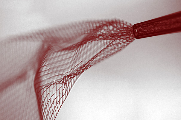
Here’s an image provided by the researchers,

Bright-field image showing the mesh electronics being injected through sub-100 micrometer inner diameter glass needle into aqueous solution. mage courtesy of Lieber Research Group, Harvard University

Bright-field image showing the mesh electronics being injected through sub-100 micrometer inner diameter glass needle into aqueous solution. mage courtesy of Lieber Research Group, Harvard University

A June 8, 2015 Harvard University new release by Peter Reuell (also on EurekAlert), which originated the news item, describes the work in more detail,

“I do feel that this has the potential to be revolutionary,” Lieber said. “This opens up a completely new frontier where we can explore the interface between electronic structures and biology. For the past thirty years, people have made incremental improvements in micro-fabrication techniques that have allowed us to make rigid probes smaller and smaller, but no one has addressed this issue – the electronics/cellular interface – at the level at which biology works.”

The idea of merging the biological with the electronic is not a new one for Lieber.

In an earlier study, scientists in Lieber’s lab demonstrated that the scaffolds could be used to create “cyborg” tissue – when cardiac or nerve cells were grown with embedded scaffolds. [emphasis mine] Researchers were then able to use the devices to record electrical signals generated by the tissues, and to measure changes in those signals as they administered cardio- or neuro-stimulating drugs.

“We were able to demonstrate that we could make this scaffold and culture cells within it, but we didn’t really have an idea how to insert that into pre-existing tissue,” Lieber said. “But if you want to study the brain or develop the tools to explore the brain-machine interface, you need to stick something into the body. When releasing the electronics scaffold completely from the fabrication substrate, we noticed that it was almost invisible and very flexible like a polymer and could literally be sucked into a glass needle or pipette. From there, we simply asked, would it be possible to deliver the mesh electronics by syringe needle injection, a process common to delivery of many species in biology and medicine – you could go to the doctor and you inject this and you’re wired up.'”

Though not the first attempts at implanting electronics into the brain – deep brain stimulation has been used to treat a variety of disorders for decades – the nano-fabricated scaffolds operate on a completely different scale.

“Existing techniques are crude relative to the way the brain is wired,” Lieber explained. “Whether it’s a silicon probe or flexible polymers…they cause inflammation in the tissue that requires periodically changing the position or the stimulation. But with our injectable electronics, it’s as if it’s not there at all. They are one million times more flexible than any state-of-the-art flexible electronics and have subcellular feature sizes. They’re what I call “neuro-philic” – they actually like to interact with neurons..”

Despite their enormous potential, the fabrication of the injectable scaffolds is surprisingly easy.

“That’s the beauty of this – it’s compatible with conventional manufacturing techniques,” Lieber said.

The process is similar to that used to etch microchips, and begins with a dissolvable layer deposited on a substrate. To create the scaffold, researchers lay out a mesh of nanowires sandwiched in layers of organic polymer. The first layer is then dissolved, leaving the flexible mesh, which can be drawn into a syringe needle and administered like any other injection.

After injection, the input/output of the mesh can be connected to standard measurement electronics so that the integrated devices can be addressed and used to stimulate or record neural activity.

“These type of things have never been done before, from both a fundamental neuroscience and medical perspective,” Lieber said. “It’s really exciting – there are a lot of potential applications.”

Going forward, Lieber said, researchers hope to better understand how the brain and other tissues react to the injectable electronics over longer periods.

Lieber’s earlier work on “cyborg tissue” was briefly mentioned here in a Feb. 20, 2014 posting.

Getting back to the most recent work, here’s a link to and a citation for the paper,

Syringe-injectable electronics by Jia Liu, Tian-Ming Fu, Zengguang Cheng, Guosong Hong, Tao Zhou, Lihua Jin, Madhavi Duvvuri, Zhe Jiang, Peter Kruskal, Chong Xie, Zhigang Suo, Ying Fang, & Charles M. Lieber. Nature Nanotechnology (2015) doi:10.1038/nnano.2015.115 Published online 08 June 2015

This paper is behind a paywall but there is a free preview via ReadCube Access.

One final note, the researchers have tested the injectable electronics (or mesh electronics) in vivo (live animals).

A more complex memristor: from two terminals to three for brain-like computing

Researchers have developed a more complex memristor device than has been the case according to an April 6, 2015 Northwestern University news release (also on EurekAlert),

Researchers are always searching for improved technologies, but the most efficient computer possible already exists. It can learn and adapt without needing to be programmed or updated. It has nearly limitless memory, is difficult to crash, and works at extremely fast speeds. It’s not a Mac or a PC; it’s the human brain. And scientists around the world want to mimic its abilities.

Both academic and industrial laboratories are working to develop computers that operate more like the human brain. Instead of operating like a conventional, digital system, these new devices could potentially function more like a network of neurons.

“Computers are very impressive in many ways, but they’re not equal to the mind,” said Mark Hersam, the Bette and Neison Harris Chair in Teaching Excellence in Northwestern University’s McCormick School of Engineering. “Neurons can achieve very complicated computation with very low power consumption compared to a digital computer.”

A team of Northwestern researchers, including Hersam, has accomplished a new step forward in electronics that could bring brain-like computing closer to reality. The team’s work advances memory resistors, or “memristors,” which are resistors in a circuit that “remember” how much current has flowed through them.

“Memristors could be used as a memory element in an integrated circuit or computer,” Hersam said. “Unlike other memories that exist today in modern electronics, memristors are stable and remember their state even if you lose power.”

Current computers use random access memory (RAM), which moves very quickly as a user works but does not retain unsaved data if power is lost. Flash drives, on the other hand, store information when they are not powered but work much slower. Memristors could provide a memory that is the best of both worlds: fast and reliable. But there’s a problem: memristors are two-terminal electronic devices, which can only control one voltage channel. Hersam wanted to transform it into a three-terminal device, allowing it to be used in more complex electronic circuits and systems.

The memristor is of some interest to a number of other parties prominent amongst them, the University of Michigan’s Professor Wei Lu and HP (Hewlett Packard) Labs, both of whom are mentioned in one of my more recent memristor pieces, a June 26, 2014 post.

Getting back to Northwestern,

Hersam and his team met this challenge by using single-layer molybdenum disulfide (MoS2), an atomically thin, two-dimensional nanomaterial semiconductor. Much like the way fibers are arranged in wood, atoms are arranged in a certain direction–called “grains”–within a material. The sheet of MoS2 that Hersam used has a well-defined grain boundary, which is the interface where two different grains come together.

“Because the atoms are not in the same orientation, there are unsatisfied chemical bonds at that interface,” Hersam explained. “These grain boundaries influence the flow of current, so they can serve as a means of tuning resistance.”

When a large electric field is applied, the grain boundary literally moves, causing a change in resistance. By using MoS2 with this grain boundary defect instead of the typical metal-oxide-metal memristor structure, the team presented a novel three-terminal memristive device that is widely tunable with a gate electrode.

“With a memristor that can be tuned with a third electrode, we have the possibility to realize a function you could not previously achieve,” Hersam said. “A three-terminal memristor has been proposed as a means of realizing brain-like computing. We are now actively exploring this possibility in the laboratory.”

Here’s a link to and a citation for the paper,

Gate-tunable memristive phenomena mediated by grain boundaries in single-layer MoS2 by Vinod K. Sangwan, Deep Jariwala, In Soo Kim, Kan-Sheng Chen, Tobin J. Marks, Lincoln J. Lauhon, & Mark C. Hersam. Nature Nanotechnology (2015) doi:10.1038/nnano.2015.56 Published online 06 April 2015

This paper is behind a paywall but there is a few preview available through ReadCube Access.

Dexter Johnson has written about this latest memristor development in an April 9, 2015 posting on his Nanoclast blog (on the IEEE [Institute for Electrical and Electronics Engineers] website) where he notes this (Note: A link has been removed),

The memristor seems to generate fairly polarized debate, especially here on this website in the comments on stories covering the technology. The controversy seems to fall along the lines that the device that HP Labs’ Stan Williams and Greg Snider developed back in 2008 doesn’t exactly line up with the original theory of the memristor proposed by Leon Chua back in 1971.

It seems the ‘debate’ has evolved from issues about how the memristor is categorized. I wonder if there’s still discussion about whether or not HP Labs is attempting to develop a patent thicket of sorts.

Brain Talks: Robotics and Rehabilitation at Vancouver (Canada) General Hospital

The latest Brain Talk will take place tomorrow, Jan. 21, 2015 at 6 pm at Vancouver (Canada) General Hospital. More logistical details follow this description of the talk (from the Robotics and Rehabilitation webpage),

Presenter:  AJung Moon, Nick Snow, and Navid Shirzad

As interactive robots become substantially more accessible to the general public in the near future, one of the main concerns for designers  is in implementing socially acceptable and ethical human-robot interaction for non-expert users.  One approach to addressing this concern is to develop a robot that can take advantage of human moral decision making – much of which are suggested to be based on intuition and strongly connected with the emotional part of the human brain, rather than the rational part of the brain.  In this talk, A. Jung will present the promising, yet cautionary, tales of the moral synergy robots and humans can create.

Speaker details:

  • AJung Moon, PhD Candidate in Mechanical Engineering, Vanier Scholar, CARIS Lab: Robo-Ethics
  • Nick Snow, Masters Candidate in Rehabilitation Sciences, Brain Behaviour Lab: Robo-Wrist Rehabilitation
  • Navid Shirzad, PhD Candidate in Biomedical Engineering, RREACH Lab: Rehabilitation Robotics

Here are the details,

6:00pm-8:00pm, Jan 21, 2015
Paetzold Auditorium, Jim Pattison Pavilion North, 899 West 12th Avenue, Vancouver, BC

Free wine and cheese reception to follow

Please RSVP here.

Maybe I’ll see you there, eh?