Tag Archives: 3D printing

Feasibility of printing ear, bone, and muscle structures

Over ten years ago I attended a show at the Vancouver (Canada) Art Gallery titled ‘Massive Change’ where I saw part of a nose or ear being grown in a petri dish (the work was from an Israeli laboratory) and that was my introduction to tissue engineering. For anyone who’s been following the tissue engineering story, 3D printers have sped up the growth process considerably. More recently, researchers at Wake Forest Baptist Medical Center (North Carolina, US) have announced another step forward for growing organs and body parts, from a Feb. 15, 2016 Wake Forest Baptist Medical Center news release on EurekAlert,

Using a sophisticated, custom-designed 3D printer, regenerative medicine scientists at Wake Forest Baptist Medical Center have proved that it is feasible to print living tissue structures to replace injured or diseased tissue in patients.

Reporting in Nature Biotechnology, the scientists said they printed ear, bone and muscle structures. When implanted in animals, the structures matured into functional tissue and developed a system of blood vessels. Most importantly, these early results indicate that the structures have the right size, strength and function for use in humans.

“This novel tissue and organ printer is an important advance in our quest to make replacement tissue for patients,” said Anthony Atala, M.D., director of the Wake Forest Institute for Regenerative Medicine (WFIRM) and senior author on the study. “It can fabricate stable, human-scale tissue of any shape. With further development, this technology could potentially be used to print living tissue and organ structures for surgical implantation.”

With funding from the Armed Forces Institute of Regenerative Medicine, a federally funded effort to apply regenerative medicine to battlefield injuries, Atala’s team aims to implant bioprinted muscle, cartilage and bone in patients in the future.

Tissue engineering is a science that aims to grow replacement tissues and organs in the laboratory to help solve the shortage of donated tissue available for transplants. The precision of 3D printing makes it a promising method for replicating the body’s complex tissues and organs. However, current printers based on jetting, extrusion and laser-induced forward transfer cannot produce structures with sufficient size or strength to implant in the body.

The Integrated Tissue and Organ Printing System (ITOP), developed over a 10-year period by scientists at the Institute for Regenerative Medicine, overcomes these challenges. The system deposits both bio-degradable, plastic-like materials to form the tissue “shape” and water-based gels that contain the cells. In addition, a strong, temporary outer structure is formed. The printing process does not harm the cells.

A major challenge of tissue engineering is ensuring that implanted structures live long enough to integrate with the body. The Wake Forest Baptist scientists addressed this in two ways. They optimized the water-based “ink” that holds the cells so that it promotes cell health and growth and they printed a lattice of micro-channels throughout the structures. These channels allow nutrients and oxygen from the body to diffuse into the structures and keep them live while they develop a system of blood vessels.

It has been previously shown that tissue structures without ready-made blood vessels must be smaller than 200 microns (0.007 inches) for cells to survive. In these studies, a baby-sized ear structure (1.5 inches) survived and showed signs of vascularization at one and two months after implantation.

“Our results indicate that the bio-ink combination we used, combined with the micro-channels, provides the right environment to keep the cells alive and to support cell and tissue growth,” said Atala.

Another advantage of the ITOP system is its ability to use data from CT and MRI scans to “tailor-make” tissue for patients. For a patient missing an ear, for example, the system could print a matching structure.

Several proof-of-concept experiments demonstrated the capabilities of ITOP. To show that ITOP can generate complex 3D structures, printed, human-sized external ears were implanted under the skin of mice. Two months later, the shape of the implanted ear was well-maintained and cartilage tissue and blood vessels had formed.

To demonstrate the ITOP can generate organized soft tissue structures, printed muscle tissue was implanted in rats. After two weeks, tests confirmed that the muscle was robust enough to maintain its structural characteristics, become vascularized and induce nerve formation.

And, to show that construction of a human-sized bone structure, jaw bone fragments were printed using human stem cells. The fragments were the size and shape needed for facial reconstruction in humans. To study the maturation of bioprinted bone in the body, printed segments of skull bone were implanted in rats. After five months, the bioprinted structures had formed vascularized bone tissue.

Ongoing studies will measure longer-term outcomes.

###

The research was supported, in part, by grants from the Armed Forces Institute of Regenerative Medicine (W81XWH-08-2-0032), the Telemedicine and Advanced Technology Research Center at the U.S. Army Medical Research and Material Command (W81XWH-07-1-0718) and the Defense Threat Reduction Agency (N66001-13-C-2027).

(Sometimes the information about the funding agencies is almost as interesting as the research.) Here’s a link to and a citation for the paper,

A 3D bioprinting system to produce human-scale tissue constructs with structural integrity by Hyun-Wook Kang, Sang Jin Lee, In Kap Ko, Carlos Kengla, James J Yoo, & Anthony Atala. Nature Biotechnology (2016)  doi:10.1038/nbt.3413 Published online 15 February 2016

This paper is behind a paywall.

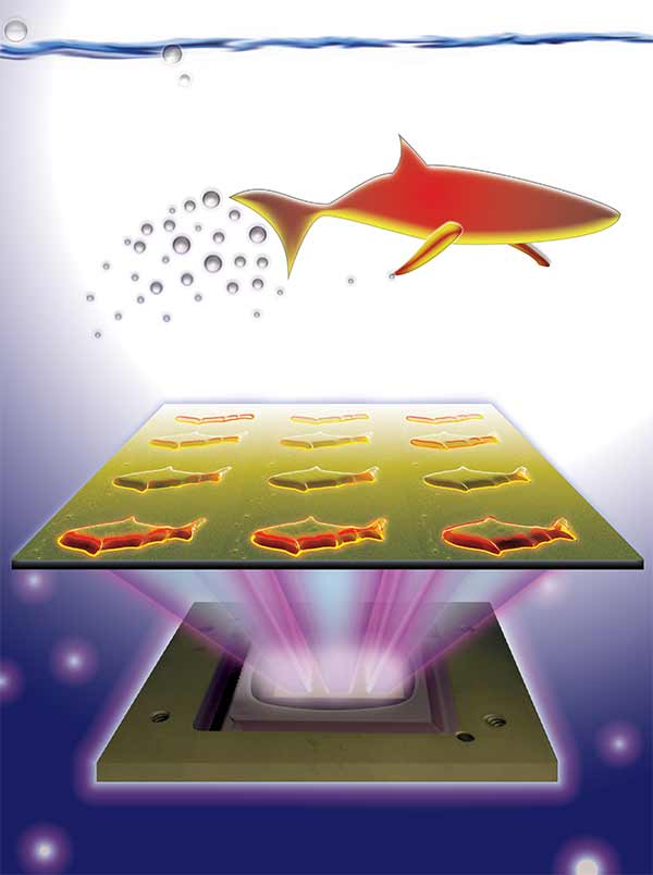
As you can see, despite being printed, this latest ear is also spending time in a dish,

WakeBaptistEar

Courtesy: Wake Forest Baptist Medical Center

Synthetic microfish (nanoengineered and 3D printed) to inspire ‘smart’ microbots

An August 26, 2015 news item on Nanowerk features some microfish (they look like sharks to me) fabricated in University of California at San Diego (UCSD) laboratories,

Nanoengineers at the University of California, San Diego used an innovative 3D printing technology they developed to manufacture multipurpose fish-shaped microrobots — called microfish — that swim around efficiently in liquids, are chemically powered by hydrogen peroxide and magnetically controlled. These proof-of-concept synthetic microfish will inspire a new generation of “smart” microrobots that have diverse capabilities such as detoxification, sensing and directed drug delivery, researchers said.

3D-printed microfish contain functional nanoparticles that enable them to be self-propelled, chemically powered and magnetically steered. The microfish are also capable of removing and sensing toxins. Image credit: J. Warner, UC San Diego Jacobs School of Engineering.

3D-printed microfish contain functional nanoparticles that enable them to be self-propelled, chemically powered and magnetically steered. The microfish are also capable of removing and sensing toxins. Image credit: J. Warner, UC San Diego Jacobs School of Engineering.

An August 25, 2015 UCSD news release (also on EurekAlert) by Liezel Labios, which originated the news item, provides more detail,

The technique used to fabricate the microfish provides numerous improvements over other methods traditionally employed to create microrobots with various locomotion mechanisms, such as microjet engines, microdrillers and microrockets. Most of these microrobots are incapable of performing more sophisticated tasks because they feature simple designs — such as spherical or cylindrical structures — and are made of homogeneous inorganic materials. In this new study, researchers demonstrated a simple way to create more complex microrobots.

By combining Chen’s 3D printing technology with Wang’s expertise in microrobots, the team was able to custom-build microfish that can do more than simply swim around when placed in a solution containing hydrogen peroxide. Nanoengineers were able to easily add functional nanoparticles into certain parts of the microfish bodies. They installed platinum nanoparticles in the tails, which react with hydrogen peroxide to propel the microfish forward, and magnetic iron oxide nanoparticles in the heads, which allowed them to be steered with magnets.

Here’s an illustration of the platinum and iron oxide microfish,

Schematic illustration of the process of functionalizing the microfish. Platinum nanoparticles are first loaded into the tail of the fish for propulsion via reaction with hydrogen peroxide. Next, iron oxide nanoparticles are loaded into the head of the fish for magnetic control. Image credit: W. Zhu and J. Li, UC San Diego Jacobs School of Engineering.

Schematic illustration of the process of functionalizing the microfish. Platinum nanoparticles are first loaded into the tail of the fish for propulsion via reaction with hydrogen peroxide. Next, iron oxide nanoparticles are loaded into the head of the fish for magnetic control. Image credit: W. Zhu and J. Li, UC San Diego Jacobs School of Engineering.

Back to the news release,

“We have developed an entirely new method to engineer nature-inspired microscopic swimmers that have complex geometric structures and are smaller than the width of a human hair. With this method, we can easily integrate different functions inside these tiny robotic swimmers for a broad spectrum of applications,” said the co-first author Wei Zhu, a nanoengineering Ph.D. student in Chen’s research group at the Jacobs School of Engineering at UC San Diego.

As a proof-of-concept demonstration, the researchers incorporated toxin-neutralizing nanoparticles throughout the bodies of the microfish. Specifically, the researchers mixed in polydiacetylene (PDA) nanoparticles, which capture harmful pore-forming toxins such as the ones found in bee venom. The researchers noted that the powerful swimming of the microfish in solution greatly enhanced their ability to clean up toxins. When the PDA nanoparticles bind with toxin molecules, they become fluorescent and emit red-colored light. The team was able to monitor the detoxification ability of the microfish by the intensity of their red glow.

“The neat thing about this experiment is that it shows how the microfish can doubly serve as detoxification systems and as toxin sensors,” said Zhu.

“Another exciting possibility we could explore is to encapsulate medicines inside the microfish and use them for directed drug delivery,” said Jinxing Li, the other co-first author of the study and a nanoengineering Ph.D. student in Wang’s research group.

For anyone curious about the new 3D printing technique, the news release provides more information about that too,

The new microfish fabrication method is based on a rapid, high-resolution 3D printing technology called microscale continuous optical printing (μCOP), which was developed in Chen’s lab. Some of the benefits of the μCOP technology are speed, scalability, precision and flexibility. Within seconds, the researchers can print an array containing hundreds of microfish, each measuring 120 microns long and 30 microns thick. This process also does not require the use of harsh chemicals. Because the μCOP technology is digitized, the researchers could easily experiment with different designs for their microfish, including shark and manta ray shapes. [emphasis mine] “With our 3D printing technology, we are not limited to just fish shapes. We can rapidly build microrobots inspired by other biological organisms such as birds,” said Zhu.

The key component of the μCOP technology is a digital micromirror array device (DMD) chip, which contains approximately two million micromirrors. Each micromirror is individually controlled to project UV light in the desired pattern (in this case, a fish shape) onto a photosensitive material, which solidifies upon exposure to UV light. The microfish are built using a photosensitive material and are constructed one layer at a time, allowing each set of functional nanoparticles to be “printed” into specific parts of the fish bodies.

“This method has made it easier for us to test different designs for these microrobots and to test different nanoparticles to insert new functional elements into these tiny structures. It’s my personal hope to further this research to eventually develop surgical microrobots that operate safer and with more precision,” said Li.

Nice to see I can recognize a shark shape when I see one. Getting back to the research, yet again, here’s a link to and a citation for the paper.

3D-Printed Artificial Microfish by Wei Zhu, Jinxing Li, Yew J. Leong, Isaac Rozen, Xin Qu, Renfeng Dong, Zhiguang Wu, Wei Gao, Peter H. Chung, Joseph Wang, and Shaochen Chen. Advanced Materials Volume 27, Issue 30, pages 4411–4417, August 12, 2015 DOI: 10.1002/adma.201501372 Article first published online: 29 JUN 2015

© 2015 WILEY-VCH Verlag GmbH & Co. KGaA, Weinheim

This paper is behind a paywall.

Canada and some graphene scene tidbits

For a long time It seemed as if every country in the world, except Canada, had some some sort of graphene event. According to a July 16, 2015 news item on Nanotechnology Now, Canada has now stepped up, albeit, in a peculiarly Canadian fashion. First the news,

Mid October [Oct. 14 -16, 2015], the Graphene & 2D Materials Canada 2015 International Conference & Exhibition (www.graphenecanada2015.com) will take place in Montreal (Canada).

I found a July 16, 2015 news release (PDF) announcing the Canadian event on the lead organizer’s (Phantoms Foundation located in Spain) website,

On the second day of the event (15th October, 2015), an Industrial Forum will bring together top industry leaders to discuss recent advances in technology developments and business opportunities in graphene commercialization.
At this stage, the event unveils 38 keynote & invited speakers. On the Industrial Forum 19 of them will present the latest in terms of Energy, Applications, Production and Worldwide Initiatives & Priorities.

Plenary:
Gary Economo (Grafoid Inc., Canada)
Khasha Ghaffarzadeh (IDTechEx, UK)
Shu-Jen Han (IBM T.J. Watson Research Center, USA)
Bor Z. Jang (Angstron Materials, USA)
Seongjun Park (Samsung Advanced Institute of Technology (SAIT), Korea)
Chun-Yun Sung (Lockheed Martin, USA)

Parallel Sessions:
Gordon Chiu (Grafoid Inc., Canada)
Jesus de la Fuente (Graphenea, Spain)
Mark Gallerneault (ALCERECO Inc., Canada)
Ray Gibbs (Haydale Graphene Industries, UK)
Masataka Hasegawa (AIST, Japan)
Byung Hee Hong (SNU & Graphene Square, Korea)
Tony Ling (Jestico + Whiles, UK)
Carla Miner (SDTC, Canada)
Gregory Pognon (THALES Research & Technology, France)
Elena Polyakova (Graphene Laboratories Inc, USA)
Federico Rosei (INRS–EMT, Université du Québec, Canada)
Aiping Yu (University of Waterloo, Canada)
Hua Zhang (MSE-NTU, Singapore)

Apart from the industrial forum, several industry-related activities will be organized:
– Extensive thematic workshops in parallel (Standardization, Materials & Devices Characterization, Bio & Health and Electronic Devices)
– An exhibition carried out with the latest graphene trends (Grafoid, RAYMOR NanoIntegris, Nanomagnetics Instruments, ICEX and Xerox Research Centre of Canada (XRCC) already confirmed)
– B2B meetings to foster technical cooperation in the field of Graphene

It’s still possible to contribute to the event with an oral presentation. The call for abstracts is open until July, 20 [2015]. [emphasis mine]

Graphene Canada 2015 is already supported by Canada’s leading graphene applications developer, Grafoid Inc., Tourisme Montréal and Université de Montréal.

This is what makes the event peculiarly Canadian: multiculturalism, anyone? From the news release,

Organisers: Phantoms Foundation www.phantomsnet.net & Grafoid Foundation (lead organizers)

CEMES/CNRS (France) | Grafoid (Canada) | Catalan Institute of Nanoscience and Nanotechnology – ICN2 (Spain) | IIT (Italy) | McGill University, Canada | Texas Instruments (USA) | Université Catholique de Louvain (Belgium) | Université de Montreal, Canada

It’s billed as a ‘Canada Graphene 2015’ and, as I recall, these types of events don’t usually have so many other countries listed as organizers. For example, UK Graphene 2015 would have mostly or all of its organizers (especially the leads) located in the UK.

Getting to the Canadian content, I wrote about Grafoid at length tracking some of its relationships to companies it owns, a business deal with Hydro Québec, and a partnership with the University of Waterloo, and a nonrepayable grant from the Canadian federal government (Sustainable Development Technology Canada [SDTC]) in a Feb. 23, 2015 posting. Do take a look at the post if you’re curious about the heavily interlinked nature of the Canadian graphene scene and take another look at the list of speakers and their agencies (Mark Gallerneault of ALCERECO [partially owned by Grafoid], Carla Miner of SDTC [Grafoid received monies from the Canadian federal department],  Federico Rosei of INRS–EMT, Université du Québec [another Quebec link], Aiping Yu, University of Waterloo [an academic partner to Grafoid]). The Canadian graphene community is a small one so it’s not surprising there are links between the Canadian speakers but it does seem odd that Lomiko Metals is not represented here. Still, new speakers have been announced since the news release (e.g., Frank Koppens of ICFO, Spain, and Vladimir Falko of Lancaster University, UK) so  time remains.

Meanwhile, Lomiko Metals has announced in a July 17, 2015 news item on Azonano that Graphene 3D labs has changed the percentage of its outstanding shares affecting the percentage that Lomiko owns, amid some production and distribution announcements. The bit about launching commercial sales of its graphene filament seems more interesting to me,

On March 16, 2015 Graphene 3D Lab (TSXV:GGG) (OTCQB:GPHBF) announced that it launched commercial sales of its Conductive Graphene Filament for 3D printing. The filament incorporates highly conductive proprietary nano-carbon materials to enhance the properties of PLA, a widely used thermoplastic material for 3D printing; therefore, the filament is compatible with most commercially available 3D printers. The conductive filament can be used to print conductive traces (similar to as used in circuit boards) within 3D printed parts for electronics.

So, that’s all I’ve got for Canada’s graphene scene.

Cosmetics giant, L’Oréal, to 3D print skin

L’Oréal, according to a May 19, 2015 BBC (British Broadcasting Corporation) online news item, has partnered with Organovo, a 3D bioprinting startup, to begin producing skin,

French cosmetics firm L’Oreal is teaming up with bio-engineering start-up Organovo to 3D-print human skin.

It said the printed skin would be used in product tests.

Organovo has already made headlines with claims that it can 3D-print a human liver but this is its first tie-up with the cosmetics industry.

Experts said the science might be legitimate but questioned why a beauty firm would want to print skin. [emphasis mine]

L’Oreal currently grows skin samples from tissues donated by plastic surgery patients. It produces more than 100,000, 0.5 sq cm skin samples per year and grows nine varieties across all ages and ethnicities.

Its statement explaining the advantage of printing skin, offered little detail: “Our partnership will not only bring about new advanced in vitro methods for evaluating product safety and performance, but the potential for where this new field of technology and research can take us is boundless.”

The beauty and cosmetics industry has a major interest in technology, especially anything to do with the skin. I’m curious as to what kind of an expert wouldn’t realize that cosmetics companies test products on skin and might like to have a ready supply. Still, I have to admit to surprise when I first (2006) started researching nanotechnology;  L’Oréal at one point was the sixth largest nanotechnology patent holder in the US (see my Nanotech Mysteries Wiki page: Marketers put the buy in nano [scroll down to Penetration subhead]). In 2008 L’Oréal company representatives were set for a discussion on their nanotechnology efforts and the precautionary principle, which was to be hosted by the Wilson Center’s Project for Emerging Nanotechnologies (PEN). The company cancelled at a rather interesting time as I had noted in my June 19, 2008 posting. (scroll down about 40% of the way until you see mention of Dr. Andrew Maynard).

Back to 3D printing technology and cosmetics giants, a May 5, 2015 Organovo/L’Oréal press release provides more detail about the deal,

L’Oreal USA, the largest subsidiary of the world’s leading beauty company, has announced a partnership with 3-D bioprinting company Organovo Holdings, Inc. (NYSE MKT: ONVO) (“Organovo”).  Developed between L’Oreal’s U.S.-based global Technology Incubator and Organovo, the collaboration will leverage Organovo’s proprietary NovoGen Bioprinting Platform and L’Oreal’s expertise in skin engineering to develop 3-D printed skin tissue for product evaluation and other areas of advanced research.

This partnership marks the first-ever application of Organovo’s groundbreaking technology within the beauty industry.

“We developed our technology incubator to uncover disruptive innovations across industries that have the potential to transform the beauty business,” said Guive Balooch, Global Vice President of L’Oreal’s Technology Incubator.  “Organovo has broken new ground with 3-D bioprinting, an area that complements L’Oreal’s pioneering work in the research and application of reconstructed skin for the past 30 years. Our partnership will not only bring about new advanced in vitro methods for evaluating product safety and performance, but the potential for where this new field of technology and research can take us is boundless.”

Organovo’s 3D bioprinting enables the reproducible, automated creation of living human tissues that mimic the form and function of native tissues in the body.

“We are excited to be partnering with L’Oreal, whose leadership in the beauty industry is rooted in scientific innovation and a deep commitment to research and development,” said Keith Murphy, Chairman and Chief Executive Officer at Organovo. “This partnership is a great next step to expand the applications of Organovo’s 3-D bioprinting technology and to create value for both L’Oreal and Organovo by building new breakthroughs in skin modeling.”

I don’t have much information about Organovo here, certainly nothing about the supposed liver (how did I miss that?), but there is a Dec. 26, 2012 posting about its deal with software giant, Autodesk.

Water-soluble 3D filament from Graphene 3D Lab

This is almost a Canadian nanotech story since one of the company’s chief investors is Canadian and the announcement is being made at an event held at Mohawk College in Hamilton, Ontario. From an April 30, 2015 news item on Azonano,

… Graphene 3D Lab Inc. will announce details of a newly-developed water-soluble 3D filament at the Canadian Manufacturers & Exporters’ (CME) Canada Makes: Additive Manufacturing Forum at Mohawk College in Hamilton, Ontario on April 30, 2015. The water-soluble filament is the latest in a line of specialty-functional filaments that Graphene 3D is introducing to the 3D printing industry.

An April 30, 2015 Graphene 3D Lab news release provides more detail,

Elena Polyakova, Chief Operating Officer of Graphene 3D, will present details on the new water-soluble filament and discuss the effect of graphene-enhanced materials for the 3D printing industry.

Water-soluble filaments are primarily used to occupy negative space during a 3D print. These filaments are essentially used to fill gaps designed in objects and allow the print to be suspended over air. Following the completion of the printing process, the object can be placed in water, and the water-soluble material will completely dissolve, leaving empty space.

Daniel Stolyarov, President and Chief Executive Officer, stated, “We are very pleased with performance characteristics that our R&D team designed into this new industry leading filament. This filament is completely environmentally-friendly, non-toxic, dissolves completely in water in approximately 1 hour, and leaves no residual material.” The water-soluble filament will work in most commercially available 3D printers and Graphene 3D expects to have them to market within 3 months. Daniel continues, “This filament reflects the core objectives of Graphene 3D. We are creating products that allow innovative designers to do more with 3D printing and are expanding the possibilities within the 3D printing environment.”

About Graphene 3D
Graphene 3D is in the business of developing, manufacturing and marketing specialty 3D printer materials, including proprietary nanocomposite materials for various types of 3D printing. The Company is also involved in the design, manufacture and marketing of 3D printers and related products for domestic and international customers.

The Graphene 3D research and production facility is located in Calverton, NY and is equipped with material processing and analytical equipment. The company has four US patent applications pending for its technology. For more information on Graphene 3D Lab Inc., visit www.graphene3dlab.com.

As for Canadian content (beyond the announcement location), the news item on Azonano notes,

Lomiko Metals 100%-owned subsidiary Lomiko Technologies Inc. has and will continue to hold its 4,396,100 share investment in Graphene 3D Lab for the long term.

Lomiko Metals is located in the Vancouver metro region of Canadian province British Columbia.

3D printing soft robots and flexible electronics with metal alloys

This research comes from Purdue University (Indiana, US) which seems to be on a publishing binge these days. From an April 7, 2015 news item on Nanowerk,

New research shows how inkjet-printing technology can be used to mass-produce electronic circuits made of liquid-metal alloys for “soft robots” and flexible electronics.

Elastic technologies could make possible a new class of pliable robots and stretchable garments that people might wear to interact with computers or for therapeutic purposes. However, new manufacturing techniques must be developed before soft machines become commercially feasible, said Rebecca Kramer, an assistant professor of mechanical engineering at Purdue University.

“We want to create stretchable electronics that might be compatible with soft machines, such as robots that need to squeeze through small spaces, or wearable technologies that aren’t restrictive of motion,” she said. “Conductors made from liquid metal can stretch and deform without breaking.”

A new potential manufacturing approach focuses on harnessing inkjet printing to create devices made of liquid alloys.

“This process now allows us to print flexible and stretchable conductors onto anything, including elastic materials and fabrics,” Kramer said.

An April 7, 2015 Purdue University news release (also on EurekAlert) by Emil Venere, which originated the news item, expands on the theme,

A research paper about the method will appear on April 18 [2015] in the journal Advanced Materials. The paper generally introduces the method, called mechanically sintered gallium-indium nanoparticles, and describes research leading up to the project. It was authored by postdoctoral researcher John William Boley, graduate student Edward L. White and Kramer.

A printable ink is made by dispersing the liquid metal in a non-metallic solvent using ultrasound, which breaks up the bulk liquid metal into nanoparticles. This nanoparticle-filled ink is compatible with inkjet printing.

“Liquid metal in its native form is not inkjet-able,” Kramer said. “So what we do is create liquid metal nanoparticles that are small enough to pass through an inkjet nozzle. Sonicating liquid metal in a carrier solvent, such as ethanol, both creates the nanoparticles and disperses them in the solvent. Then we can print the ink onto any substrate. The ethanol evaporates away so we are just left with liquid metal nanoparticles on a surface.”

After printing, the nanoparticles must be rejoined by applying light pressure, which renders the material conductive. This step is necessary because the liquid-metal nanoparticles are initially coated with oxidized gallium, which acts as a skin that prevents electrical conductivity.

“But it’s a fragile skin, so when you apply pressure it breaks the skin and everything coalesces into one uniform film,” Kramer said. “We can do this either by stamping or by dragging something across the surface, such as the sharp edge of a silicon tip.”

The approach makes it possible to select which portions to activate depending on particular designs, suggesting that a blank film might be manufactured for a multitude of potential applications.

“We selectively activate what electronics we want to turn on by applying pressure to just those areas,” said Kramer, who this year was awarded an Early Career Development award from the National Science Foundation, which supports research to determine how to best develop the liquid-metal ink.

The process could make it possible to rapidly mass-produce large quantities of the film.

Future research will explore how the interaction between the ink and the surface being printed on might be conducive to the production of specific types of devices.

“For example, how do the nanoparticles orient themselves on hydrophobic versus hydrophilic surfaces? How can we formulate the ink and exploit its interaction with a surface to enable self-assembly of the particles?” she said.

The researchers also will study and model how individual particles rupture when pressure is applied, providing information that could allow the manufacture of ultrathin traces and new types of sensors.

Here’s a link to and a citation for the paper,

Nanoparticles: Mechanically Sintered Gallium–Indium Nanoparticles by John William Boley, Edward L. White and Rebecca K. Kramer. Advanced Materials Volume 27, Issue 14, page 2270, April 8, 2015 DOI: 10.1002/adma.201570094 Article first published online: 7 APR 2015

© 2015 WILEY-VCH Verlag GmbH & Co. KGaA, Weinheim

This article is behind a paywall.

Projecting beams of light from contact lenses courtesy of Princeton University (US)

Princeton University’s 3D printed contact lenses with LED (light-emitting diodes) included are not meant for use by humans or other living beings but they are a flashy demonstration. From a Dec. 10, 2014 news item on phys.org,

As part of a project demonstrating new 3-D printing techniques, Princeton researchers have embedded tiny light-emitting diodes into a standard contact lens, allowing the device to project beams of colored light.

Michael McAlpine, the lead researcher, cautioned that the lens is not designed for actual use—for one, it requires an external power supply. Instead, he said the team created the device to demonstrate the ability to “3-D print” electronics into complex shapes and materials.

“This shows that we can use 3-D printing to create complex electronics including semiconductors,” said McAlpine, an assistant professor of mechanical and aerospace engineering. “We were able to 3-D print an entire device, in this case an LED.”

A Dec. 9, 2014 Princeton University news release by John Sullivan, which originated the news item, describes the 3D lens, the objectives for this project, and an earlier project involving a ‘bionic ear’ in more detail (Note: Links have been removed),

The hard contact lens is made of plastic. The researchers used tiny crystals, called quantum dots, to create the LEDs that generated the colored light. Different size dots can be used to generate various colors.

“We used the quantum dots [also known as nanoparticles] as an ink,” McAlpine said. “We were able to generate two different colors, orange and green.”

The contact lens is also part of an ongoing effort to use 3-D printing to assemble diverse, and often hard-to-combine, materials into functioning devices. In the recent past, a team of Princeton professors including McAlpine created a bionic ear out of living cells with an embedded antenna that could receive radio signals.

Yong Lin Kong, a researcher on both projects, said the bionic ear presented a different type of challenge.

“The main focus of the bionic ear project was to demonstrate the merger of electronics and biological materials,” said Kong, a graduate student in mechanical and aerospace engineering.

Kong, the lead author of the Oct. 31 [2014] article describing the current work in the journal Nano Letters, said that the contact lens project, on the other hand, involved the printing of active electronics using diverse materials. The materials were often mechanically, chemically or thermally incompatible — for example, using heat to shape one material could inadvertently destroy another material in close proximity. The team had to find ways to handle these incompatibilities and also had to develop new methods to print electronics, rather than use the techniques commonly used in the electronics industry.

“For example, it is not trivial to pattern a thin and uniform coating of nanoparticles and polymers without the involvement of conventional microfabrication techniques, yet the thickness and uniformity of the printed films are two of the critical parameters that determine the performance and yield of the printed active device,” Kong said.

To solve these interdisciplinary challenges, the researchers collaborated with Ian Tamargo, who graduated this year with a bachelor’s degree in chemistry; Hyoungsoo Kim, a postdoctoral research associate and fluid dynamics expert in the mechanical and aerospace engineering department; and Barry Rand, an assistant professor of electrical engineering and the Andlinger Center for Energy and the Environment.

McAlpine said that one of 3-D printing’s greatest strengths is its ability to create electronics in complex forms. Unlike traditional electronics manufacturing, which builds circuits in flat assemblies and then stacks them into three dimensions, 3-D printers can create vertical structures as easily as horizontal ones.

“In this case, we had a cube of LEDs,” he said. “Some of the wiring was vertical and some was horizontal.”

To conduct the research, the team built a new type of 3-D printer that McAlpine described as “somewhere between off-the-shelf and really fancy.” Dan Steingart, an assistant professor of mechanical and aerospace engineering and the Andlinger Center, helped design and build the new printer, which McAlpine estimated cost in the neighborhood of $20,000.

McAlpine said that he does not envision 3-D printing replacing traditional manufacturing in electronics any time soon; instead, they are complementary technologies with very different strengths. Traditional manufacturing, which uses lithography to create electronic components, is a fast and efficient way to make multiple copies with a very high reliability. Manufacturers are using 3-D printing, which is slow but easy to change and customize, to create molds and patterns for rapid prototyping.

Prime uses for 3-D printing are situations that demand flexibility and that need to be tailored to a specific use. For example, conventional manufacturing techniques are not practical for medical devices that need to be fit to a patient’s particular shape or devices that require the blending of unusual materials in customized ways.

“Trying to print a cellphone is probably not the way to go,” McAlpine said. “It is customization that gives the power to 3-D printing.”

In this case, the researchers were able to custom 3-D print electronics on a contact lens by first scanning the lens, and feeding the geometric information back into the printer. This allowed for conformal 3-D printing of an LED on the contact lens.

Here’s what the contact lens looks like,

Michael McAlpine, an assistant professor of mechanical and aerospace engineering at Princeton, is leading a research team that uses 3-D printing to create complex electronics devices such as this light-emitting diode printed in a plastic contact lens. (Photos by Frank Wojciechowski)

Michael McAlpine, an assistant professor of mechanical and aerospace engineering at Princeton, is leading a research team that uses 3-D printing to create complex electronics devices such as this light-emitting diode printed in a plastic contact lens. (Photos by Frank Wojciechowski)

Also, here’s a link to and a citation for the research paper,

3D Printed Quantum Dot Light-Emitting Diodes by Yong Lin Kong, Ian A. Tamargo, Hyoungsoo Kim, Blake N. Johnson, Maneesh K. Gupta, Tae-Wook Koh, Huai-An Chin, Daniel A. Steingart, Barry P. Rand, and Michael C. McAlpine. Nano Lett., 2014, 14 (12), pp 7017–7023 DOI: 10.1021/nl5033292 Publication Date (Web): October 31, 2014

Copyright © 2014 American Chemical Society

This paper is behind a paywall.

I’m always a day behind for Dexter Johnson’s postings on the Nanoclast blog (located on the IEEE [institute of Electrical and Electronics Engineers]) so I didn’t see his Dec. 11, 2014 post about these 3Dprinted LED[embedded contact lenses until this morning (Dec. 12, 2014). In any event, I’m excerpting his very nice description of quantum dots,

The LED was made out of the somewhat exotic nanoparticles known as quantum dots. Quantum dots are a nanocrystal that have been fashioned out of semiconductor materials and possess distinct optoelectronic properties, most notably fluorescence, which makes them applicable in this case for the LEDs of the contact lens.

“We used the quantum dots [also known as nanoparticles] as an ink,” McAlpine said. “We were able to generate two different colors, orange and green.”

I encourage you to read Dexter’s post as he provides additional insights based on his long-standing membership within the nanotechnology community.

Let’s make our turbine blades really big (greater than 75 metres) with new nanocomposite

The is a story about balsa wood, wind farms, turbine blades, and nanocomposites according to a June 25, 2014 news item on ScienceDaily,

In wind farms across North America and Europe, sleek turbines equipped with state-of-the-art technology convert wind energy into electric power. But tucked inside the blades of these feats of modern engineering is a decidedly low-tech core material: balsa wood.

Like other manufactured products that use sandwich panel construction to achieve a combination of light weight and strength, turbine blades contain carefully arrayed strips of balsa wood from Ecuador, which provides 95 percent of the world’s supply.

For centuries, the fast-growing balsa tree has been prized for its light weight and stiffness relative to density. But balsa wood is expensive and natural variations in the grain can be an impediment to achieving the increasingly precise performance requirements of turbine blades and other sophisticated applications.

As turbine makers produce ever-larger blades — the longest now measure 75 meters, almost matching the wingspan of an Airbus A380 jetliner — they must be engineered to operate virtually maintenance-free for decades. In order to meet more demanding specifications for precision, weight, and quality consistency, manufacturers are searching for new sandwich construction material options.

Now, using a cocktail of fiber-reinforced epoxy-based thermosetting resins and 3D extrusion printing techniques, materials scientists at the Harvard School of Engineering and Applied Sciences (SEAS) and the Wyss Institute for Biologically Inspired Engineering have developed cellular composite materials of unprecedented light weight and stiffness.

A June 25, 2014 Harvard University news release (also on EurekAlert), which originated the news item, goes on to describe the new technology in more detail while throwing 3D printing into the mix,

Until now, 3D printing has been developed for thermo plastics and UV-curable resins—materials that are not typically considered as engineering solutions for structural applications. “By moving into new classes of materials like epoxies, we open up new avenues for using 3D printing to construct lightweight architectures,” says principal investigator Jennifer A. Lewis, the Hansjörg Wyss Professor of Biologically Inspired Engineering at Harvard SEAS. “Essentially, we are broadening the materials palate for 3D printing.”

“Balsa wood has a cellular architecture that minimizes its weight since most of the space is empty and only the cell walls carry the load. It therefore has a high specific stiffness and strength,” explains Lewis, who in addition to her role at Harvard SEAS is also a Core Faculty Member at the Wyss Institute. “We’ve borrowed this design concept and mimicked it in an engineered composite.”

Lewis and Brett G. Compton, a former postdoctoral fellow in her group, developed inks of epoxy resins, spiked with viscosity-enhancing nanoclay platelets and a compound called dimethyl methylphosphonate, and then added two types of fillers: tiny silicon carbide “whiskers” and discrete carbon fibers. Key to the versatility of the resulting fiber-filled inks is the ability to control the orientation of the fillers.

The direction that the fillers are deposited controls the strength of the materials (think of the ease of splitting a piece of firewood lengthwise versus the relative difficulty of chopping on the perpendicular against the grain).

Lewis and Compton have shown that their technique yields cellular composites that are as stiff as wood, 10 to 20 times stiffer than commercial 3D-printed polymers, and twice as strong as the best printed polymer composites. The ability to control the alignment of the fillers means that fabricators can digitally integrate the composition, stiffness, and toughness of an object with its design.

“This paper demonstrates, for the first time, 3D printing of honeycombs with fiber-reinforced cell walls,” said Lorna Gibson, a professor of materials science and mechanical engineering at the Massachusetts Institute of Technology and one of world’s leading experts in cellular composites, who was not involved in this research. “Of particular significance is the way that the fibers can be aligned, through control of the fiber aspect ratio—the length relative to the diameter—and the nozzle diameter. This marks an important step forward in designing engineering materials that mimic wood, long known for its remarkable mechanical properties for its weight.”

“As we gain additional levels of control in filler alignment and learn how to better integrate that orientation into component design, we can further optimize component design and improve materials efficiency,” adds Compton, who is now a staff scientist in additive manufacturing at Oak Ridge National Laboratory. “Eventually, we will be able to use 3D printing technology to change the degree of fiber filler alignment and local composition on the fly.”

The work could have applications in many fields, including the automotive industry where lighter materials hold the key to achieving aggressive government-mandated fuel economy standards. According to one estimate, shedding 110 pounds from each of the 1 billion cars on the road worldwide could produce $40 billion in annual fuel savings.

3D printing has the potential to radically change manufacturing in other ways too. Lewis says the next step will be to test the use of thermosetting resins to create different kinds of architectures, especially by exploiting the technique of blending fillers and precisely aligning them. This could lead to advances not only in structural materials, but also in conductive composites.

Previously, Lewis has conducted groundbreaking research in the 3D printing of tissue constructs with vasculature and lithium-ion microbatteries.

Here’s a link to and citation for the paper,

3D-Printing of Lightweight Cellular Composites by Brett G. Compton and Jennifer A. Lewis. Advanced Materials DOI: 10.1002/adma.201401804 Article first published online: 18 JUN 2014

© 2014 WILEY-VCH Verlag GmbH & Co. KGaA, Weinheim

This paper is behind a paywall.

Sound speakers—shaped like rubber duckies or any other way you like them—from Disney Research

An April 28, 2014 Disney Research news release on EurekAlert describes a technology that allows 3D printing of speakers in any shape you’d like,

Forget everything you know about what a loudspeaker should look like. Scientists at Disney Research, Pittsburgh have developed methods using a 3D printer to produce electrostatic loudspeakers that can take the shape of anything, from a rubber ducky to an abstract spiral.

The simple speakers require little assembly, but even those few manual steps might be eliminated in the future, said Yoshio Ishiguro, a Disney Research, Pittsburgh post-doctoral associate. “In five to 10 years, a 3D printer capable of using conductive materials could create the entire piece,” he predicted.

The speaker technology could be used to add sound to any number of toys or other objects. Because the same speakers that produce audible sound also can produce inaudible ultrasound, the objects can be identified and tracked so that they can be integrated into games and other interactive systems. The objects can be touched or held in a user’s hand without a noticeable decrease in sound quality, so simple tactile feedback may also be possible.

The technology has its roots in the 1930s (from the news release),

The speakers are based on electrostatic speaker technology that was first explored in the early 1930s, but never widely adopted. This type of speaker is simpler than conventional electromagnetic speakers and includes no moving parts, which makes it suitable for producing with a 3D printer.

An electrostatic speaker consists of a thin, conductive diaphragm and an electrode plate, separated by a layer of air. An audio signal is amplified to high voltage and applied to the electrode; as the electrode charges, an electrostatic force develops between it and the diaphragm, causing the diaphragm to deform and produce sound as the audio signal changes.

This type of speaker has relatively little bass response, but does a good job of producing high-frequency sounds, such as chirping birds, computer-generated blips and even the human voice. Sound reproduction of up to 60 decibels is possible – an appropriate level for small objects.

The speakers have some advantages (other than decorative) over the standard type (from the news release),

“What’s more, it can generate sound across the entire face of the speaker,” Ishiguro noted. That makes it possible to not only produce directional, cone-shaped speakers but also omnidirectional speakers in which the entire 3D surface emits sound.

Also, the speakers can be built with any number or configuration of electrodes; placing multiple electrodes in a curved speaker, for instance, makes it possible to vary the direction of the sound emitted.

Ishiguro and Poupyrev created conductive surfaces by spraying a nickel-based conductive paint and developed a method for making full-body compliant diaphragms using negative molds produced by 3D printing and spraying them with the conductive paint and with a polyethylene coating. Once multi-material 3D printers are developed that can print functional electrical circuits and electrodes, these manual steps could be eliminated.

The Disney Corporation has produced a video featuring the speakers,

A paper about this work was presented at the 2014 CHI conference (ACM CHI Conference on Human Factors in Computing Systems, the premier international conference of Human-Computer Interaction) held April 26 – May 1, 2014 in Toronto, Ontario, Canada.

 Yoshio Ishiguro (Disney Research Pittsburgh), Ivan Poupyrev (Disney Research Pittsburgh)
ACM Conference on Human Factors in Computing Systems (CHI), 2014
Paper [PDF, 2.3MB]

Replacement cartilage grown on laboratory chip

Most of us don’t think too much about cartilage (soft, flexible connective tissue found in the body) unless it’s damaged in which case it’s importance becomes immediately apparent. There is no substitute for cartilage although scientists are working on that problem and it seems that one team may have made a significant breakthrough according to an April 27, 2014 news item on ScienceDaily,

In a significant step toward reducing the heavy toll of osteoarthritis around the world, scientists have created the first example of living human cartilage grown on a laboratory chip. The researchers ultimately aim to use their innovative 3-D printing approach to create replacement cartilage for patients with osteoarthritis or soldiers with battlefield injuries.

“Osteoarthritis has a severe impact on quality of life, and there is an urgent need to understand the origin of the disease and develop effective treatments” said Rocky Tuan, Ph.D., director of the Center for Cellular and Molecular Engineering at the University of Pittsburgh School of Medicine, member of the American Association of Anatomists and the study’s senior investigator. “We hope that the methods we’re developing will really make a difference, both in the study of the disease and, ultimately, in treatments for people with cartilage degeneration or joint injuries.”

Osteoarthritis is marked by a gradual disintegration of cartilage, a flexible tissue that provides padding where bones come together in a joint. Causing severe pain and loss of mobility in joints such as knees and fingers, osteoarthritis is one of the leading causes of physical disability in the United States. It is estimated that up to 1 in 2 Americans will develop some form of the disease in their lifetime.

Although some treatments can help relieve arthritis symptoms, there is no cure. Many patients with severe arthritis ultimately require a joint replacement.

An April 27,2014 Experimental Biology (EB) 2014 news release provides more insight,

Tuan said artificial cartilage built using a patient’s own stem cells could offer enormous therapeutic potential. “Ideally we would like to be able to regenerate this tissue so people can avoid having to get a joint replacement, which is a pretty drastic procedure and is unfortunately something that some patients have to go through multiple times,” said Tuan.

In addition to offering relief for people with osteoarthritis, Tuan said replacement cartilage could also be a game-changer for people with debilitating joint injuries, such as soldiers with battlefield injuries. “We really want these technologies to help wounded warriors return to service or pursue a meaningful post-combat life,” said Tuan, who co-directs the Armed Forces Institute of Regenerative Medicine, a national consortium focused on developing regenerative therapies for injured soldiers. “We are on a mission.”

Creating artificial cartilage requires three main elements: stem cells, biological factors to make the cells grow into cartilage, and a scaffold to give the tissue its shape. Tuan’s 3-D printing approach achieves all three by extruding thin layers of stem cells embedded in a solution that retains its shape and provides growth factors. “We essentially speed up the development process by giving the cells everything they need, while creating a scaffold to give the tissue the exact shape and structure that we want,” said Tuan.

The ultimate vision is to give doctors a tool they can thread through a catheter to print new cartilage right where it’s needed in the patient’s body. Although other researchers have experimented with 3-D printing approaches for cartilage, Tuan’s method represents a significant step forward because it uses visible light, while others have required UV light, which can be harmful to living cells.

In another significant step, Tuan has successfully used the 3-D printing method to produce the first “tissue-on-a-chip” replica of the bone-cartilage interface. Housing 96 blocks of living human tissue 4 millimeters across by 8 millimeters deep, the chip could serve as a test-bed for researchers to learn about how osteoarthritis develops and develop new drugs. “With more testing, I think we’ll be able to use our platform to simulate osteoarthritis, which would be extremely useful since scientists really know very little about how the disease develops,” said Tuan.

As a next step, the team is working to combine their 3-D printing method with a nanofiber spinning technique they developed previously. They hope combining the two methods will provide a more robust scaffold and allow them to create artificial cartilage that even more closely resembles natural cartilage.

Rocky Tuan presented the research during the Experimental Biology 2014 meeting on Sunday, April 27 [2014].

I haven’t been able to find any papers published on this work but you can find Rocky Tuan’s faculty page (along with a list of publications) here and you may have more luck with the EB 2014 conference website than I did.