Monthly Archives: September 2015

Structural memory of water and the picosecond timescale

Water is a unique liquid and researchers from Germany and the Netherlands can detail at least part of why that’s so according to a Sept. 18, 2015 news item on Nanowerk,

A team of scientists from the Max Planck Institute for Polymer Research (MPI-P) in Mainz, Germany and FOM Institute AMOLF in the Netherlands have characterized the local structural dynamics of liquid water, i.e. how quickly water molecules change their binding state. Using innovative ultrafast vibrational spectroscopies, the researchers show why liquid water is so unique compared to other molecular liquids. …

With the help of a novel combination of ultrafast laser experiments, the scientists found that local structures persist in water for longer than a picosecond, a picosecond (ps) being one thousandth of one billionth of a second ((1012 s). This observation changes the general perception of water as a solvent.

A Sept. 18, 2015 Max Planck Institute for Polymer Research press release (also on EurekAlert), which originated the news item, details the research,

… “71% of the earth’s surface is covered with water. As most chemical and biological reactions on earth occur in water or at the air water interface in oceans or in clouds, the details of how water behaves at the molecular level are crucial. Our results show that water cannot be treated as a continuum, but that specific local structures exist and are likely very important” says Mischa Bonn, director at the MPI-P.

Water is a very special liquid with extremely fast dynamics. Water molecules wiggle and jiggle on sub-picosecond timescales, which make them undistinguishable on this timescale. While the existence of very short-lived local structures – e.g. two water molecules that are very close to one another, or are very far apart from each other – is known to occur, it was commonly believed that they lose the memory of their local structure within less than 0.1 picoseconds.

The proof for relatively long-lived local structures in liquid water was obtained by measuring the vibrations of the Oxygen-Hydrogen (O-H) bonds in water. For this purpose the team of scientists used ultrafast infrared spectroscopy, particularly focusing on water molecules that are weakly (or strongly) hydrogen-bonded to their neighboring water molecules. The scientists found that the vibrations live much longer (up to about 1 ps) for water molecules with a large separation, than for those that are very close (down to 0.2 ps). In other words, the weakly bound water molecules remain weakly bound for a remarkably long time.

Here’s a link to and a citation for the study,

Strong frequency dependence of vibrational relaxation in bulk and surface water reveals sub-picosecond structural heterogeneity by Sietse T. van der Post, Cho-Shuen Hsieh, Masanari Okuno, Yuki Nagata, Huib J. Bakker, Mischa Bonn & Johannes Hunger. Nature Communications 6, Article number: 8384 doi:10.1038/ncomms9384 Published 18 September 2015

This is an open access paper,

Carbyne: 40x stiffer than diamond

A material that’s tougher than diamond is the object of interest for researchers at the US Department of Energy’s Lawrence Livermore National Laboratory (LLNL) according to a Sept. 18, 2015 news item by Beth Ellison on Azonano (Note: A link has been removed),

Researchers at Lawrence Livermore National Laboratory (LLNL) have explored a method that uses laser-melted graphite to develop linear chains of carbon atoms.

This material, referred to as carbyne, could possess numerous unique properties, such as modification of the quantity of electrical current passing through a circuit according to the needs of a user. This research could probably lead to the creation of tiny electronics capable of turning on and off at an atomic scale.

A Sept. 17, 2015 LLNL news release (also on EurekAlert) details the research (Note: A link has been removed),

Carbyne is the subject of intense research because of its presence in astrophysical bodies, as well as its potential use in nanoelectronic devices and superhard materials. Its linear shape gives it unique electrical properties that are sensitive to stretching and bending, and it is 40 times stiffer than diamond. It also was found in the Murchison and Allende meteorites and could be an ingredient of interstellar dust.

Using computer simulations, LLNL scientist Nir Goldman and colleague Christopher Cannella, an undergraduate summer researcher from Caltech, initially intended to study the properties of liquid carbon as it evaporates, after being formed by shining a laser beam on the surface of graphite. The laser can heat the graphite surface to a few thousands of degrees, which then forms a fairly volatile droplet. To their surprise, as the liquid droplet evaporated and cooled in their simulations, it formed bundles of linear chains of carbon atoms.

“There’s been a lot of speculation about how to make carbyne and how stable it is,” Goldman said. “We showed that laser melting of graphite is one viable avenue for its synthesis. If you regulate carbyne synthesis in a controlled way, it could have applications as a new material for a number of different research areas, including as a tunable semiconductor or even for hydrogen storage.

“Our method shows that carbyne can be formed easily in the laboratory or otherwise. The process also could occur in astrophysical bodies or in the interstellar medium, where carbon-containing material can be exposed to relatively high temperatures and carbon can liquefy.”

Goldman’s study and computational models allow for direct comparison with experiments and can help determine parameters for synthesis of carbon-based materials with potentially exotic properties.

“Our simulations indicate a possible mechanism for carbyne fiber synthesis that confirms previous experimental observation of its formation,” Goldman said. “These results help determine one set of thermodynamic conditions for its synthesis and could account for its detection in meteorites resulting from high-pressure conditions due to impact.”

Here’s a link to and a citation for the paper,

Carbyne Fiber Synthesis within Evaporating Metallic Liquid Carbon by Christopher B. Cannella and Nir Goldman. J. Phys. Chem. C, 2015, 119 (37), pp 21605–21611 DOI: 10.1021/acs.jpcc.5b03781 Publication Date (Web): July 9, 2015 (print): Sept. 17, 2015

Copyright © 2015 American Chemical Society

This paper is behind a paywall.

Dexter Johnson in a Sept. 18, 2015 posting about the latest carbyne developments on his Nanoclast blog (on the IEEE [Institute for Electrical and Electronics Engineers] website) provides a little history (Note: Links have been removed),

A couple of years ago, a material dubbed carbyne—which is a chain of carbon atoms held together by either double or alternating single and triple atomic bonds—was awarded the title of the world’s strongest material. Later, scientists also demonstrated that it has the unusual property of being able to change from being a conductor to an insulator when it’s stretched by as little as 3 percent.

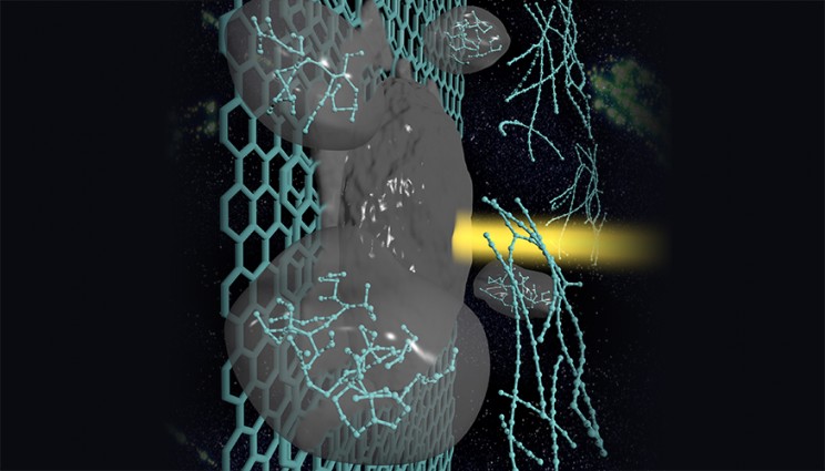
Here’s an image illustrating the process,

A carbyne strand forms in laser-melted graphite. Carbyne is found in astrophysical bodies and has the potential to be used in nanoelectronic devices and superhard materials. Image by Liam Krauss/LLNL

A carbyne strand forms in laser-melted graphite. Carbyne is found in astrophysical bodies and has the potential to be used in nanoelectronic devices and superhard materials. Image by Liam Krauss/LLNL

Self-assembly for stunning structural colour

Researchers from McGill University (Montréal, Canada) have developed a computational model which they believe explains how nature achieves structural colo(u)r as exemplified by this tulip,

 Caption: The Queen of the Night tulip displays an iridescent shimmer caused by microscopic ridges on its petals that diffract light. Credit: S. Vignolini/


Caption: The Queen of the Night tulip displays an iridescent shimmer caused by microscopic ridges on its petals that diffract light.
Credit: S. Vignolini/

A Sept. 16, 2015 news item on phys.org describes the phenomenon,

The tulip called Queen of the Night has a fitting name. Its petals are a lush, deep purple that verges on black. An iridescent shimmer dances on top of the nighttime hues, almost like moonlight glittering off regal jewels.

Certain rainforest plants in Malaysia demonstrate an even more striking color feature: Their iridescent blue leaves turn green when dunked in water.

Both the tulip’s rainbow sparkle and the Malaysian plants’ color change are examples of structural color—an optical effect that is produced by a physical structure, instead of a chemical pigment.

Now researchers have shown how plant cellulose can self-assemble [emphasis mine] into wrinkled surfaces that give rise to effects like iridescence and color change. Their findings provide a foundation to understand structural color in nature, as well as yield insights that could guide the design of devices like optical humidity sensors. …

A Sept. 15, 2015 American Institute of Physics news release on EurekAlert, which originated the news item, describes the research into cellulose and structural colour in more detail,

Cellulose is one of the most abundant organic materials on Earth. It forms a key part of the cell wall of green plants, where the cellulose fibers are found in layers. The fibers in a single layer tend to align in a single direction. However, when you move up or down a layer the axis of orientation of the fibers can shift. If you imagined an arrow pointing in the direction of the fiber alignment, it would often spin in a circle as you moved through the layers of cellulose. This twisting pattern is called a cholesteric phase, because it was first observed while studying cholesterol molecules.

Scientists think that cellulose twists mainly to provide strength. “The fibers reinforce in the direction they are oriented,” said Alejandro Rey, a chemical engineer at McGill University in Montreal, Canada. “When the orientation rotates you get multi-directional stiffness.”

Rey and his colleagues, however, weren’t primarily interested in cellulose’s mechanical properties. Instead, they wondered if the twisting structure could produce striking optical effects, as seen in plants like iridescent tulips.

The team constructed a computational model to examine the behavior of cholesteric phase cellulose. In the model, the axis of twisting runs parallel to the surface of the cellulose. The researchers found that subsurface helices naturally caused the surface to wrinkle. The tiny ridges had a height range in the nanoscale and were spaced apart on the order of microns.

The pattern of parallel ridges resembled the microscopic pattern on the petals of the Queen of the Night tulip. The ridges split white light into its many colored components and create an iridescent sheen — a process called diffraction. The effect can also be observed when light hits the microscopic grooves in a CD.

The researchers also experimented with how the amount of water in the cellulose layers affected the optical properties. More water made the layers twist less tightly, which in turn made the ridges farther apart. How tightly the cellulose helices twist is called the pitch. The team found that a surface with spatially varying pitch (in which some areas were more hydrated than others) was less iridescent and reflected a longer primary wavelength of light than surfaces with a constant pitch. The wavelength shift from around 460 nm (visible blue light) to around 520 nm (visible green light) could explain some plants’ color changing properties, Rey said.

Insights into Nature and Inspiration for New Technologies

Although proving that diffractive surfaces in nature form in the same way will require further work, the model does offer a good foundation to further explore structural color, the researchers said. They imagine the model could also guide the design of new optical devices, for example sensors that change color to indicate a change in humidity.

“The results show the optics [of cholesteric cellulose] are just as exciting as the mechanical properties,” Rey said. He said scientists tend to think of the structures as biological armor, because of their reinforcing properties. “We’ve shown this armor can also have striking colors,” he said.

Here’s a link to and a citation for the paper,

Tunable nano-wrinkling of chiral surfaces: Structure and diffraction optics by P. Rofouie, D. Pasini, and A. D. Rey. J. Chem. Phys. 143, 114701 (2015); http://dx.doi.org/10.1063/1.4929337

This is an open access paper.

Of Canadian 2015 election science debates and science weeks

You’d think science and technology might rate a mention in a debate focused on the economy but according to all accounts, that wasn’t the case last night in a Sept. 17, 2015 Canadian federal election debate featuring three party leaders, Justin Trudeau of the Liberal Party, Thomas Mulcair of the New Democratic Party (NDP), and Stephen Harper, Prime Minister and leader of the Conservative Party. BTW, Elizabeth May, leader of the Green Party, was not invited but managed to participate by tweeting video responses to the debate questions. For one of the more amusing and, in its way, insightful commentaries on the debate, there’s a Sept. 17, 2015 blog posting on CBC [Canadian Broadcasting Corporation] News titled: ‘Old stock Canadians,’ egg timer, creepy set top debate’s odd moments; Moderator David Walmsley’s Irish accent and a ringing bell get reaction on social media.

As for science and the 2015 Canadian federal election, Science Borealis has compiled an informal resource list in a Sept. 18, 2015 posting and while I’ve excerpted the resources where you can find suggested questions for candidates, there’s much more to be found there,


Interestingly, the journal Nature has published a Sept. 17, 2015 article (h/t @CBC Quirks) by Nicola Jones featuring the Canadian election and science concerns and the impact science concerns have had on opposition party platforms (Note: Links have been removed),

Canadians will head to the polls on 19 October [2015], in a federal election that many scientists hope will mark a turning point after years of declining research budgets and allegations of government censorship.

In an unprecedented move, the Professional Institute of the Public Service of Canada — a union in Ottawa that represents more than 57,000 government scientists and other professionals — is campaigning in a federal race. “Here’s how we do things in the Harper government,” declares one of the union’s radio advertisements. “We muzzle scientists, we cut research and we ignore anyone who doesn’t tell us what we want to hear.”

Science advocates see little chance that their issues will be aired during a 17 September [2015] debate in Calgary that will pit Harper against NDP [New Democratic Party] leader Thomas Mulcair and Liberal leader Justin Trudeau. But concerns about the state of Canadian science have nevertheless influenced party platforms.

The middle-left Liberal Party has made scientific integrity part of its election campaign, proposing the creation of a central public portal to disseminate government-funded research. The party seeks to appoint a chief science officer to ensure the free flow of information.

Similarly, the NDP has called for a parliamentary science officer, a position that would be independent of the majority party or coalition leading the government.

Adding to the concern about the practice of science in Canada is the delayed release of a biennial report from the government’s Science, Technology and Innovation Council (STIC). Paul Wells in a June 26, 2015 article for Maclean’s Magazine discusses the situation (Note: Links have been removed),

It is distressing when organizations with no partisan role play the sort of games partisans want. The latest example is the advisory board that the Harper government created to tell it how Canada is doing in science.

I have written about the Science, Technology and Innovation Council every two years since it produced its first major report, in May 2009. STIC, as it’s known, is not some fringe group of pinko malcontents trying to stir up trouble and turn the people against their right and proper governing party. It was conceived by the Harper government (in 2007), appointed by the Harper government (in bits ever since), and it consists, in part, of senior officials who work with the Harper government every day. …

This group gives the feds the best advice they can get about how Canada is faring against other countries in its science, research and technology efforts. Its reports have been increasingly discouraging.

Perhaps you wonder: What’s the situation now? Keep wondering. Every previous STIC biennial report was released in the spring. This winter, I met a STIC member, who told me the next report would come out in May 2015 and that it would continue most of the declining trend lines established by the first three reports. I wrote to the STIC to ascertain the status of the latest report. Here’s the answer I received:

“Thank you for your interest. STIC’s next State of the Nation report will be released later in the Fall. We will be happy to inform you of the precise date and release details when they have been confirmed.”

There is no reason this year’s report was not released in the spring, as every previous report was. None except the approach of a federal election.

Getting back to a national science debate, I have written about a proposed debate to be held on the CBC Quirks and Quarks radio programme here in a Sept. 3, 2015 posting which also features a local upcoming (on Weds., Sept. 23, 2015) election science and technology debate amongst  federal candidates in Victoria, BC. I cannot find anything more current about the proposed national science debate other than the CBC radio producer’s claim that it would occur in early October. Earlier today (Sept. 18, 2015) I checked their Twitter feed (https://twitter.com/CBCQuirks) and their website (http://www.cbc.ca/radio/quirks). I wonder what’s taking so long for an announcement. In the space of a few hours, I managed to get Ted Hsu and Lynne Quarmby, science shadow ministers for the Liberal and Green parties, respectively, to express interest in participating.

Well, whether or not there is a 2015 national science debate, I find the level of interest, in contrast to the 2011 election, exciting and affirming.

In the midst of all this election and science discussion, there are some big Canadian science events on the horizon. First and technically speaking not on the horizon, there’s Beakerhead (a smashup of art, science, and engineering) in Calgary, Alberta which runs from Sept. 16 – 20, 2015. Here are a few of the exhibits and installations you can find should you get to Calgary in time (from a Sept. 16, 2015 Beakerhead news release),

The five days of Beakerhead officially get rolling today with the world’s largest pop-up gallery, called a String (Theory) of Incredible Encounters, with a circumference of five kilometres around downtown Calgary.

The series of public art installations is an exploration in creativity at the crossroads of art, science and engineering, and can be seen by touring Calgary’s neighbourhoods, from Inglewood to East Village to Victoria Park, 17th Ave and Kensington. The home base or hub for Beakerhead this year is at Station B (the Beakerhead moniker for installations at Fort Calgary).

Station B is home to two other massive firsts – a 30-foot high version of the arcade claw game, and a 6,400 square foot sandbox – all designed to inspire human ingenuity.

Beakerhead 2015 event will erupt on the streets and venues of Calgary from September 16 to 20, and includes more than 160 collaborators and 60 public events, ranging from theatre where the audience is dining as part of the show to installations where you walk through a human nose. More than 25,000 students will be engaged in Beakerhead through field trips, classroom visits and ingenuity challenges.

Just as Beakerhead ends, Canada’s 2015 Science Literacy Week opens Sept. 21 – 27, 2015. Here’s more about the week from a Sept. 18, 2015 article by Natalie Samson for University Affairs,

On Nov. 12 last year [2014], the European Space Agency landed a robot on a comet. It was a remarkable moment in the history of space exploration and scientific inquiry. The feat amounted to “trying to throw a dart and hit a fly 10 miles away,” said Jesse Hildebrand, a science educator and communicator. “The math and the physics behind that is mindboggling.”

Imagine Mr. Hildebrand’s disappointment then, as national news programs that night spent about half as much time reporting on the comet landing as they did covering Barack Obama’s gum-chewing faux pas in China. For Mr. Hildebrand, the incident perfectly illustrates why he founded Science Literacy Week, a Canada-wide public education campaign celebrating all things scientific.

From Sept. 21 to 27 [2015], several universities, libraries and museums will highlight the value of science in our contemporary world by hosting events and exhibits on topics ranging from the lifecycle of a honeybee to the science behind Hollywood films like Jurassic World and Contact.

Mr. Hildebrand began developing the campaign last year, shortly after graduating from the University of Toronto with a bachelor’s degree in ecology and evolutionary biology. He approached the U of T Libraries for support and “it really snowballed from there,” the 23-year-old said.

Though Mr. Hildebrand said Science Literacy Week wasn’t inspired by public criticism against the federal government’s approach to scientific research and communication, he admitted that it makes the campaign seem that much more important. “I’ve always wanted to shout from the rooftops how cool science is. This is my way of shouting from the rooftops,” he said.

In the lead-up to Science Literacy Week, museum scientists with the Alliance of Natural History Museums of Canada have been posting videos of what they do and why it’s important under the hashtag #canadalovesscience. The end of the campaign will coincide with a lunar eclipse and will see several universities and observatories hosting stargazing parties.

You can find out more about this year’s events on the Science Literacy Week website. Here are a few of the BC events I found particularly intriguing,

UBC Botanical Garden – Jointly run as part of National Forest Week/Organic Week

September 20th, 10 a.m-12 p.m – A Walk in the Woods

Come discover the forest above, below and in between on our guided forest tour! Explore and connect with trees that hold up our 300-metre long canopy walkway. [emphasis mine] Meet with grand Firs, Douglas Firs and Western Red Cedars and learn about the importance of forests to biodiversity, climate change and our lives.

September 24th, 7:30-11 P.M – Food Garden Tour and Outdoor Movie Night

What better way to celebrate Organic Week than to hear about our exciting plans for the UBC Food Garden? Tour renewed garden beds to see what’s been growing. Learn about rootstocks, cultivars, training techniques and tree forms for fruit trees in this area.  Then make your way to out enchanting outdoor Ampitheatre and watch Symphony of the Soil, a film celebrated by the UN for 2015, the International Year of the Soil.

I highlighted the UBC Botanical Garden canopy walkway because you really do walk high up in the forest as you can see in this image of the walkway,

[downloaded from http://www.familyfuncanada.com/vancouver/canopy-walk-ubc-botanical-garden/]

[downloaded from http://www.familyfuncanada.com/vancouver/canopy-walk-ubc-botanical-garden/]

This image is from an undated article by Lindsay Follett for Family Fun Vancouver.

While it’s still a month away, there is Canada’s upcoming 2015 National Science and Technology Week, which will run from Oct. 16 – 25. To date, they do not have any events listed for this year’s week but they do invite you to submit your planned event for inclusion in their 2015 event map and list of events.
Observing nanoparticle therapeutics interact with blood in real time

Sadly, there are no images showing nanoparticle therapeutics interacting with blood or anything else for that matter to illustrate this story but perhaps the insights offered should suffice. From Sept. 15, 2015 news item on Nanowerk,

Researchers at the National University of Singapore (NUS) have developed a technique to observe, in real time, how individual blood components interact and modify advanced nanoparticle therapeutics. The method, developed by an interdisciplinary team consisting clinician-scientist Assistant Professor Chester Lee Drum of the Department of Medicine at the NUS Yong Loo Lin School of Medicine, Professor T. Venky Venkatesan, Director of NUS Nanoscience and Nanotechnology Institute, and Assistant Professor James Kah of the Department of Biomedical Engineering at the NUS Faculty of Engineering, helps guide the design of future nanoparticles to interact in concert with human blood components, thus avoiding unwanted side effects.

A Sept. 15, 2015 NUS press release, which originated the news item, describes the research in more specific detail,

With their small size and multiple functionalities, nanoparticles have attracted intense attention as both diagnostic and drug delivery systems. However, within minutes of being delivered into the bloodstream, nanoparticles are covered with a shell of serum proteins, also known as a protein ‘corona’.

“The binding of serum proteins can profoundly change the behaviour of nanoparticles, at times leading to rapid clearance by the body and a diminished clinical outcome,” said Asst Prof Kah.

Existing methods such as mass spectroscopy and diffusional radius estimation, although useful for studying important nanoparticle parameters, are unable to provide detailed, real-time binding kinetics.

Novel method to understand nano-bio interactions

The NUS team, together with external collaborator Professor Bo Liedberg from the Nanyang Technological University, showed highly reproducible kinetics for the binding between gold nanoparticles and the four most common serum proteins: human serum albumin, fibrinogen, apolipoprotein A-1, and polyclonal IgG.

“What was remarkable about this project was the initiative taken by Abhijeet Patra, my graduate student from NUS Graduate School for Integrative Sciences and Engineering, in conceptualising the problem, and bringing together the various teams in NUS and beyond to make this a successful programme,” said Prof Venkatesan. “The key development is the use of a new technique using surface plasmon resonance (SPR) technology to measure the protein corona formed when common proteins in the bloodstream bind to nanoparticles,” he added.

The researchers first immobilised the gold nanoparticles to the surface of a SPR sensor chip with a linker molecule. The chip was specially modified with an alginate polymer layer which both provided a negative charge and active sites for ligand immobilisation, and prevented non-specific binding. Using a 6 x 6 microfluidic channel array, they studied up to 36 nanoparticle-protein interactions in a single experiment, running test samples alongside experimental controls.

“Reproducibility and reliability have been a bottleneck in the studies of protein coronas,” said Mr Abhijeet Patra. “The quality and reliability of the data depends most importantly upon the design of good control experiments. Our multiplexed SPR setup was therefore key to ensuring the reliability of our data.”

Testing different concentrations of each of the four proteins, the team found that apolipoprotein A-1 had the highest binding affinity for the gold nanoparticle surface, with an association constant almost 100 times that of the lowest affinity protein, polyclonal IgG.

“Our results show that the rate of association, rather than dissociation, is the main determinant of binding with the tested blood components,” said Asst Prof Drum.

The multiplex SPR system was also used to study the effect of modification with polyethylene (PEG), a synthetic polymer commonly used in nanoparticle formulations to prevent protein accumulation. The researchers found that shorter PEG chains (2-10 kilodaltons) are about three to four times more effective than longer PEG chains (20-30 kilodaltons) at preventing corona formation.

“The modular nature of our protocol allows us to study any nanoparticle which can be chemically tethered to the sensing surface,” explained Asst Prof Drum. “Using our technique, we can quickly evaluate a series of nanoparticle-based drug formulations before conducting in vivo studies, thereby resulting in savings in time and money and a reduction of in vivo testing,” he added.

The researchers plan to use the technology to quantitatively study protein corona formation for a variety of nanoparticle formulations, and rationally design nanomedicines for applications in cardiovascular diseases and cancer.

Here’s a link to and a citation for the paper,

Component-Specific Analysis of Plasma Protein Corona Formation on Gold Nanoparticles Using Multiplexed Surface Plasmon Resonance by Abhijeet Patra, Tao Ding, Gokce Engudar, Yi Wang, Michal Marcin Dykas, Bo Liedberg, James Chen Yong Kah, Thirumalai Venkatesan, and Chester Lee Drum. Small  DOI: 10.1002/smll.201501603 Article first published online: 10 SEP 2015

© 2015 WILEY-VCH Verlag GmbH & Co. KGaA, Weinheim

This paper is behind a paywall.

$81M for US National Nanotechnology Coordinated Infrastructure (NNCI)

Academics, small business, and industry researchers are the big winners in a US National Science Foundation bonanza according to a Sept. 16, 2015 news item on Nanowerk,

To advance research in nanoscale science, engineering and technology, the National Science Foundation (NSF) will provide a total of $81 million over five years to support 16 sites and a coordinating office as part of a new National Nanotechnology Coordinated Infrastructure (NNCI).

The NNCI sites will provide researchers from academia, government, and companies large and small with access to university user facilities with leading-edge fabrication and characterization tools, instrumentation, and expertise within all disciplines of nanoscale science, engineering and technology.

A Sept. 16, 2015 NSF news release provides a brief history of US nanotechnology infrastructures and describes this latest effort in slightly more detail (Note: Links have been removed),

The NNCI framework builds on the National Nanotechnology Infrastructure Network (NNIN), which enabled major discoveries, innovations, and contributions to education and commerce for more than 10 years.

“NSF’s long-standing investments in nanotechnology infrastructure have helped the research community to make great progress by making research facilities available,” said Pramod Khargonekar, assistant director for engineering. “NNCI will serve as a nationwide backbone for nanoscale research, which will lead to continuing innovations and economic and societal benefits.”

The awards are up to five years and range from $500,000 to $1.6 million each per year. Nine of the sites have at least one regional partner institution. These 16 sites are located in 15 states and involve 27 universities across the nation.

Through a fiscal year 2016 competition, one of the newly awarded sites will be chosen to coordinate the facilities. This coordinating office will enhance the sites’ impact as a national nanotechnology infrastructure and establish a web portal to link the individual facilities’ websites to provide a unified entry point to the user community of overall capabilities, tools and instrumentation. The office will also help to coordinate and disseminate best practices for national-level education and outreach programs across sites.

New NNCI awards:

Mid-Atlantic Nanotechnology Hub for Research, Education and Innovation, University of Pennsylvania with partner Community College of Philadelphia, principal investigator (PI): Mark Allen
Texas Nanofabrication Facility, University of Texas at Austin, PI: Sanjay Banerjee

Northwest Nanotechnology Infrastructure, University of Washington with partner Oregon State University, PI: Karl Bohringer

Southeastern Nanotechnology Infrastructure Corridor, Georgia Institute of Technology with partners North Carolina A&T State University and University of North Carolina-Greensboro, PI: Oliver Brand

Midwest Nano Infrastructure Corridor, University of  Minnesota Twin Cities with partner North Dakota State University, PI: Stephen Campbell

Montana Nanotechnology Facility, Montana State University with partner Carlton College, PI: David Dickensheets
Soft and Hybrid Nanotechnology Experimental Resource,

Northwestern University with partner University of Chicago, PI: Vinayak Dravid

The Virginia Tech National Center for Earth and Environmental Nanotechnology Infrastructure, Virginia Polytechnic Institute and State University, PI: Michael Hochella

North Carolina Research Triangle Nanotechnology Network, North Carolina State University with partners Duke University and University of North Carolina-Chapel Hill, PI: Jacob Jones

San Diego Nanotechnology Infrastructure, University of California, San Diego, PI: Yu-Hwa Lo

Stanford Site, Stanford University, PI: Kathryn Moler

Cornell Nanoscale Science and Technology Facility, Cornell University, PI: Daniel Ralph

Nebraska Nanoscale Facility, University of Nebraska-Lincoln, PI: David Sellmyer

Nanotechnology Collaborative Infrastructure Southwest, Arizona State University with partners Maricopa County Community College District and Science Foundation Arizona, PI: Trevor Thornton

The Kentucky Multi-scale Manufacturing and Nano Integration Node, University of Louisville with partner University of Kentucky, PI: Kevin Walsh

The Center for Nanoscale Systems at Harvard University, Harvard University, PI: Robert Westervelt

The universities are trumpeting this latest nanotechnology funding,

NSF-funded network set to help businesses, educators pursue nanotechnology innovation (North Carolina State University, Duke University, and University of North Carolina at Chapel Hill)

Nanotech expertise earns Virginia Tech a spot in National Science Foundation network

ASU [Arizona State University] chosen to lead national nanotechnology site

UChicago, Northwestern awarded $5 million nanotechnology infrastructure grant

That is a lot of excitement.

University of Vermont and the ‘excitons’ of an electron superhighway

This story starts off with one of the current crazes, folding and bendable electronics, before heading off onto the ‘electron highway’. From a Sept. 14, 2015 news item on ScienceDaily (Note: Links have been removed),

TV screens that roll up. Roofing tiles that double as solar panels. Sun-powered cell phone chargers woven into the fabric of backpacks. A new generation of organic semiconductors may allow these kinds of flexible electronics to be manufactured at low cost, says University of Vermont physicist and materials scientist Madalina Furis.

But the basic science of how to get electrons to move quickly and easily in these organic materials remains murky.

To help, Furis and a team of UVM materials scientists have invented a new way to create what they are calling “an electron superhighway” in one of these materials — a low-cost blue dye called phthalocyanine — that promises to allow electrons to flow faster and farther in organic semiconductors.

A Sept. 14, 2015 University of Vermont news release (also on EurekAlert) by Joshua E. Brown, which originated the news item, describes the problem the researches were trying to solve and the solution they found,

Hills and potholes

Many of these types of flexible electronic devices will rely on thin films of organic materials that catch sunlight and convert the light into electric current using excited states in the material called “excitons.” Roughly speaking, an exciton is a displaced electron bound together with the hole it left behind. Increasing the distance these excitons can diffuse — before they reach a juncture where they’re broken apart to produce electrical current — is essential to improving the efficiency of organic semiconductors.

Using a new imaging technique, the UVM team was able to observe nanoscale defects and boundaries in the crystal grains in the thin films of phthalocyanine — roadblocks in the electron highway. “We have discovered that we have hills that electrons have to go over and potholes that they need to avoid,” Furis explains.

To find these defects, the UVM team — with support from the National Science Foundation — built a scanning laser microscope, “as big as a table” Furis says. The instrument combines a specialized form of linearly polarized light and photoluminescence to optically probe the molecular structure of the phthalocyanine crystals.

“Marrying these two techniques together is new; it’s never been reported anywhere,” says Lane Manning ’08 a doctoral student in Furis’ lab and co-author on the new study.

The new technique allows the scientists a deeper understanding of how the arrangement of molecules and the boundaries in the crystals influence the movement of excitons. It’s these boundaries that form a “barrier for exciton diffusion,” the team writes.

And then, with this enhanced view, “this energy barrier can be entirely eliminated,” the team writes. The trick: very carefully controlling how the thin films are deposited. Using a novel “pen-writing” technique with a hollow capillary, the team worked in the lab of UVM physics and materials science professor Randy Headrick to successfully form films with jumbo-sized crystal grains and “small angle boundaries.” Think of these as easy-on ramps onto a highway — instead of an awkward stop sign at the top of a hill — that allow excitons to move far and fast.

Better solar cells

Though the Nature Communications study focused on just one organic material, phthalocyanine, the new research provides a powerful way to explore many other types of organic materials, too — with particular promise for improved solar cells. A recent U.S. Department of Energy report identified one of the fundamental bottlenecks to improved solar power technologies as “determining the mechanisms by which the absorbed energy (exciton) migrates through the system prior to splitting into charges that are converted to electricity.”

The new UVM study — led by two of Furis’ students, Zhenwen Pan G’12, and Naveen Rawat G’15 — opens a window to view how increasing “long-range order” in the organic semiconductor films is a key mechanism that allows excitons to migrate farther. “The molecules are stacked like dishes in a dish rack,” Furis explains, “these stacked molecules — this dish rack — is the electron superhighway.”

Though excitons are neutrally charged — and can’t be pushed by voltage like the electrons flowing in a light bulb — they can, in a sense, bounce from one of these tightly stacked molecules to the next. This allows organic thin films to carry energy along this molecular highway with relative ease, though no net electrical charge is transported.

“One of today’s big challenges is how to make better photovoltaics and solar technologies,” says Furis, who directs UVM’s program in materials science, “and to do that we need a deeper understanding of exciton diffusion. That’s what this research is about.”

Here’s a link to and a citation for the paper,

Polarization-resolved spectroscopy imaging of grain boundaries and optical excitations in crystalline organic thin films by Z. Pan, N. Rawat, I. Cour, L. Manning, R. L. Headrick, & M. Furis. Nature Communications 6, Article number: 8201 doi:10.1038/ncomms9201 Published 14 September 2015

This is an open access article.

Complex networks to provide ‘grand unified theory’

Trying to mesh classical physics and quantum physics together in one theory which accounts for behaviour on the macro and quantum scales has occupied scientists for decades and it seems that mathematicians have discovered a clue so solving the mystery. A Sept. 13, 2015 news item on Nanotechnology Now describes the findings,

Mathematicians investigating one of science’s great questions — how to unite the physics of the very big with that of the very small — have discovered that when the understanding of complex networks such as the brain or the Internet is applied to geometry the results match up with quantum behavior.

A Sept. 9, 2015 Queen Mary University of London press release, which originated the news item, describes the collaboration between Queen Mary and Karlsruhe Institute of Technology mathematicians,

The findings, published today (Thursday) in Scientific Reports, by researchers from Queen Mary University of London and Karlsruhe Institute of Technology, could explain one of the great problems in modern physics.

Currently ideas of gravity, developed by Einstein and Newton, explain how physics operates on a very large scale, but do not work at the sub-atomic level. Conversely, quantum mechanics works on the very small scale but does not explain the interactions of larger objects like stars. Scientists are looking for a so called ‘grand unified theory’ that joins the two, known as quantum gravity.

Several models have been proposed for how different quantum spaces are linked but most assume that the links between quantum spaces are fairly uniform, with little deviation from the average number of links between each space. The new model, which applies ideas from the theory of complex networks, has found that some quantum spaces might actually include hubs, i.e. nodes with significantly more links than others, like a particularly popular Facebook user.

Calculations run with this model show that these spaces are described by well-known quantum Fermi-Dirac, and Bose-Einstein statistics, used in quantum mechanics, indicating that they could be useful to physicists working on quantum gravity.

Dr Ginestra Bianconi, from Queen Mary University of London, and lead author of the paper, said:

“We hope that by applying our understanding of complex networks to one of the fundamental questions in physics we might be able to help explain how discrete quantum spaces emerge.

“What we can see is that space-time at the quantum-scale might be networked in a very similar way to things we are starting to understand very well like biological networks in cells, our brains and online social networks.”

Here’s a link to and a citation for the paper,

Complex Quantum Network Manifolds in Dimension d > 2 are Scale-Free by Ginestra Bianconi & Christoph Rahmede. Scientific Reports 5, Article number: 13979 (2015) doi:10.1038/srep13979 Published online: 10 September 2015

This is an open access paper.

Characterizing anatase titanium dixoide at the nanoscale

An international collaboration of researchers combined atomic force microscopy (AFM) and scanning tunneling microscopy (STM) to characterize anatase titanium dixoxide. From a Sept. 14, 2015 news item on Azonano,

A [Japan National Institute for Materials Science] NIMS research team successfully identified the atoms and common defects existing at the most stable surface of the anatase form of titanium dioxide by characterizing this material at the atomic scale with scanning probe microscopy. This work was published under open access policy in the online version of Nature Communications on June 29, 2015.

A June 29, 2015 NIMS press release, which originated the news item, includes the paper’s abstract in numbered point form,

  1. The research team consisting of Oscar Custance and Tomoko Shimizu, group leader and senior scientist, respectively, at the Atomic Force Probe Group, NIMS, Daisuke Fujita and Keisuke Sagisaka, group leader and senior researcher, respectively, at the Surface Characterization Group, NIMS, and scientists at Charles University in the Czech Republic, Autonomous University of Madrid in Spain, and other organizations combined simultaneous atomic force microscopy (AFM) and scanning tunneling microscopy (STM) measurements with first-principles calculations for the unambiguous identification of the atomic species at the most stable surface of the anatase form of titanium dioxide (hereinafter referred to as anatase) and its most common defects.
  2. In recent years, anatase has attracted considerable attention, because it has become a pivotal material in devices for photo-catalysis and for the conversion of solar energy to electricity. It is extremely challenging to grow large single crystals of anatase, and most of the applications of this material are in the form of nano crystals. To enhance the catalytic reactivity of anatase and the efficiency of devices for solar energy conversion based on anatase, it is critical to gain in-depth understanding and control of the reactions taking place at the surface of this material down to the atomic level. Only a few research groups worldwide possess the technology to create proper test samples and to make in-situ atomic-level observations of anatase surfaces.
  3. In this study, the research team used samples obtained from anatase natural single crystals extracted from naturally occurring anatase rocks. The team characterized the (101) surface of anatase at atomic level by means of simultaneous AFM and STM. Using single water molecules as atomic markers, the team successfully identified the atomic species of this surface; result that was additionally confirmed by the comparison of simultaneous AFM and STM measurements with the outcomes of first-principles calculations.
  4. In regular STM, in which an atomically sharp probe is scanned over the surface by keeping constant an electrical current flowing between them, it is difficult to stably image anatase surfaces as this material presents poor electrical conductivity over some of the atomic positions of the surface. However, simultaneous operation of AFM and STM allowed imaging the surface with atomic resolution even within the materials band gap (a region where the flow of current between the probe and the surface is, in principle, prohibited). Here, the detection of inter-atomic forces between the last atom of the atomically sharp probe and the atoms of the surface by AFM was of crucial importance. By regulating the probe-surface distance using AFM, it was possible to image the surface at atomic-scale while collecting STM data over both conductive and not conductive areas of the surface. By comparing simultaneous AFM and STM measurements with theoretical simulations, the team was not only able to discern which atomic species were contributing to the AFM and the STM images but also to identify the most common defects found at the surface.
  5. In the future, based on the information gained from this study, the NIMS research team will conduct research on molecules of technologically relevance that adsorb on anatase and characterize these hybrid systems by using simultaneous AFM and STM. Their ultimate goal is to formulate novel approaches for the development of photo-catalysts and solar cell materials and devices.

Here’s a link to and a citation for the paper,

Atomic species identification at the (101) anatase surface by simultaneous scanning tunnelling and atomic force microscopy by Oleksandr Stetsovych, Milica Todorović, Tomoko K. Shimizu, César Moreno, James William Ryan, Carmen Pérez León, Keisuke Sagisaka, Emilio Palomares, Vladimír Matolín, Daisuke Fujita, Ruben Perez, & Oscar Custance. Nature Communications 6, Article number: 7265 doi:10.1038/ncomms8265 Published 29 June 2015

This is an open access paper.