Tag Archives: tissue engineering

Cellulose biosensor heralds new bioimaging approach to tissue engineering

I keep an eye on how nanocellulose is being used in various applications and I’m not sure that this cellulose biosensor quite fits the bill as nanocellulose, nonetheless, it’s interesting and that’s enough for me. From a December 12, 2018 Sechenov University (Russia) press release on EurekAlert,

I.M. Sechenov First Moscow State Medical University teamed up together with Irish colleagues to develop a new imaging approach for tissue engineering. The team produced so-called ‘hybrid biosensor’ scaffold materials, which are based on cellulose matrices labeled with pH- and calcium-sensitive fluorescent proteins. These materials enable visualization of the metabolism and other important biomarkers in the engineered artificial tissues by microscopy. The results of the work were published in the Acta Biomaterialia journal.
The success of tissue engineering is based on the use of scaffold matrices – materials that support the viability and direct the growth of cells, tissues, and organoids. Scaffolds are important for basic and applied biomedical research, tissue engineering and regenerative medicine, and are promising for development of new therapeutics. However, the ability ‘to see’ what happens within the scaffolds during the tissue growth poses a significant research challenge

“We developed a new approach allowing visualization of scaffold-grown tissue and cells by using labeling with biosensor fluorescent proteins. Due to the high specificity of labeling and the use of fluorescence microscopy FLIM, we can quantify changes in pH and calcium in the vicinity of cells,” says Dr. Ruslan Dmitriev, Group Leader at the University College Cork and the Institute for Regenerative Medicine (I.M. Sechenov First Moscow State Medical University).
To achieve the specific labeling of cellulose matrices, researchers used well-known cellulose-binding proteins. The use of extracellular pH- and calcium-sensitive biosensors allow for analysis of cell metabolism: indeed, the extracellular acidification is directly associated with the balance of cell energy production pathways and the glycolytic flux (release of lactate). It is also a frequent hallmark of cancer and transformed cell types. On the other hand, calcium plays a key role in the extra- and intracellular signaling affecting cell growth and differentiation.

The approach was tested on different types of cellulose matrices (bacterial and produced from decellularised plant tissues) using 3D culture of human colon cancer cells and stem-cell derived mouse small intestinal organoids. The scaffolds informed on changes in the extracellular acidification and were used together with the analysis of real-time oxygenation of intestinal organoids. The resulting data can be presented in the form of colour maps, corresponding to the areas of cell growth within different microenvironments.

“Our results open new prospects in the imaging of tissue-engineered constructs for regenerative medicine. They enable deeper understanding of tissue metabolism in 3D and are also highly promising for commercialisation,” concludes Dr. Dmitriev.

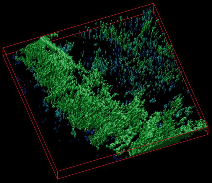
The researchers have provided an image to illustrate their work,

Caption: A 3D reconstruction of a cellulose matrix stained with a pH-sensitive biosensor. Credit: Dr. R. Dmitriev

Here’s a link to and a citation for the paper,

Cellulose-based scaffolds for fluorescence lifetime imaging-assisted tissue engineering by Neil O’Donnell, Irina A. Okkelman, Peter Timashev, Tatyana I.Gromovykh, Dmitri B. Papkovsky, Ruslan I.Dmitriev. Acta Biomaterialia Volume 80, 15 October 2018, Pages 85-96 DOI: https://doi.org/10.1016/j.actbio.2018.09.034


This paper is behind a paywall.

Periodic table of nanomaterials

This charming illustration is the only pictorial representation i’ve seen for Kyoto University’s (Japan) proposed periodic table of nanomaterials, (By the way, 2019 is UNESCO’s [United Nations Educational, Scientific and Cultural Organization] International Year of the Periodic Table of Elements, an event recognizing the table’s 150th anniversary. See my January 8, 2019 posting for information about more events.)

Caption: Molecules interact and align with each other as they self-assemble. This new simulation enables to find what molecules best interact with each other to build nanomaterials, such as materials that work as a nano electrical wire.
Credit Illustration by Izumi Mindy Takamiya

A July 23, 2018 news item on Nanowerk announces the new periodic table (Note: A link has been removed),

The approach was developed by Daniel Packwood of Kyoto University’s Institute for Integrated Cell-Material Sciences (iCeMS) and Taro Hitosugi of the Tokyo Institute of Technology (Nature Communications, “Materials informatics for self-assembly of functionalized organic precursors on metal surfaces”). It involves connecting the chemical properties of molecules with the nanostructures that form as a result of their interaction. A machine learning technique generates data that is then used to develop a diagram that categorizes different molecules according to the nano-sized shapes they form.

This approach could help materials scientists identify the appropriate molecules to use in order to synthesize target nanomaterials.

A July 23, 2018 Kyoto University press release on EurekAlert, which originated the news item, explains further about the computer simulations run by the scientists in pursuit of their specialized periodic table,

Fabricating nanomaterials using a bottom-up approach requires finding ‘precursor molecules’ that interact and align correctly with each other as they self-assemble. But it’s been a major challenge knowing how precursor molecules will interact and what shapes they will form.

Bottom-up fabrication of graphene nanoribbons is receiving much attention due to their potential use in electronics, tissue engineering, construction, and bio-imaging. One way to synthesise them is by using bianthracene precursor molecules that have bromine ‘functional’ groups attached to them. The bromine groups interact with a copper substrate to form nano-sized chains. When these chains are heated, they turn into graphene nanoribbons.

Packwood and Hitosugi tested their simulator using this method for building graphene nanoribbons.

Data was input into the model about the chemical properties of a variety of molecules that can be attached to bianthracene to ‘functionalize’ it and facilitate its interaction with copper. The data went through a series of processes that ultimately led to the formation of a ‘dendrogram’.

This showed that attaching hydrogen molecules to bianthracene led to the development of strong one-dimensional nano-chains. Fluorine, bromine, chlorine, amidogen, and vinyl functional groups led to the formation of moderately strong nano-chains. Trifluoromethyl and methyl functional groups led to the formation of weak one-dimensional islands of molecules, and hydroxide and aldehyde groups led to the formation of strong two-dimensional tile-shaped islands.

The information produced in the dendogram changed based on the temperature data provided. The above categories apply when the interactions are conducted at -73°C. The results changed with warmer temperatures. The researchers recommend applying the data at low temperatures where the effect of the functional groups’ chemical properties on nano-shapes are most clear.

The technique can be applied to other substrates and precursor molecules. The researchers describe their method as analogous to the periodic table of chemical elements, which groups atoms based on how they bond to each other. “However, in order to truly prove that the dendrograms or other informatics-based approaches can be as valuable to materials science as the periodic table, we must incorporate them in a real bottom-up nanomaterial fabrication experiment,” the researchers conclude in their study published in the journal xxx. “We are currently pursuing this direction in our laboratories.”

Here’s a link to and a citation for the paper,

Materials informatics for self-assembly of functionalized organic precursors on metal surfaces by Daniel M. Packwood & Taro Hitosugi. Nature Communicationsvolume 9, Article number: 2469 (2018)DOI: https://doi.org/10.1038/s41467-018-04940-z Published 25 June 2018

This paper is open access.

Injectable bandages for internal bleeding and hydrogel for the brain

This injectable bandage could be a gamechanger (as they say) if it can be taken beyond the ‘in vitro’ (i.e., petri dish) testing stage. A May 22, 2018 news item on Nanowerk makes the announcement (Note: A link has been removed),

While several products are available to quickly seal surface wounds, rapidly stopping fatal internal bleeding has proven more difficult. Now researchers from the Department of Biomedical Engineering at Texas A&M University are developing an injectable hydrogel bandage that could save lives in emergencies such as penetrating shrapnel wounds on the battlefield (Acta Biomaterialia, “Nanoengineered injectable hydrogels for wound healing application”).

A May 22, 2018 US National Institute of Biomedical Engineering and Bioengiineering news release, which originated the news item, provides more detail (Note: Links have been removed),

The researchers combined a hydrogel base (a water-swollen polymer) and nanoparticles that interact with the body’s natural blood-clotting mechanism. “The hydrogel expands to rapidly fill puncture wounds and stop blood loss,” explained Akhilesh Gaharwar, Ph.D., assistant professor and senior investigator on the work. “The surface of the nanoparticles attracts blood platelets that become activated and start the natural clotting cascade of the body.”

Enhanced clotting when the nanoparticles were added to the hydrogel was confirmed by standard laboratory blood clotting tests. Clotting time was reduced from eight minutes to six minutes when the hydrogel was introduced into the mixture. When nanoparticles were added, clotting time was significantly reduced, to less than three minutes.

In addition to the rapid clotting mechanism of the hydrogel composite, the engineers took advantage of special properties of the nanoparticle component. They found they could use the electric charge of the nanoparticles to add growth factors that efficiently adhered to the particles. “Stopping fatal bleeding rapidly was the goal of our work,” said Gaharwar. “However, we found that we could attach growth factors to the nanoparticles. This was an added bonus because the growth factors act to begin the body’s natural wound healing process—the next step needed after bleeding has stopped.”

The researchers were able to attach vascular endothelial growth factor (VEGF) to the nanoparticles. They tested the hydrogel/nanoparticle/VEGF combination in a cell culture test that mimics the wound healing process. The test uses a petri dish with a layer of endothelial cells on the surface that create a solid skin-like sheet. The sheet is then scratched down the center creating a rip or hole in the sheet that resembles a wound.

When the hydrogel containing VEGF bound to the nanoparticles was added to the damaged endothelial cell wound, the cells were induced to grow back and fill-in the scratched region—essentially mimicking the healing of a wound.

“Our laboratory experiments have verified the effectiveness of the hydrogel for initiating both blood clotting and wound healing,” said Gaharwar. “We are anxious to begin tests in animals with the hope of testing and eventual use in humans where we believe our formulation has great potential to have a significant impact on saving lives in critical situations.”

The work was funded by grant EB023454 from the National Institute of Biomedical Imaging and Bioengineering (NIBIB), and the National Science Foundation. The results were reported in the February issue of the journal Acta Biomaterialia.

The paper was published back in April 2018 and there was an April 2, 2018 Texas A&M University news release on EurekAlert making the announcement (and providing a few unique details),

A penetrating injury from shrapnel is a serious obstacle in overcoming battlefield wounds that can ultimately lead to death.Given the high mortality rates due to hemorrhaging, there is an unmet need to quickly self-administer materials that prevent fatality due to excessive blood loss.

With a gelling agent commonly used in preparing pastries, researchers from the Inspired Nanomaterials and Tissue Engineering Laboratory have successfully fabricated an injectable bandage to stop bleeding and promote wound healing.

In a recent article “Nanoengineered Injectable Hydrogels for Wound Healing Application” published in Acta Biomaterialia, Dr. Akhilesh K. Gaharwar, assistant professor in the Department of Biomedical Engineering at Texas A&M University, uses kappa-carrageenan and nanosilicates to form injectable hydrogels to promote hemostasis (the process to stop bleeding) and facilitate wound healing via a controlled release of therapeutics.

“Injectable hydrogels are promising materials for achieving hemostasis in case of internal injuries and bleeding, as these biomaterials can be introduced into a wound site using minimally invasive approaches,” said Gaharwar. “An ideal injectable bandage should solidify after injection in the wound area and promote a natural clotting cascade. In addition, the injectable bandage should initiate wound healing response after achieving hemostasis.”

The study uses a commonly used thickening agent known as kappa-carrageenan, obtained from seaweed, to design injectable hydrogels. Hydrogels are a 3-D water swollen polymer network, similar to Jell-O, simulating the structure of human tissues.

When kappa-carrageenan is mixed with clay-based nanoparticles, injectable gelatin is obtained. The charged characteristics of clay-based nanoparticles provide hemostatic ability to the hydrogels. Specifically, plasma protein and platelets form blood adsorption on the gel surface and trigger a blood clotting cascade.

“Interestingly, we also found that these injectable bandages can show a prolonged release of therapeutics that can be used to heal the wound” said Giriraj Lokhande, a graduate student in Gaharwar’s lab and first author of the paper. “The negative surface charge of nanoparticles enabled electrostatic interactions with therapeutics thus resulting in the slow release of therapeutics.”

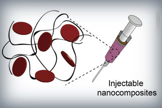
Nanoparticles that promote blood clotting and wound healing (red discs), attached to the wound-filling hydrogel component (black) form a nanocomposite hydrogel. The gel is designed to be self-administered to stop bleeding and begin wound-healing in emergency situations. Credit: Lokhande, et al. 1

Here’s a link to and a citation for the paper,

Nanoengineered injectable hydrogels for wound healing application by Giriraj Lokhande, James K. Carrow, Teena Thakur, Janet R. Xavier, Madasamy Parani, Kayla J. Bayless, Akhilesh K. Gaharwar. Acta Biomaterialia Volume 70, 1 April 2018, Pages 35-47
https://doi.org/10.1016/j.actbio.2018.01.045

This paper is behind a paywall.

Hydrogel and the brain

It’s been an interesting week for hydrogels. On May 21, 2018 there was a news item on ScienceDaily about a bioengineered hydrogel which stimulated brain tissue growth after a stroke (mouse model),

In a first-of-its-kind finding, a new stroke-healing gel helped regrow neurons and blood vessels in mice with stroke-damaged brains, UCLA researchers report in the May 21 issue of Nature Materials.

“We tested this in laboratory mice to determine if it would repair the brain in a model of stroke, and lead to recovery,” said Dr. S. Thomas Carmichael, Professor and Chair of neurology at UCLA. “This study indicated that new brain tissue can be regenerated in what was previously just an inactive brain scar after stroke.”

The brain has a limited capacity for recovery after stroke and other diseases. Unlike some other organs in the body, such as the liver or skin, the brain does not regenerate new connections, blood vessels or new tissue structures. Tissue that dies in the brain from stroke is absorbed, leaving a cavity, devoid of blood vessels, neurons or axons, the thin nerve fibers that project from neurons.

After 16 weeks, stroke cavities in mice contained regenerated brain tissue, including new neural networks — a result that had not been seen before. The mice with new neurons showed improved motor behavior, though the exact mechanism wasn’t clear.

Remarkable stuff.

New wound dressings with nanofibres for tissue regeneration

The Rotary Jet-Spinning manufacturing system was developed specifically as a therapeutic for the wounds of war. The dressings could be a good option for large wounds, such as burns, as well as smaller wounds on the face and hands, where preventing scarring is important. Illustration courtesy of Michael Rosnach/Harvard University

This image really gets the idea of regeneration across to the viewer while also informing you that this is medicine that comes from the military. A March 19,2018 news item on phys.org announces the work,

Researchers from the Harvard John A. Paulson School of Engineering and Applied Sciences (SEAS) and the Wyss Institute for Biologically Inspired Engineering have developed new wound dressings that dramatically accelerate healing and improve tissue regeneration. The two different types of nanofiber dressings, described in separate papers, use naturally-occurring proteins in plants and animals to promote healing and regrow tissue.

Our fiber manufacturing system was developed specifically for the purpose of developing therapeutics for the wounds of war,” said Kit Parker, the Tarr Family Professor of Bioengineering and Applied Physics at SEAS and senior author of the research. “As a soldier in Afghanistan, I witnessed horrible wounds and, at times, the healing process for those wounds was a horror unto itself. This research is a years-long effort by many people on my team to help with these problems.”

Parker is also a Core Faculty Member of the Wyss Institute.

The most recent paper, published in Biomaterials, describes a wound dressing inspired by fetal tissue.

A March 19, 2018 Harvard University John A. Paulson School of Engineering and Applied Science news release by Leah Burrows (also on EurekAlert), which originated the news item, provides some background information before launching into more detail about this latest work,

In the late 1970s, when scientists first started studying the wound-healing process early in development, they discovered something unexpected: Wounds incurred before the third trimester left no scars. This opened a range of possibilities for regenerative medicine. But for decades, researchers have struggled to replicate those unique properties of fetal skin.

Unlike adult skin, fetal skin has high levels of a protein called fibronectin, which assembles into the extracellular matrix and promotes cell binding and adhesion. Fibronectin has two structures: globular, which is found in blood, and fibrous, which is found in tissue. Even though fibrous fibronectin holds the most promise for wound healing, previous research focused on the globular structure, in part because manufacturing fibrous fibronectin was a major engineering challenge.

But Parker and his team are pioneers in the field of nanofiber engineering.

The researchers made fibrous fibronectin using a fiber-manufacturing platform called Rotary Jet-Spinning (RJS), developed by Parker’s Disease Biophysics Group. RJS works likes a cotton-candy machine — a liquid polymer solution, in this case globular fibronectin dissolved in a solvent, is loaded into a reservoir and pushed out through a tiny opening by centrifugal force as the device spins. As the solution leaves the reservoir, the solvent evaporates and the polymers solidify. The centrifugal force unfolds the globular protein into small, thin fibers. These fibers — less than one micrometer in diameter — can be collected to form a large-scale wound dressing or bandage.

“The dressing integrates into the wound and acts like an instructive scaffold, recruiting different stem cells that are relevant for regeneration and assisting in the healing process before being absorbed into the body,” said Christophe Chantre, a graduate student in the Disease Biophysics Group and first author of the paper.

In in vivo testing, the researchers found that wounds treated with the fibronectin dressing showed 84 percent tissue restoration within 20 days, compared with 55.6 percent restoration in wounds treated with a standard dressing.

The researchers also demonstrated that wounds treated with the fibronectin dressing had almost normal epidermal thickness and dermal architecture, and even regrew hair follicles — often considered one of the biggest challenges in the field of wound healing.

“This is an important step forward,” said Chantre. “Most work done on skin regeneration to date involves complex treatments combining scaffolds, cells, and even growth factors. Here we were able to demonstrate tissue repair and hair follicle regeneration using an entirely material approach. This has clear advantages for clinical translation.”

In another paper published in Advanced Healthcare Materials, the Disease Biophysics Group demonstrated a soy-based nanofiber that also enhances and promotes wound healing.

Soy protein contains both estrogen-like molecules — which have been shown to accelerate wound healing — and bioactive molecules similar to those that build and support human cells.

“Both the soy- and fibronectin-fiber technologies owe their success to keen observations in reproductive medicine,” said Parker. “During a woman’s cycle, when her estrogen levels go high, a cut will heal faster. If you do a surgery on a baby still in the womb, they have scar-less wound healing. Both of these new technologies are rooted in the most fascinating of all the topics in human biology — how we reproduce.”

In a similar way to fibronectin fibers, the research team used RJS to spin ultrathin soy fibers into wound dressings. In experiments, the soy- and cellulose-based dressing demonstrated a 72 percent increase in healing over wounds with no dressing and a 21 percent increase in healing over wounds dressed without soy protein.

“These findings show the great promise of soy-based nanofibers for wound healing,” said Seungkuk Ahn, a graduate student in the Disease Biophysics Group and first author of the paper. “These one-step, cost-effective scaffolds could be the next generation of regenerative dressings and push the envelope of nanofiber technology and the wound-care market.”

Both kinds of dressing, according to researchers, have advantages in the wound-healing space. The soy-based nanofibers — consisting of cellulose acetate and soy protein hydrolysate — are inexpensive, making them a good option for large-scale use, such as on burns. The fibronectin dressings, on the other hand, could be used for smaller wounds on the face and hands, where preventing scarring is important.

Here’s are links and citations for both papers mentioned in the news release,

Soy Protein/Cellulose Nanofiber Scaffolds Mimicking Skin Extracellular Matrix for Enhanced Wound Healing by Seungkuk Ahn, Christophe O. Chantre, Alanna R. Gannon, Johan U. Lind, Patrick H. Campbell, Thomas Grevesse, Blakely B. O’Connor, Kevin Kit Parker. Advanced Healthcare Materials https://doi.org/10.1002/adhm.201701175 First published: 23 January 2018

Production-scale fibronectin nanofibers promote wound closure and tissue repair in a dermal mouse model by Christophe O. Chantre, Patrick H. Campbell, Holly M. Golecki, Adrian T. Buganza, Andrew K. Capulli, Leila F. Deravi, Stephanie Dauth, Sean P. Sheehy, Jeffrey A.Paten. KarlGledhill, Yanne S. Doucet, Hasan E.Abaci, Seungkuk Ahn, Benjamin D.Pope, Jeffrey W.Ruberti, Simon P.Hoerstrup, Angela M.Christiano, Kevin Kit Parker. Biomaterials Volume 166, June 2018, Pages 96-108 https://doi.org/10.1016/j.biomaterials.2018.03.006 Available online 5 March 2018

Both papers are behind paywalls although you may want to check with ResearchGate where many researchers make their papers available for free.

One last comment, I noticed this at the end of Burrows’ news release,

The Harvard Office of Technology Development has protected the intellectual property relating to these projects and is exploring commercialization opportunities.

It reminded me of the patent battle between the Broad Institute (a Harvard University and Massachusetts Institute of Technology joint venture) and the University of California at Berkeley over CRISPR (clustered regularly interspaced short palindromic repeats) technology. (My March 15, 2017 posting describes the battle’s outcome.)

Lest we forget, there could be major financial rewards from this work.

Tractor beams for artificial cells

This particular piece has videos of cells moving around. I won’t be including all of them but they are weirdly fascinating. First, a May 14, 2018 news item on Nanowerk announces the latest in tractor beam news from the Imperial College London (ICL; UK),

Researchers have used lasers to connect, arrange and merge artificial cells, paving the way for networks of artificial cells that act like tissues.

The team say that by altering artificial cell membranes they can now get the cells to stick together like ‘stickle bricks’ – allowing them to be arranged into whole new structures.

Biological cells can perform complex functions, but are difficult to controllably engineer.

Artificial cells, however, can in principle be made to order. Now, researchers from Imperial College London and Loughborough University have demonstrated a new level of complexity with artificial cells by arranging them into basic tissue structures with different types of connectivity.

These structures could be used to perform functions like initiating chemical reactions or moving chemicals around networks of artificial and biological cells. This could be useful in carrying out chemical reactions in ultra-small volumes, in studying the mechanisms through which cells communicate with one another, and in the development of a new generation of smart biomaterials.

A May 14, 2018 ICL press release by Hayley Dunning , which originated the news item, provides more detail,

Cells are the basic units of biology, which are capable of working together as a collective when arranged into tissues. In order to do this, cells must be connected and be capable of exchanging materials with one another.

The team were able to link up artificial cells into a range of new architectures, the results of which are published today in Nature Communications.

The artificial cells have a membrane-like layer as their shell, which the researchers engineered to ‘stick’ to each other. In order to get the cells to come close enough, the team first had to manipulate the cells with ‘optical tweezers’ that act like mini ‘tractor beams’ dragging and dropping cells into any position. Once connected in this way the cells can be moved as one unit.

Lead researcher Dr Yuval Elani, an EPSRC Research Fellow from the Department of Chemistry at Imperial, said: “Artificial cell membranes usually bounce off each other like rubber balls. By altering the biophysics of the membranes in our cells, we got them instead to stick to each other like stickle bricks.

“With this, we were able to form networks of cells connected by ‘biojunctions’. By reinserting biological components such as proteins in the membrane, we could get the cells to communicate and exchange material with one another. This mimics what is seen in nature, so it’s a great step forward in creating biological-like artificial cell tissues.”

Building up complexity

The team were also able to engineer a ‘tether’ between two cells. Here the membranes are not stuck together, but a tendril of membrane material links them so that they can be moved together.

Once they had perfected the cell-sticking process, the team were able to build up more complex arrangements. These include lines of cells, 2D shapes like squares, and 3D shapes like pyramids. Once the cells are stuck together, they can be rearranged, and also pulled by the laser beam as an ensemble

Finally, the team were also able to connect two cells, and then make them merge into one larger cell. This was achieved by coating the membranes with gold nanoparticles.

When the laser beam at the heart of the ‘optical tweezer’ technology was concentrated at the junction between the two cells, the nanoparticles resonated, breaking the membranes at that point. The membrane then reforms as a whole.

Merging cells in this way allowed whatever chemicals they were carrying to mix within the new, larger cell, kicking off chemical reactions. This could be useful, for example, for delivering materials such as drugs into cells, and in changing the composition of cells in real time, getting them to adopt new functions.

Professor Oscar Ces, also from the Department of Chemistry at Imperial, said: “Connecting artificial cells together is a valuable technology in the wider toolkit we are assembling for creating these biological systems using bottom-up approaches.

“We can now start to scale up basic cell technologies into larger tissue-scale networks, with precise control over the kind of architecture we create.”

Here’s one of the videos that has been embedded with ICL press release,

You can see the whole series if you go to the May 14, 2018 ICL press release.

Here’s a link to and a citation for the paper,

Sculpting and fusing biomimetic vesicle networks using optical tweezers by Guido Bolognesi, Mark S. Friddin, Ali Salehi-Reyhani, Nathan E. Barlow, Nicholas J. Brooks, Oscar Ces, & Yuval Elani. Nature Communicationsvolume 9, Article number: 1882 (2018) doi:10.1038/s41467-018-04282-w Published: 14 May 2018

This paper is open access.

Robotics where and how you don’t expect them: a wearable robot and a robot implant for regeneration

Generally I  expect robots to be machines that are external to my body but recently there were two news bits about some different approaches. First, the wearable robot.

A robot that supports your hip

A January 10, 2018 news item on ScienceDaily describes research into muscles that can be worn,

Scientists are one step closer to artificial muscles. Orthotics have come a long way since their initial wood and strap designs, yet innovation lapsed when it came to compensating for muscle power — until now.

A collaborative research team has designed a wearable robot to support a person’s hip joint while walking. The team, led by Minoru Hashimoto, a professor of textile science and technology at Shinshu University in Japan, published the details of their prototype in Smart Materials and Structures, a journal published by the Institute of Physics.

A January 9, 2018 Shinshu University press release on EurekAlert, which originated the news item, provides more detail,

“With a rapidly aging society, an increasing number of elderly people require care after suffering from stroke, and other-age related disabilities. Various technologies, devices, and robots are emerging to aid caretakers,” wrote Hashimoto, noting that several technologies meant to assist a person with walking are often cumbersome to the user. “[In our] current study, [we] sought to develop a lightweight, soft, wearable assist wear for supporting activities of daily life for older people with weakened muscles and those with mobility issues.”

The wearable system consists of plasticized polyvinyl chloride (PVC) gel, mesh electrodes, and applied voltage. The mesh electrodes sandwich the gel, and when voltage is applied, the gel flexes and contracts, like a muscle. It’s a wearable actuator, the mechanism that causes movement.

“We thought that the electrical mechanical properties of the PVC gel could be used for robotic artificial muscles, so we started researching the PVC gel,” said Hashimoto. “The ability to add voltage to PVC gel is especially attractive for high speed movement, and the gel moves with high speed with just a few hundred volts.”

In a preliminary evaluation, a stroke patient with some paralysis on one side of his body walked with and without the wearable system.

“We found that the assist wear enabled natural movement, increasing step length and decreasing muscular activity during straight line walking,” wrote Hashimoto. The researchers also found that adjusting the charge could change the level of assistance the actuator provides.

The robotic system earned first place in demonstrations with their multilayer PVC gel artificial muscle at the, “24th International Symposium on Smart Structures and Materials & Nondestructive Evaluation and Health Monitoring” for SPIE the international society for optics and photonics.

Next, the researchers plan to create a string actuator using the PVC gel, which could potentially lead to the development of fabric capable of providing more manageable external muscular support with ease.

Here’s a link to and a citation for the paper,

PVC gel soft actuator-based wearable assist wear for hip joint support during walking by Yi Li and Minoru Hashimoto. Smart Materials and Structures, Volume 26, Number 12 DOI: 10.1088/1361-665X/aa9315 Published 30 October 2017

© 2017 IOP Publishing Ltd

This paper is behind a paywall and I see it was published in the Fall of 2017. Either they postponed the publicity or this is the second wave. In any event, it was timely as it allowed me to post this along with the robotic research on regeneration.

Robotic implants and tissue regeneration

Boston Children’s Hospital in a January 10, 2018 news release on EurekAlert describes a new (to me) method for tissue regeneration,

An implanted, programmable medical robot can gradually lengthen tubular organs by applying traction forces — stimulating tissue growth in stunted organs without interfering with organ function or causing apparent discomfort, report researchers at Boston Children’s Hospital.

The robotic system, described today in Science Robotics, induced cell proliferation and lengthened part of the esophagus in a large animal by about 75 percent, while the animal remained awake and mobile. The researchers say the system could treat long-gap esophageal atresia, a rare birth defect in which part of the esophagus is missing, and could also be used to lengthen the small intestine in short bowel syndrome.

The most effective current operation for long-gap esophageal atresia, called the Foker process, uses sutures anchored on the patient’s back to gradually pull on the esophagus. To prevent the esophagus from tearing, patients must be paralyzed in a medically induced coma and placed on mechanical ventilation in the intensive care unit for one to four weeks. The long period of immobilization can also cause medical complications such as bone fractures and blood clots.

“This project demonstrates proof-of-concept that miniature robots can induce organ growth inside a living being for repair or replacement, while avoiding the sedation and paralysis currently required for the most difficult cases of esophageal atresia,” says Russell Jennings, MD, surgical director of the Esophageal and Airway Treatment Center at Boston Children’s Hospital, and a co-investigator on the study. “The potential uses of such robots are yet to be fully explored, but they will certainly be applied to many organs in the near future.”

The motorized robotic device is attached only to the esophagus, so would allow a patient to move freely. Covered by a smooth, biocompatible, waterproof “skin,” it includes two attachment rings, placed around the esophagus and sewn into place with sutures. A programmable control unit outside the body applies adjustable traction forces to the rings, slowly and steadily pulling the tissue in the desired direction.

The device was tested in the esophagi of pigs (five received the implant and three served as controls). The distance between the two rings (pulling the esophagus in opposite directions) was increased by small, 2.5-millimeter increments each day for 8 to 9 days. The animals were able to eat normally even with the device applying traction to its esophagus, and showed no sign of discomfort.

On day 10, the segment of esophagus had increased in length by 77 percent on average. Examination of the tissue showed a proliferation of the cells that make up the esophagus. The organ also maintained its normal diameter.

“This shows we didn’t simply stretch the esophagus — it lengthened through cell growth,” says Pierre Dupont, PhD, the study’s senior investigator and Chief of Pediatric Cardiac Bioengineering at Boston Children’s.

The research team is now starting to test the robotic system in a large animal model of short bowel syndrome. While long-gap esophageal atresia is quite rare, the prevalence of short bowel syndrome is much higher. Short bowel can be caused by necrotizing enterocolitis in the newborn, Crohn’s disease in adults, or a serious infection or cancer requiring a large segment of intestine to be removed.

“Short bowel syndrome is a devastating illness requiring patients to be fed intravenously,” says gastroenterologist Peter Ngo, MD, a coauthor on the study. “This, in turn, can lead to liver failure, sometimes requiring a liver or multivisceral (liver-intestine) transplant, outcomes that are both devastating and costly.”

The team hopes to get support to continue its tests of the device in large animal models, and eventually conduct clinical trials. They will also test other features.

“No one knows the best amount of force to apply to an organ to induce growth,” explains Dupont. “Today, in fact, we don’t even know what forces we are applying clinically. It’s all based on surgeon experience. A robotic device can figure out the best forces to apply and then apply those forces precisely.”

Here’s a link to and a citation for the paper,

In vivo tissue regeneration with robotic implants by Dana D. Damian, Karl Price, Slava Arabagi, Ignacio Berra, Zurab Machaidze, Sunil Manjila, Shogo Shimada, Assunta Fabozzo, Gustavo Arnal, David Van Story, Jeffrey D. Goldsmith, Agoston T. Agoston, Chunwoo Kim, Russell W. Jennings, Peter D. Ngo, Michael Manfredi, and Pierre E. Dupont. Science Robotics 10 Jan 2018: Vol. 3, Issue 14, eaaq0018 DOI: 10.1126/scirobotics.aaq0018

This paper is behind a paywall.
A transatlantic report highlighting the risks and opportunities associated with synthetic biology and bioengineering

I love e-Life, the open access journal where its editors noted that a submitted synthetic biology and bioengineering report was replete with US and UK experts (along with a European or two) but no expert input from other parts of the world. In response the authors added ‘transatlantic’ to the title. It was a good decision since it was too late to add any new experts if the authors planned to have their paper published in the foreseeable future.

I’ve commented many times here when panels of experts include only Canadian, US, UK, and, sometimes, European or Commonwealth (Australia/New Zealand) experts that we need to broaden our perspectives and now I can add: or at least acknowledge (e.g. transatlantic) that the perspectives taken are reflective of a rather narrow range of countries.

Now getting to the report, here’s more from a November 21, 2017 University of Cambridge press release,

Human genome editing, 3D-printed replacement organs and artificial photosynthesis – the field of bioengineering offers great promise for tackling the major challenges that face our society. But as a new article out today highlights, these developments provide both opportunities and risks in the short and long term.

Rapid developments in the field of synthetic biology and its associated tools and methods, including more widely available gene editing techniques, have substantially increased our capabilities for bioengineering – the application of principles and techniques from engineering to biological systems, often with the goal of addressing ‘real-world’ problems.

In a feature article published in the open access journal eLife, an international team of experts led by Dr Bonnie Wintle and Dr Christian R. Boehm from the Centre for the Study of Existential Risk at the University of Cambridge, capture perspectives of industry, innovators, scholars, and the security community in the UK and US on what they view as the major emerging issues in the field.

Dr Wintle says: “The growth of the bio-based economy offers the promise of addressing global environmental and societal challenges, but as our paper shows, it can also present new kinds of challenges and risks. The sector needs to proceed with caution to ensure we can reap the benefits safely and securely.”

The report is intended as a summary and launching point for policy makers across a range of sectors to further explore those issues that may be relevant to them.

Among the issues highlighted by the report as being most relevant over the next five years are:

Artificial photosynthesis and carbon capture for producing biofuels

If technical hurdles can be overcome, such developments might contribute to the future adoption of carbon capture systems, and provide sustainable sources of commodity chemicals and fuel.

Enhanced photosynthesis for agricultural productivity

Synthetic biology may hold the key to increasing yields on currently farmed land – and hence helping address food security – by enhancing photosynthesis and reducing pre-harvest losses, as well as reducing post-harvest and post-consumer waste.

Synthetic gene drives

Gene drives promote the inheritance of preferred genetic traits throughout a species, for example to prevent malaria-transmitting mosquitoes from breeding. However, this technology raises questions about whether it may alter ecosystems [emphasis mine], potentially even creating niches where a new disease-carrying species or new disease organism may take hold.

Human genome editing

Genome engineering technologies such as CRISPR/Cas9 offer the possibility to improve human lifespans and health. However, their implementation poses major ethical dilemmas. It is feasible that individuals or states with the financial and technological means may elect to provide strategic advantages to future generations.

Defence agency research in biological engineering

The areas of synthetic biology in which some defence agencies invest raise the risk of ‘dual-use’. For example, one programme intends to use insects to disseminate engineered plant viruses that confer traits to the target plants they feed on, with the aim of protecting crops from potential plant pathogens – but such technologies could plausibly also be used by others to harm targets.

In the next five to ten years, the authors identified areas of interest including:

Regenerative medicine: 3D printing body parts and tissue engineering

While this technology will undoubtedly ease suffering caused by traumatic injuries and a myriad of illnesses, reversing the decay associated with age is still fraught with ethical, social and economic concerns. Healthcare systems would rapidly become overburdened by the cost of replenishing body parts of citizens as they age and could lead new socioeconomic classes, as only those who can pay for such care themselves can extend their healthy years.

Microbiome-based therapies

The human microbiome is implicated in a large number of human disorders, from Parkinson’s to colon cancer, as well as metabolic conditions such as obesity and type 2 diabetes. Synthetic biology approaches could greatly accelerate the development of more effective microbiota-based therapeutics. However, there is a risk that DNA from genetically engineered microbes may spread to other microbiota in the human microbiome or into the wider environment.

Intersection of information security and bio-automation

Advancements in automation technology combined with faster and more reliable engineering techniques have resulted in the emergence of robotic ‘cloud labs’ where digital information is transformed into DNA then expressed in some target organisms. This opens the possibility of new kinds of information security threats, which could include tampering with digital DNA sequences leading to the production of harmful organisms, and sabotaging vaccine and drug production through attacks on critical DNA sequence databases or equipment.

Over the longer term, issues identified include:

New makers disrupt pharmaceutical markets

Community bio-labs and entrepreneurial startups are customizing and sharing methods and tools for biological experiments and engineering. Combined with open business models and open source technologies, this could herald opportunities for manufacturing therapies tailored to regional diseases that multinational pharmaceutical companies might not find profitable. But this raises concerns around the potential disruption of existing manufacturing markets and raw material supply chains as well as fears about inadequate regulation, less rigorous product quality control and misuse.

Platform technologies to address emerging disease pandemics

Emerging infectious diseases—such as recent Ebola and Zika virus disease outbreaks—and potential biological weapons attacks require scalable, flexible diagnosis and treatment. New technologies could enable the rapid identification and development of vaccine candidates, and plant-based antibody production systems.

Shifting ownership models in biotechnology

The rise of off-patent, generic tools and the lowering of technical barriers for engineering biology has the potential to help those in low-resource settings, benefit from developing a sustainable bioeconomy based on local needs and priorities, particularly where new advances are made open for others to build on.

Dr Jenny Molloy comments: “One theme that emerged repeatedly was that of inequality of access to the technology and its benefits. The rise of open source, off-patent tools could enable widespread sharing of knowledge within the biological engineering field and increase access to benefits for those in developing countries.”

Professor Johnathan Napier from Rothamsted Research adds: “The challenges embodied in the Sustainable Development Goals will require all manner of ideas and innovations to deliver significant outcomes. In agriculture, we are on the cusp of new paradigms for how and what we grow, and where. Demonstrating the fairness and usefulness of such approaches is crucial to ensure public acceptance and also to delivering impact in a meaningful way.”

Dr Christian R. Boehm concludes: “As these technologies emerge and develop, we must ensure public trust and acceptance. People may be willing to accept some of the benefits, such as the shift in ownership away from big business and towards more open science, and the ability to address problems that disproportionately affect the developing world, such as food security and disease. But proceeding without the appropriate safety precautions and societal consensus—whatever the public health benefits—could damage the field for many years to come.”

The research was made possible by the Centre for the Study of Existential Risk, the Synthetic Biology Strategic Research Initiative (both at the University of Cambridge), and the Future of Humanity Institute (University of Oxford). It was based on a workshop co-funded by the Templeton World Charity Foundation and the European Research Council under the European Union’s Horizon 2020 research and innovation programme.

Here’s a link to and a citation for the paper,

A transatlantic perspective on 20 emerging issues in biological engineering by Bonnie C Wintle, Christian R Boehm, Catherine Rhodes, Jennifer C Molloy, Piers Millett, Laura Adam, Rainer Breitling, Rob Carlson, Rocco Casagrande, Malcolm Dando, Robert Doubleday, Eric Drexler, Brett Edwards, Tom Ellis, Nicholas G Evans, Richard Hammond, Jim Haseloff, Linda Kahl, Todd Kuiken, Benjamin R Lichman, Colette A Matthewman, Johnathan A Napier, Seán S ÓhÉigeartaigh, Nicola J Patron, Edward Perello, Philip Shapira, Joyce Tait, Eriko Takano, William J Sutherland. eLife; 14 Nov 2017; DOI: 10.7554/eLife.30247

This paper is open access and the editors have included their notes to the authors and the authors’ response.

You may have noticed that I highlighted a portion of the text concerning synthetic gene drives. Coincidentally I ran across a November 16, 2017 article by Ed Yong for The Atlantic where the topic is discussed within the context of a project in New Zealand, ‘Predator Free 2050’ (Note: A link has been removed),

Until the 13th century, the only land mammals in New Zealand were bats. In this furless world, local birds evolved a docile temperament. Many of them, like the iconic kiwi and the giant kakapo parrot, lost their powers of flight. Gentle and grounded, they were easy prey for the rats, dogs, cats, stoats, weasels, and possums that were later introduced by humans. Between them, these predators devour more than 26 million chicks and eggs every year. They have already driven a quarter of the nation’s unique birds to extinction.

Many species now persist only in offshore islands where rats and their ilk have been successfully eradicated, or in small mainland sites like Zealandia where they are encircled by predator-proof fences. The songs in those sanctuaries are echoes of the New Zealand that was.

But perhaps, they also represent the New Zealand that could be.

In recent years, many of the country’s conservationists and residents have rallied behind Predator-Free 2050, an extraordinarily ambitious plan to save the country’s birds by eradicating its invasive predators. Native birds of prey will be unharmed, but Predator-Free 2050’s research strategy, which is released today, spells doom for rats, possums, and stoats (a large weasel). They are to die, every last one of them. No country, anywhere in the world, has managed such a task in an area that big. The largest island ever cleared of rats, Australia’s Macquarie Island, is just 50 square miles in size. New Zealand is 2,000 times bigger. But, the country has committed to fulfilling its ecological moonshot within three decades.

In 2014, Kevin Esvelt, a biologist at MIT, drew a Venn diagram that troubles him to this day. In it, he and his colleagues laid out several possible uses for gene drives—a nascent technology for spreading designer genes through groups of wild animals. Typically, a given gene has a 50-50 chance of being passed to the next generation. But gene drives turn that coin toss into a guarantee, allowing traits to zoom through populations in just a few generations. There are a few natural examples, but with CRISPR, scientists can deliberately engineer such drives.

Suppose you have a population of rats, roughly half of which are brown, and the other half white. Now, imagine there is a gene that affects each rat’s color. It comes in two forms, one leading to brown fur, and the other leading to white fur. A male with two brown copies mates with a female with two white copies, and all their offspring inherit one of each. Those offspring breed themselves, and the brown and white genes continue cascading through the generations in a 50-50 split. This is the usual story of inheritance. But you can subvert it with CRISPR, by programming the brown gene to cut its counterpart and replace it with another copy of itself. Now, the rats’ children are all brown-furred, as are their grandchildren, and soon the whole population is brown.

Forget fur. The same technique could spread an antimalarial gene through a mosquito population, or drought-resistance through crop plants. The applications are vast, but so are the risks. In theory, gene drives spread so quickly and relentlessly that they could rewrite an entire wild population, and once released, they would be hard to contain. If the concept of modifying the genes of organisms is already distasteful to some, gene drives magnify that distaste across national, continental, and perhaps even global scales.

These excerpts don’t do justice to this thought-provoking article. If you have time, I recommend reading it in its entirety  as it provides some insight into gene drives and, with some imagination on the reader’s part, the potential for the other technologies discussed in the report.

One last comment, I notice that Eric Drexler is cited as on the report’s authors. He’s familiar to me as K. Eric Drexler, the author of the book that popularized nanotechnology in the US and other countries, Engines of Creation (1986) .

A cheaper way to make artificial organs

In the quest to develop artificial organs, the University of British Columbia (UBC) is the not the first research institution that comes to my mind. It seems I may need to reevaluate now that UBC (Okanagan) has announced some work on bio-inks and artificial organs in a Sept. 12, 2017 news  release (also on EurekAlert) by Patty Wellborn,,

A new bio-ink that may support a more efficient and inexpensive fabrication of human tissues and organs has been created by researchers at UBC’s Okanagan campus.

Keekyoung Kim, an assistant professor at UBC Okanagan’s School of Engineering, says this development can accelerate advances in regenerative medicine.

Using techniques like 3D printing, scientists are creating bio-material products that function alongside living cells. These products are made using a number of biomaterials including gelatin methacrylate (GelMA), a hydrogel that can serve as a building block in bio-printing. This type of bio-material—called bio-ink—are made of living cells, but can be printed and molded into specific organ or tissue shapes.

The UBC team analyzed the physical and biological properties of three different GelMA hydrogels—porcine skin, cold-water fish skin and cold-soluble gelatin. They found that hydrogel made from cold-soluble gelatin (gelatin which dissolves without heat) was by far the best performer and a strong candidate for future 3D organ printing.

“A big drawback of conventional hydrogel is its thermal instability. Even small changes in temperature cause significant changes in its viscosity or thickness,” says Kim. “This makes it problematic for many room temperature bio-fabrication systems, which are compatible with only a narrow range of hydrogel viscosities and which must generate products that are as uniform as possible if they are to function properly.”

Kim’s team created two new hydrogels—one from fish skin, and one from cold-soluble gelatin—and compared their properties to those of porcine skin GelMA. Although fish skin GelMA had some benefits, cold-soluble GelMA was the top overall performer. Not only could it form healthy tissue scaffolds, allowing cells to successfully grow and adhere to it, but it was also thermally stable at room temperature.

The UBC team also demonstrated that cold-soluble GelMA produces consistently uniform droplets at temperatures, thus making it an excellent choice for use in 3D bio-printing.

“We hope this new bio-ink will help researchers create improved artificial organs and lead to the development of better drugs, tissue engineering and regenerative therapies,” Kim says. “The next step is to investigate whether or not cold-soluble GelMA-based tissue scaffolds are can be used long-term both in the laboratory and in real-world transplants.”

Three times cheaper than porcine skin gelatin, cold-soluble gelatin is used primarily in culinary applications.

Here’s a link to and a citation for the paper,

Comparative study of gelatin methacrylate hydrogels from different sources for biofabrication applications by Zongjie Wang, Zhenlin Tian, Fredric Menard, and Keekyoung Kim. Biofabrication, Volume 9, Number 4 Special issue on Bioinks https://doi.org/10.1088/1758-5090/aa83cf Published 21 August 2017

© 2017 IOP Publishing Ltd

This paper is behind a paywall.

‘Origami organs’ for tissue engineering

This is a different approach to tissue engineering and its the consequence of a serendipitous accident.  From an Aug. 7, 2017 Northwestern University news release (also on EurekAlert),

Northwestern Medicine scientists and engineers have invented a range of bioactive “tissue papers” made of materials derived from organs that are thin and flexible enough to even fold into an origami bird. The new biomaterials can potentially be used to support natural hormone production in young cancer patients and aid wound healing.

The tissue papers are made from structural proteins excreted by cells that give organs their form and structure. The proteins are combined with a polymer to make the material pliable.

In the study, individual types of tissue papers were made from ovarian, uterine, kidney, liver, muscle or heart proteins obtained by processing pig and cow organs. Each tissue paper had specific cellular properties of the organ from which it was made.

The article describing the tissue paper and its function will be published Aug. 7 in the journal Advanced Functional Materials.

“This new class of biomaterials has potential for tissue engineering and regenerative medicine as well as drug discovery and therapeutics,” corresponding author Ramille Shah said. “It’s versatile and surgically friendly.”

Shah is an assistant professor of surgery at the Feinberg School of Medicine and an assistant professor of materials science and engineering at McCormick School of Engineering. She also is a member of the Simpson Querrey Institute for BioNanotechnology.

For wound healing, Shah thinks the tissue paper could provide support and the cell signaling needed to help regenerate tissue to prevent scarring and accelerate healing.

The tissue papers are made from natural organs or tissues. The cells are removed, leaving the natural structural proteins – known as the extracellular matrix – that then are dried into a powder and processed into the tissue papers. Each type of paper contains residual biochemicals and protein architecture from its original organ that can stimulate cells to behave in a certain way.

In the lab of reproductive scientist Teresa Woodruff, the tissue paper made from a bovine ovary was used to grow ovarian follicles when they were cultured in vitro. The follicles (eggs and hormone-producing cells) grown on the tissue paper produced hormones necessary for proper function and maturation.

“This could provide another option to restore normal hormone function to young cancer patients who often lose their hormone function as a result of chemotherapy and radiation,” Woodruff, a study coauthor, said.

A strip of the ovarian paper with the follicles could be implanted under the arm to restore hormone production for cancer patients or even women in menopause.

Woodruff is the director of the Oncofertility Consortium and the Thomas J. Watkins Memorial Professor of Obstetrics and Gynecology at Feinberg.

In addition, the tissue paper made from various organs separately supported the growth of adult human stem cells. Scientists placed human bone marrow stem cells on the tissue paper, and all the stem cells attached and multiplied over four weeks.

“That’s a good sign that the paper supports human stem cell growth,” said first author Adam Jakus, who developed the tissue papers. “It’s an indicator that once we start using tissue paper in animal models it will be biocompatible.”

The tissue papers feel and behave much like standard office paper when they are dry, Jakus said. Jakus simply stacks them in a refrigerator or a freezer. He even playfully folded them into an origami bird.

“Even when wet, the tissue papers maintain their mechanical properties and can be rolled, folded, cut and sutured to tissue,” he said.

Jakus was a Hartwell postdoctoral fellow in Shah’s lab for the study and is now chief technology officer and cofounder of the startup company Dimension Inx, LLC, which was also cofounded by Shah. The company will develop, produce and sell 3-D printable materials primarily for medical applications. The Intellectual Property is owned by Northwestern University and will be licensed to Dimension Inx.

An Accidental Spill Sparked Invention

An accidental spill of 3-D printing ink in Shah’s lab by Jakus sparked the invention of the tissue paper. Jakus was attempting to make a 3-D printable ovary ink similar to the other 3-D printable materials he previously developed to repair and regenerate bone, muscle and nerve tissue. When he went to wipe up the spill, the ovary ink had already formed a dry sheet.

“When I tried to pick it up, it felt strong,” Jakus said. “I knew right then I could make large amounts of bioactive materials from other organs. The light bulb went on in my head. I could do this with other organs.”

“It is really amazing that meat and animal by-products like a kidney, liver, heart and uterus can be transformed into paper-like biomaterials that can potentially regenerate and restore function to tissues and organs,” Jakus said. “I’ll never look at a steak or pork tenderloin the same way again.”

For those who like their news in a video,

As someone who once made baklava, that does not look like filo pastry, where an individual sheet is quite thin and rips easily. Enough said.

Here’s a link to and a citation for the paper,

“Tissue Papers” from Organ-Specific Decellularized Extracellular Matrices by Adam E. Jakus, Monica M. Laronda, Alexandra S. Rashedi, Christina M. Robinson, Chris Lee, Sumanas W. Jordan, Kyle E. Orwig, Teresa K. Woodruff, and Ramille N. Shah. Advnaced Functional Materials DOI: 10.1002/adfm.201700992 Version of Record online: 7 AUG 2017

© 2017 WILEY-VCH Verlag GmbH & Co. KGaA, Weinheim

This paper is behind a paywall.

Mussels to muscles for biocompatible fibres

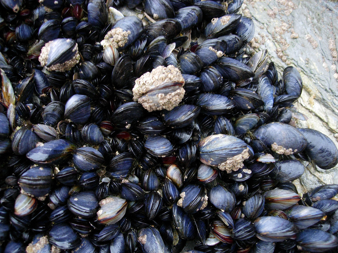
Mussels and barnacles in the intertidal near Newquay, Cornwall, England.
Wilson44691 at English Wikipedia – Photograph taken by Mark A. Wilson (Department of Geology, The College of Wooster

I love puns and word play so I was happy to see this in a June 9, 2017 news item on ScienceDaily,

Rice University chemists can thank the mussel for putting the muscle into their new macroscale scaffold fibers.

A June 9, 2017 Rice University news release (also on EurekAlert), which originated the news item, provides more details about the research,

The Rice lab of chemist Jeffrey Hartgerink had already figured out how to make biocompatible nanofibers out of synthetic peptides. In new work, the lab is using an amino acid found in the sticky feet of mussels to make those fibers line up into strong hydrogel strings.

Hartgerink and Rice graduate student I-Che Li introduced their room-temperature method this month in an open-access paper in the Journal of the American Chemical Society.

The hydrogel strings can be picked up and moved with tweezers, and Li said he expects they will help labs gain better control over the growth of cell cultures.

“Usually when cells grow on a surface, they spread randomly,” he said. “There are a lot of biomaterials we want to grow in a specific direction. With the hydrogel scaffold aligned, we can expect cells to grow the way we want them to. One example would be neuron cells, which we want to grow head-to-tail to aid nerve regeneration.

“Basically, this could allow us to direct cell growth from here to there,” he said. “That’s why this material is so exciting.”

In previous research Hartgerink’s lab had developed synthetic hydrogels that could be injected into the body to serve as scaffolds for tissue growth. The hydrogels contained hydrophobic peptides that self-assembled into fibers about 6 nanometers wide and up to several microns long. However, because the fibers did not interact with one other, they generally appeared in microscope images as a tangled mass.

Experiments showed the fibers could be coaxed into alignment with the application of shear forces, in the same way that playing cards are aligned during shuffling by pushing on both the top and bottom of the deck.

Hartgerink and Li decided to try pushing the fibers through a needle to force them into alignment, a process that would be easier if the material was water soluble. So they added a chain of amino acids known as DOPA to the sides of the fibers to allow them to remain water-soluble in the syringe, Li said.

DOPA — short for 3,4-dihydroxyphenylalanine — is the compound that lets mussels stick to just about anything. Hartgerink and Li found that the combination of DOPA and shear stress from passing through the needle prompted the fibers to form visible, rope-like bundles.

They also found that DOPA promoted chemical cross-linking reactions that helped the bundles hold their shape. “DOPA is really sensitive to oxidizing agents,” Li said. “Even exposing DOPA to air oxidizes it, and that aids in cross-linking the fibers.”

As a bonus, the aligned fibers also proved to have a curious and useful optical property called “uniform birefringence,” or double-refraction. Li said this could allow researchers to use polarized light to see exactly where the aligned fibers are, even if they’re covered by cells.

“This will be an important technique for us to make sure of the long-range order of fiber alignment when we are testing directed cell growth,” he said.

The researchers expect the aligned fibers can be used for macroscale medical applications but with nanoscale control over the structures.

“Self-assembly is basically the ability of a molecule to make ordered structure from chaos, and what I-Che has done is push this organization to a new level with his aligned strings,” said Hartgerink, a professor of chemistry and of bioengineering. “With this material, we are excited to see if we can impose this organization onto the growth of cells that interact with it.”

Here’s a link to and a citation for the paper,

Covalent Capture of Aligned Self-Assembling Nanofibers by I-Che Li and Jeffrey D. Hartgerink. J. Am. Chem. Soc., Article ASAP DOI: 10.1021/jacs.7b04655 Publication Date (Web): June 5, 2017

Copyright © 2017 American Chemical Society

This paper is open access.