Tag Archives: neurons

Nanotubes tunnel between neurons in Parkinson’s disease

An Aug. 22, 2016 news item on ScienceDaily describes how scientists from the Institut Pasteur (France) have developed insight into one of the processes in Parkinson’s disease,

Scientists have demonstrated the role of lysosomal vesicles in transporting alpha-synuclein aggregates, responsible for Parkinson’s and other neurodegenerative diseases, between neurons. These proteins move from one neuron to the next in lysosomal vesicles which travel along the ‘tunneling nanotubes’ between cells.

An Aug. 22, 2016 Institut Pasteur press release (also on EurekAlert), expands on the theme,

Synucleinopathies, a group of neurodegenerative diseases including Parkinson’s disease, are characterized by the pathological deposition of aggregates of the misfolded α-synuclein protein into inclusions throughout the central and peripheral nervous system. Intercellular propagation (from one neuron to the next) of α-synuclein aggregates contributes to the progression of the neuropathology, but little was known about the mechanism by which spread occurs.

In this study, scientists from the Membrane Traffic and Pathogenesis Unit, directed by Chiara Zurzolo at the Institut Pasteur, used fluorescence microscopy to demonstrate that pathogenic α-synuclein fibrils travel between neurons in culture, inside lysosomal vesicles through tunneling nanotubes (TNTs), a new mechanism of intercellular communication.

After being transferred via TNTs, α-synuclein fibrils are able to recruit and induce aggregation of the soluble α-synuclein protein in the cytosol of cells receiving the fibrils, thus explaining the propagation of the disease. The scientists propose that cells overloaded with α-synuclein aggregates in lysosomes dispose of this material by hijacking TNT-mediated intercellular trafficking. However, this results in the disease being spread to naive neurons.

This study demonstrates that TNTs play a significant part in the intercellular transfer of α-synuclein fibrils and reveals the specific role of lysosomes in this process. This represents a major breakthrough in understanding the mechanisms underlying the progression of synucleinopathies.

These compelling findings, together with previous reports from the same team, point to the general role of TNTs in the propagation of prion-like proteins in neurodegenerative diseases and identify TNTs as a new therapeutic target to combat the progression of these incurable diseases.

Here’s a link to and a citation for the paper,

Tunneling nanotubes spread fibrillar α‐synuclein by intercellular trafficking of lysosomes by Saïda Abounit, Luc Bousset, Frida Loria, Seng Zhu, Fabrice de Chaumont, Laura Pieri, Jean-Christophe Olivo-Marin, Ronald Melki, Chiara Zurzolo. The EMBO Journal (2016) e201593411 DOI 10.15252/embj.201593411 Published online 22.08.2016

This paper is behind a paywall.

US white paper on neuromorphic computing (or the nanotechnology-inspired Grand Challenge for future computing)

The US has embarked on a number of what is called “Grand Challenges.” I first came across the concept when reading about the Bill and Melinda Gates (of Microsoft fame) Foundation. I gather these challenges are intended to provide funding for research that advances bold visions.

There is the US National Strategic Computing Initiative established on July 29, 2015 and its first anniversary results were announced one year to the day later. Within that initiative a nanotechnology-inspired Grand Challenge for Future Computing was issued and, according to a July 29, 2016 news item on Nanowerk, a white paper on the topic has been issued (Note: A link has been removed),

Today [July 29, 2016), Federal agencies participating in the National Nanotechnology Initiative (NNI) released a white paper (pdf) describing the collective Federal vision for the emerging and innovative solutions needed to realize the Nanotechnology-Inspired Grand Challenge for Future Computing.

The grand challenge, announced on October 20, 2015, is to “create a new type of computer that can proactively interpret and learn from data, solve unfamiliar problems using what it has learned, and operate with the energy efficiency of the human brain.” The white paper describes the technical priorities shared by the agencies, highlights the challenges and opportunities associated with these priorities, and presents a guiding vision for the research and development (R&D) needed to achieve key technical goals. By coordinating and collaborating across multiple levels of government, industry, academia, and nonprofit organizations, the nanotechnology and computer science communities can look beyond the decades-old approach to computing based on the von Neumann architecture and chart a new path that will continue the rapid pace of innovation beyond the next decade.

A July 29, 2016 US National Nanotechnology Coordination Office news release, which originated the news item, further and succinctly describes the contents of the paper,

“Materials and devices for computing have been and will continue to be a key application domain in the field of nanotechnology. As evident by the R&D topics highlighted in the white paper, this challenge will require the convergence of nanotechnology, neuroscience, and computer science to create a whole new paradigm for low-power computing with revolutionary, brain-like capabilities,” said Dr. Michael Meador, Director of the National Nanotechnology Coordination Office. …

The white paper was produced as a collaboration by technical staff at the Department of Energy, the National Science Foundation, the Department of Defense, the National Institute of Standards and Technology, and the Intelligence Community. …

The white paper titled “A Federal Vision for Future Computing: A Nanotechnology-Inspired Grand Challenge” is 15 pp. and it offers tidbits such as this (Note: Footnotes not included),

A new materials base may be needed for future electronic hardware. While most of today’s electronics use silicon, this approach is unsustainable if billions of disposable and short-lived sensor nodes are needed for the coming Internet-of-Things (IoT). To what extent can the materials base for the implementation of future information technology (IT) components and systems support sustainability through recycling and bio-degradability? More sustainable materials, such as compostable or biodegradable systems (polymers, paper, etc.) that can be recycled or reused,  may play an important role. The potential role for such alternative materials in the fabrication of integrated systems needs to be explored as well. [p. 5]

The basic architecture of computers today is essentially the same as those built in the 1940s—the von Neumann architecture—with separate compute, high-speed memory, and high-density storage components that are electronically interconnected. However, it is well known that continued performance increases using this architecture are not feasible in the long term, with power density constraints being one of the fundamental roadblocks.7 Further advances in the current approach using multiple cores, chip multiprocessors, and associated architectures are plagued by challenges in software and programming models. Thus,  research and development is required in radically new and different computing architectures involving processors, memory, input-output devices, and how they behave and are interconnected. [p. 7]

Neuroscience research suggests that the brain is a complex, high-performance computing system with low energy consumption and incredible parallelism. A highly plastic and flexible organ, the human brain is able to grow new neurons, synapses, and connections to cope with an ever-changing environment. Energy efficiency, growth, and flexibility occur at all scales, from molecular to cellular, and allow the brain, from early to late stage, to never stop learning and to act with proactive intelligence in both familiar and novel situations. Understanding how these mechanisms work and cooperate within and across scales has the potential to offer tremendous technical insights and novel engineering frameworks for materials, devices, and systems seeking to perform efficient and autonomous computing. This research focus area is the most synergistic with the national BRAIN Initiative. However, unlike the BRAIN Initiative, where the goal is to map the network connectivity of the brain, the objective here is to understand the nature, methods, and mechanisms for computation,  and how the brain performs some of its tasks. Even within this broad paradigm,  one can loosely distinguish between neuromorphic computing and artificial neural network (ANN) approaches. The goal of neuromorphic computing is oriented towards a hardware approach to reverse engineering the computational architecture of the brain. On the other hand, ANNs include algorithmic approaches arising from machinelearning,  which in turn could leverage advancements and understanding in neuroscience as well as novel cognitive, mathematical, and statistical techniques. Indeed, the ultimate intelligent systems may as well be the result of merging existing ANN (e.g., deep learning) and bio-inspired techniques. [p. 8]

As government documents go, this is quite readable.

For anyone interested in learning more about the future federal plans for computing in the US, there is a July 29, 2016 posting on the White House blog celebrating the first year of the US National Strategic Computing Initiative Strategic Plan (29 pp. PDF; awkward but that is the title).

Memory material with functions resembling synapses and neurons in the brain

This work comes from the University of Twente’s MESA+ Institute for Nanotechnology according to a July 8, 2016 news item on ScienceDaily,

Our brain does not work like a typical computer memory storing just ones and zeroes: thanks to a much larger variation in memory states, it can calculate faster consuming less energy. Scientists of the MESA+ Institute for Nanotechnology of the University of Twente (The Netherlands) now developed a ferro-electric material with a memory function resembling synapses and neurons in the brain, resulting in a multistate memory. …

A July 8, 2016 University of Twente press release, which originated the news item, provides more technical detail,

The material that could be the basic building block for ‘brain-inspired computing’ is lead-zirconium-titanate (PZT): a sandwich of materials with several attractive properties. One of them is that it is ferro-electric: you can switch it to a desired state, this state remains stable after the electric field is gone. This is called polarization: it leads to a fast memory function that is non-volatile. Combined with processor chips, a computer could be designed that starts much faster, for example. The UT scientists now added a thin layer of zinc oxide to the PZT, 25 nanometer thickness. They discovered that switching from one state to another not only happens from ‘zero’ to ‘one’ vice versa. It is possible to control smaller areas within the crystal: will they be polarized (‘flip’) or not?

In a PZT layer without zinc oxide (ZnO) there are basically two memorystates. Adding a nano layer of ZnO, every state in between is possible as well.

Multistate

By using variable writing times in those smaller areas, the result is that many states can be stored anywhere between zero and one. This resembles the way synapses and neurons ‘weigh’ signals in our brain. Multistate memories, coupled to transistors, could drastically improve the speed of pattern recognition, for example: our brain performs this kind of tasks consuming only a fraction of the energy a computer system needs. Looking at the graphs, the writing times seem quite long compared to nowaday’s processor speeds, but it is possible to create many memories in parallel. The function of the brain has already been mimicked in software like neurale networks, but in that case conventional digital hardware is still a limitation. The new material is a first step towards electronic hardware with a brain-like memory. Finding solutions for combining PZT with semiconductors, or even developing new kinds of semiconductors for this, is one of the next steps.

Here’s a link to and a citation for the paper,

Multistability in Bistable Ferroelectric Materials toward Adaptive Applications by Anirban Ghosh, Gertjan Koster, and Guus Rijnders. Advanced Functional Materials DOI: 10.1002/adfm.201601353 Version of Record online: 4 JUL 2016

© 2016 WILEY-VCH Verlag GmbH & Co. KGaA, Weinheim

This paper is behind a paywall.

3D brain-on-a-chip from the University of Twente

Dutch researchers have developed a 3D brain-on-a-chip according to a June 23, 2016 news item on Nanowerk,

To study brain cell’s operation and test the effect of medication on individual cells, the conventional Petri dish with flat electrodes is not sufficient. For truly realistic studies, cells have to flourish within three-dimensional surroundings.

Bart Schurink, researcher at University of Twente’s MESA+ Institute for Nanotechnology, has developed a sieve with 900 openings, each of which has the shape of an inverted pyramid. On top of this array of pyramids, a micro-reactor takes care of cell growth. Schurink defends his PhD thesis June 23 [2016].

A June 23, 2016 University of Twente press release, which originated the news item, provides more detail,

A brain-on-a-chip demands more than a series of electrodes in 2D, on which brain cells can be cultured. To mimic the brain in a realistic way, you need facilities for fluid flow, and the cells need some freedom for themselves even when they are kept at predefined spaces. Schurink therefore developed a micro sieve structure with hundreds of openings on a 2 by 2 mm surface. Each of these holes has the shape of  an inverted pyramid. Each pyramid, in turn, is equipped with an electrode, for measuring electrical signals or sending stimuli to the network. At the same time, liquids can flow through tiny holes, needed to capture the cells and for sending nutrients or medication to a single cell.

NEURONAL NETWORK

After neurons have been placed inside all the pyramids, they will start to form a network. This is not just a 2D network between the holes: by placing a micro reactor on top of the sieve, a neuron network can develop in the vertical direction as well. Growth and electrical activity can be monitored subsequently: each individual cell can be identified by the pyramid it is in. Manufacturing this system, demands a lot of both the production facilities at UT’s NanoLab and of creative solutions the designers come up with. For example, finding the proper way of guaranteeing  the same dimensions for every hole, is quite challenging.

Schurink’s new µSEA (micro sieve electrode array) has been tested with living cells, from the brains of laboratory rats. Both the positioning of the cells and neuronal network growth have been tested. The result of this PhD research is a fully new research platform for performing research on the brain, diseases and effects of medication.

Schurink (1982) has conducted his research within the group Meso Scale Chemical Systems, of Prof Han Gardeniers. The group is part of the MESA+ Institute for Nanotechnology of the University of Twente. Schurink’s thesis is titled ‘Microfabrication and microfluidics for 3D brain-on-chip’ …

I have written about one other piece about a ‘3D’ organ-on-a-chip project in China (my Jan. 29, 2016 posting).

Small, soft, and electrically functional: an injectable biomaterial

This development could be looked at as a form of synthetic biology without the genetic engineering. From a July 1, 2016 news item on ScienceDaily,

Ideally, injectable or implantable medical devices should not only be small and electrically functional, they should be soft, like the body tissues with which they interact. Scientists from two UChicago labs set out to see if they could design a material with all three of those properties.

The material they came up with, published online June 27, 2016, in Nature Materials, forms the basis of an ingenious light-activated injectable device that could eventually be used to stimulate nerve cells and manipulate the behavior of muscles and organs.

“Most traditional materials for implants are very rigid and bulky, especially if you want to do electrical stimulation,” said Bozhi Tian, an assistant professor in chemistry whose lab collaborated with that of neuroscientist Francisco Bezanilla on the research.

The new material, in contrast, is soft and tiny — particles just a few micrometers in diameter (far less than the width of a human hair) that disperse easily in a saline solution so they can be injected. The particles also degrade naturally inside the body after a few months, so no surgery would be needed to remove them.

A July 1, 2016 University of Chicago news release (also on EurekAlert) by , which originated the news item, provides more detail,

Each particle is built of two types of silicon that together form a structure full of nano-scale pores, like a tiny sponge. And like a sponge, it is squishy — a hundred to a thousand times less rigid than the familiar crystalline silicon used in transistors and solar cells. “It is comparable to the rigidity of the collagen fibers in our bodies,” said Yuanwen Jiang, Tian’s graduate student. “So we’re creating a material that matches the rigidity of real tissue.”

The material constitutes half of an electrical device that creates itself spontaneously when one of the silicon particles is injected into a cell culture, or, eventually, a human body. The particle attaches to a cell, making an interface with the cell’s plasma membrane. Those two elements together — cell membrane plus particle — form a unit that generates current when light is shined on the silicon particle.

“You don’t need to inject the entire device; you just need to inject one component,” João L. Carvalho-de-Souza , Bezanilla’s postdoc said. “This single particle connection with the cell membrane allows sufficient generation of current that could be used to stimulate the cell and change its activity. After you achieve your therapeutic goal, the material degrades naturally. And if you want to do therapy again, you do another injection.”

The scientists built the particles using a process they call nano-casting. They fabricate a silicon dioxide mold composed of tiny channels — “nano-wires” — about seven nanometers in diameter (less than 10,000 times smaller than the width of a human hair) connected by much smaller “micro-bridges.” Into the mold they inject silane gas, which fills the pores and channels and decomposes into silicon.

And this is where things get particularly cunning. The scientists exploit the fact the smaller an object is, the more the atoms on its surface dominate its reactions to what is around it. The micro-bridges are minute, so most of their atoms are on the surface. These interact with oxygen that is present in the silicon dioxide mold, creating micro-bridges made of oxidized silicon gleaned from materials at hand. The much larger nano-wires have proportionately fewer surface atoms, are much less interactive, and remain mostly pure silicon. [I have a note regarding ‘micro’ and ‘nano’ later in this posting.]

“This is the beauty of nanoscience,” Jiang said. “It allows you to engineer chemical compositions just by manipulating the size of things.”

Web-like nanostructure

Finally, the mold is dissolved. What remains is a web-like structure of silicon nano-wires connected by micro-bridges of oxidized silicon that can absorb water and help increase the structure’s softness. The pure silicon retains its ability to absorb light.

Transmission electron microscopy image shows an ordered nanowire array. The 100-nanometer scale bar is 1,000 times narrower than a hair. Courtesy of Tian Lab

Transmission electron microscopy image shows an ordered nanowire array. The 100-nanometer scale bar is 1,000 times narrower than a hair. Courtesy of
Tian Lab

The scientists have added the particles onto neurons in culture in the lab, shone light on the particles, and seen current flow into the neurons which activates the cells. The next step is to see what happens in living animals. They are particularly interested in stimulating nerves in the peripheral nervous system that connect to organs. These nerves are relatively close to the surface of the body, so near-infra-red wavelength light can reach them through the skin.

Tian imagines using the light-activated devices to engineer human tissue and create artificial organs to replace damaged ones. Currently, scientists can make engineered organs with the correct form but not the ideal function.

To get a lab-built organ to function properly, they will need to be able to manipulate individual cells in the engineered tissue. The injectable device would allow a scientist to do that, tweaking an individual cell using a tightly focused beam of light like a mechanic reaching into an engine and turning a single bolt. The possibility of doing this kind of synthetic biology without genetic engineering [emphasis mine] is enticing.

“No one wants their genetics to be altered,” Tian said. “It can be risky. There’s a need for a non-genetic system that can still manipulate cell behavior. This could be that kind of system.”

Tian’s graduate student Yuanwen Jiang did the material development and characterization on the project. The biological part of the collaboration was done in the lab of Francisco Bezanilla, the Lillian Eichelberger Cannon Professor of Biochemistry and Molecular Biology, by postdoc João L. Carvalho-de-Souza. They were, said Tian, the “heroes” of the work.

I was a little puzzled about the use of the word ‘micro’ in a context suggesting it was smaller than something measured at the nanoscale. Dr. Tian very kindly cleared up my confusion with this response in a July 4, 2016 email,

In fact, the definition of ‘micro’ and ’nano’ have been quite ambiguous in literature. For example, microporous materials (e.g., zeolite) usually refer to materials with pore sizes of less than 2 nm — this is defined based on IUPAC [International Union of Pure and Applied Chemistry] definition (http://goldbook.iupac.org/M03853.html). We used ‘micro-bridges’ because they come from the ‘micropores’ in the original template.

Thank you Dr. Tian for that very clear reply and Steve Koppes for forwarding my request to Dr. Tian!

Here’s a link to and a citation for the paper,

Heterogeneous silicon mesostructures for lipid-supported bioelectric interfaces by Yuanwen Jiang, João L. Carvalho-de-Souza, Raymond C. S. Wong, Zhiqiang Luo, Dieter Isheim, Xiaobing Zuo, Alan W. Nicholls, Il Woong Jung, Jiping Yue, Di-Jia Liu, Yucai Wang, Vincent De Andrade, Xianghui Xiao, Luizetta Navrazhnykh, Dara E. Weiss, Xiaoyang Wu, David N. Seidman, Francisco Bezanilla, & Bozhi Tian. Nature Materials (2016)  doi:10.1038/nmat4673 Published online 27 June 2016

This paper is behind a paywall.

I gather animal testing will be the next step as they continue to develop this exciting technology. Good luck!

Replicating brain’s neural networks with 3D nanoprinting

An announcement about European Union funding for a project to reproduce neural networks by 3D nanoprinting can be found in a June 10, 2016 news item on Nanowerk,

The MESO-BRAIN consortium has received a prestigious award of €3.3million in funding from the European Commission as part of its Future and Emerging Technology (FET) scheme. The project aims to develop three-dimensional (3D) human neural networks with specific biological architecture, and the inherent ability to interrogate the network’s brain-like activity both electrophysiologically and optically. It is expected that the MESO-BRAIN will facilitate a better understanding of human disease progression, neuronal growth and enable the development of large-scale human cell-based assays to test the modulatory effects of pharmacological and toxicological compounds on neural network activity. The use of more physiologically relevant human models will increase drug screening efficiency and reduce the need for animal testing.

A June 9, 2016 Institute of Photonic Sciences (ICFO) press release (also on EurekAlert), which originated the news item, provides more detail,

About the MESO-BRAIN project

The MESO-BRAIN project’s cornerstone will use human induced pluripotent stem cells (iPSCs) that have been differentiated into neurons upon a defined and reproducible 3D scaffold to support the development of human neural networks that emulate brain activity. The structure will be based on a brain cortical module and will be unique in that it will be designed and produced using nanoscale 3D-laser-printed structures incorporating nano-electrodes to enable downstream electrophysiological analysis of neural network function. Optical analysis will be conducted using cutting-edge light sheet-based, fast volumetric imaging technology to enable cellular resolution throughout the 3D network. The MESO-BRAIN project will allow for a comprehensive and detailed investigation of neural network development in health and disease.

Prof Edik Rafailov, Head of the MESO-BRAIN project (Aston University) said: “What we’re proposing to achieve with this project has, until recently, been the stuff of science fiction. Being able to extract and replicate neural networks from the brain through 3D nanoprinting promises to change this. The MESO-BRAIN project has the potential to revolutionise the way we are able to understand the onset and development of disease and discover treatments for those with dementia or brain injuries. We cannot wait to get started!”

The MESO-BRAIN project will launch in September 2016 and research will be conducted over three years.

About the MESO-BRAIN consortium

Each of the consortium partners have been chosen for the highly specific skills & knowledge that they bring to this project. These include technologies and expertise in stem cells, photonics, physics, 3D nanoprinting, electrophysiology, molecular biology, imaging and commercialisation.

Aston University (UK) Aston Institute of Photonic Technologies (School of Engineering and Applied Science) is one of the largest photonic groups in UK and an internationally recognised research centre in the fields of lasers, fibre-optics, high-speed optical communications, nonlinear and biomedical photonics. The Cell & Tissue Biomedical Research Group (Aston Research Centre for Healthy Ageing) combines collective expertise in genetic manipulation, tissue engineering and neuronal modelling with the electrophysiological and optical analysis of human iPSC-derived neural networks. Axol Bioscience Ltd. (UK) was founded to fulfil the unmet demand for high quality, clinically relevant human iPSC-derived cells for use in biomedical research and drug discovery. The Laser Zentrum Hannover (Germany) is a leading research organisation in the fields of laser development, material processing, laser medicine, and laser-based nanotechnologies. The Neurophysics Group (Physics Department) at University of Barcelona (Spain) are experts in combing experiments with theoretical and computational modelling to infer functional connectivity in neuronal circuits. The Institute of Photonic Sciences (ICFO) (Spain) is a world-leading research centre in photonics with expertise in several microscopy techniques including light sheet imaging. KITE Innovation (UK) helps to bridge the gap between the academic and business sectors in supporting collaboration, enterprise, and knowledge-based business development.

For anyone curious about the FET funding scheme, there’s this from the press release,

Horizon 2020 aims to ensure Europe produces world-class science by removing barriers to innovation through funding programmes such as the FET. The FET (Open) funds forward-looking collaborations between advanced multidisciplinary science and cutting-edge engineering for radically new future technologies. The published success rate is below 1.4%, making it amongst the toughest in the Horizon 2020 suite of funding schemes. The MESO-BRAIN proposal scored a perfect 5/5.

You can find out more about the MESO-BRAIN project on its ICFO webpage.

They don’t say anything about it but I can’t help wondering if the scientists aren’t also considering the possibility of creating an artificial brain.

Memristor-based electronic synapses for neural networks

Caption: Neuron connections in biological neural networks. Credit: MIPT press office

Caption: Neuron connections in biological neural networks. Credit: MIPT press office

Russian scientists have recently published a paper about neural networks and electronic synapses based on ‘thin film’ memristors according to an April 19, 2016 news item on Nanowerk,

A team of scientists from the Moscow Institute of Physics and Technology (MIPT) have created prototypes of “electronic synapses” based on ultra-thin films of hafnium oxide (HfO2). These prototypes could potentially be used in fundamentally new computing systems.

An April 20, 2016 MIPT press release (also on EurekAlert), which originated the news item (the date inconsistency likely due to timezone differences) explains the connection between thin films and memristors,

The group of researchers from MIPT have made HfO2-based memristors measuring just 40×40 nm2. The nanostructures they built exhibit properties similar to biological synapses. Using newly developed technology, the memristors were integrated in matrices: in the future this technology may be used to design computers that function similar to biological neural networks.

Memristors (resistors with memory) are devices that are able to change their state (conductivity) depending on the charge passing through them, and they therefore have a memory of their “history”. In this study, the scientists used devices based on thin-film hafnium oxide, a material that is already used in the production of modern processors. This means that this new lab technology could, if required, easily be used in industrial processes.

“In a simpler version, memristors are promising binary non-volatile memory cells, in which information is written by switching the electric resistance – from high to low and back again. What we are trying to demonstrate are much more complex functions of memristors – that they behave similar to biological synapses,” said Yury Matveyev, the corresponding author of the paper, and senior researcher of MIPT’s Laboratory of Functional Materials and Devices for Nanoelectronics, commenting on the study.

The press release offers a description of biological synapses and their relationship to learning and memory,

A synapse is point of connection between neurons, the main function of which is to transmit a signal (a spike – a particular type of signal, see fig. 2) from one neuron to another. Each neuron may have thousands of synapses, i.e. connect with a large number of other neurons. This means that information can be processed in parallel, rather than sequentially (as in modern computers). This is the reason why “living” neural networks are so immensely effective both in terms of speed and energy consumption in solving large range of tasks, such as image / voice recognition, etc.

Over time, synapses may change their “weight”, i.e. their ability to transmit a signal. This property is believed to be the key to understanding the learning and memory functions of thebrain.

From the physical point of view, synaptic “memory” and “learning” in the brain can be interpreted as follows: the neural connection possesses a certain “conductivity”, which is determined by the previous “history” of signals that have passed through the connection. If a synapse transmits a signal from one neuron to another, we can say that it has high “conductivity”, and if it does not, we say it has low “conductivity”. However, synapses do not simply function in on/off mode; they can have any intermediate “weight” (intermediate conductivity value). Accordingly, if we want to simulate them using certain devices, these devices will also have to have analogous characteristics.

The researchers have provided an illustration of a biological synapse,

Fig.2 The type of electrical signal transmitted by neurons (a “spike”). The red lines are various other biological signals, the black line is the averaged signal. Source: MIPT press office

Fig.2 The type of electrical signal transmitted by neurons (a “spike”). The red lines are various other biological signals, the black line is the averaged signal. Source: MIPT press office

Now, the press release ties the memristor information together with the biological synapse information to describe the new work at the MIPT,

As in a biological synapse, the value of the electrical conductivity of a memristor is the result of its previous “life” – from the moment it was made.

There is a number of physical effects that can be exploited to design memristors. In this study, the authors used devices based on ultrathin-film hafnium oxide, which exhibit the effect of soft (reversible) electrical breakdown under an applied external electric field. Most often, these devices use only two different states encoding logic zero and one. However, in order to simulate biological synapses, a continuous spectrum of conductivities had to be used in the devices.

“The detailed physical mechanism behind the function of the memristors in question is still debated. However, the qualitative model is as follows: in the metal–ultrathin oxide–metal structure, charged point defects, such as vacancies of oxygen atoms, are formed and move around in the oxide layer when exposed to an electric field. It is these defects that are responsible for the reversible change in the conductivity of the oxide layer,” says the co-author of the paper and researcher of MIPT’s Laboratory of Functional Materials and Devices for Nanoelectronics, Sergey Zakharchenko.

The authors used the newly developed “analogue” memristors to model various learning mechanisms (“plasticity”) of biological synapses. In particular, this involved functions such as long-term potentiation (LTP) or long-term depression (LTD) of a connection between two neurons. It is generally accepted that these functions are the underlying mechanisms of  memory in the brain.

The authors also succeeded in demonstrating a more complex mechanism – spike-timing-dependent plasticity, i.e. the dependence of the value of the connection between neurons on the relative time taken for them to be “triggered”. It had previously been shown that this mechanism is responsible for associative learning – the ability of the brain to find connections between different events.

To demonstrate this function in their memristor devices, the authors purposefully used an electric signal which reproduced, as far as possible, the signals in living neurons, and they obtained a dependency very similar to those observed in living synapses (see fig. 3).

Fig.3. The change in conductivity of memristors depending on the temporal separation between "spikes"(rigth) and thr change in potential of the neuron connections in biological neural networks. Source: MIPT press office

Fig.3. The change in conductivity of memristors depending on the temporal separation between “spikes”(rigth) and thr change in potential of the neuron connections in biological neural networks. Source: MIPT press office

These results allowed the authors to confirm that the elements that they had developed could be considered a prototype of the “electronic synapse”, which could be used as a basis for the hardware implementation of artificial neural networks.

“We have created a baseline matrix of nanoscale memristors demonstrating the properties of biological synapses. Thanks to this research, we are now one step closer to building an artificial neural network. It may only be the very simplest of networks, but it is nevertheless a hardware prototype,” said the head of MIPT’s Laboratory of Functional Materials and Devices for Nanoelectronics, Andrey Zenkevich.

Here’s a link to and a citation for the paper,

Crossbar Nanoscale HfO2-Based Electronic Synapses by Yury Matveyev, Roman Kirtaev, Alena Fetisova, Sergey Zakharchenko, Dmitry Negrov and Andrey Zenkevich. Nanoscale Research Letters201611:147 DOI: 10.1186/s11671-016-1360-6

Published: 15 March 2016

This is an open access paper.

Graphene Flagship high points

The European Union’s Graphene Flagship project has provided a series of highlights in place of an overview for the project’s ramp-up phase (in 2013 the Graphene Flagship was announced as one of two winners of a science competition, the other winner was the Human Brain Project, with two prizes of 1B Euros for each project). Here are the highlights from the April 19, 2016 Graphene Flagship press release,

Graphene and Neurons – the Best of Friends

Flagship researchers have shown that it is possible to interface untreated graphene with neuron cells whilst maintaining the integrity of these vital cells [1]. This result is a significant first step towards using graphene to produce better deep brain implants which can both harness and control the brain.

Graphene and Neurons
 

This paper emerged from the Graphene Flagship Work Package Health and Environment. Prof. Prato, the WP leader from the University of Trieste in Italy, commented that “We are currently involved in frontline research in graphene technology towards biomedical applications, exploring the interactions between graphene nano- and micro-sheets with the sophisticated signalling machinery of nerve cells. Our work is a first step in that direction.”

[1] Fabbro A., et al., Graphene-Based Interfaces do not Alter Target Nerve Cells. ACS Nano, 10 (1), 615 (2016).

Pressure Sensing with Graphene: Quite a Squeeze

The Graphene Flagship developed a small, robust, highly efficient squeeze film pressure sensor [2]. Pressure sensors are present in most mobile handsets and by replacing current sensor membranes with a graphene membrane they allow the sensor to decrease in size and significantly increase its responsiveness and lifetime.

Discussing this work which emerged from the Graphene Flagship Work Package Sensors is the paper’s lead author, Robin Dolleman from the Technical University of Delft in The Netherlands “After spending a year modelling various systems the idea of the squeeze-film pressure sensor was formed. Funding from the Graphene Flagship provided the opportunity to perform the experiments and we obtained very good results. We built a squeeze-film pressure sensor from 31 layers of graphene, which showed a 45 times higher response than silicon based devices, while reducing the area of the device by a factor of 25. Currently, our work is focused on obtaining similar results on monolayer graphene.”


[2] Dolleman R. J. et al., Graphene Squeeze-Film Pressure Sensors. Nano Lett., 16, 568 (2016)

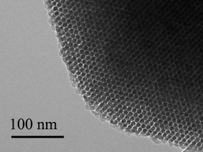
Frictionless Graphene


Image caption: A graphene nanoribbon was anchored at the tip of a atomic force microscope and dragged over a gold surface. The observed friction force was extremely low.

Image caption: A graphene nanoribbon was anchored at the tip of a atomic force microscope and dragged over a gold surface. The observed friction force was extremely low.

Research done within the Graphene Flagship, has observed the onset of superlubricity in graphene nanoribbons sliding on a surface, unravelling the role played by ribbon size and elasticity [3]. This important finding opens up the development potential of nanographene frictionless coatings. This research lead by the Graphene Flagship Work Package Nanocomposites also involved researchers from Work Package Materials and Work Package Health and the Environment, a shining example of the inter-disciplinary, cross-collaborative approach to research undertaken within the Graphene Flagship. Discussing this further is the Work Package Nanocomposites Leader, Dr Vincenzo Palermo from CNR National Research Council, Italy “Strengthening the collaboration and interactions with other Flagship Work Packages created added value through a strong exchange of materials, samples and information”.

[3] Kawai S., et al., Superlubricity of graphene nanoribbons on gold surfaces. Science. 351, 6276, 957 (2016) 

​Graphene Paddles Forward

Work undertaken within the Graphene Flagship saw Spanish automotive interiors specialist, and Flagship partner, Grupo Antolin SA work in collaboration with Roman Kayaks to develop an innovative kayak that incorporates graphene into its thermoset polymeric matrices. The use of graphene and related materials results in a significant increase in both impact strength and stiffness, improving the resistance to breakage in critical areas of the boat. Pushing the graphene canoe well beyond the prototype demonstration bubble, Roman Kayaks chose to use the K-1 kayak in the Canoe Marathon World Championships held in September in Gyor, Hungary where the Graphene Canoe was really put through its paces.

Talking further about this collaboration from the Graphene Flagship Work Package Production is the WP leader, Dr Ken Teo from Aixtron Ltd., UK “In the Graphene Flagship project, Work Package Production works as a technology enabler for real-world applications. Here we show the worlds first K-1 kayak (5.2 meters long), using graphene related materials developed by Grupo Antolin. We are very happy to see that graphene is creating value beyond traditional industries.” 

​Graphene Production – a Kitchen Sink Approach

Researchers from the Graphene Flagship have devised a way of producing large quantities of graphene by separating graphite flakes in liquids with a rotating tool that works in much the same way as a kitchen blender [4]. This paves the way to mass production of high quality graphene at a low cost.

The method was produced within the Graphene Flagship Work Package Production and is talked about further here by the WP deputy leader, Prof. Jonathan Coleman from Trinity College Dublin, Ireland “This technique produced graphene at higher rates than most other methods, and produced sheets of 2D materials that will be useful in a range of applications, from printed electronics to energy generation.” 

[4] Paton K.R., et al., Scalable production of large quantities of defect-free few-layer graphene by shear exfoliation in liquids. Nat. Mater. 13, 624 (2014).

Flexible Displays – Rolled Up in your Pocket

Working with researchers from the Graphene Flagship the Flagship partner, FlexEnable, demonstrated the world’s first flexible display with graphene incorporated into its pixel backplane. Combined with an electrophoretic imaging film, the result is a low-power, durable display suitable for use in many and varied environments.

Emerging from the Graphene Flagship Work Package Flexible Electronics this illustrates the power of collaboration.  Talking about this is the WP leader Dr Henrik Sandberg from the VTT Technical Research Centre of Finland Ltd., Finland “Here we show the power of collaboration. To deliver these flexible demonstrators and prototypes we have seen materials experts working together with components manufacturers and system integrators. These devices will have a potential impact in several emerging fields such as wearables and the Internet of Things.”

​Fibre-Optics Data Boost from Graphene

A team of researches from the Graphene Flagship have demonstrated high-performance photo detectors for infrared fibre-optic communication systems based on wafer-scale graphene [5]. This can increase the amount of information transferred whilst at the same time make the devises smaller and more cost effective.

Discussing this work which emerged from the Graphene Flagship Work Package Optoelectronics is the paper’s lead author, Daniel Schall from AMO, Germany “Graphene has outstanding properties when it comes to the mobility of its electric charge carriers, and this can increase the speed at which electronic devices operate.”

[5] Schall D., et al., 50 GBit/s Photodetectors Based on Wafer-Scale Graphene for Integrated Silicon Photonic Communication Systems. ACS Photonics. 1 (9), 781 (2014)

​Rechargeable Batteries with Graphene

A number of different research groups within the Graphene Flagship are working on rechargeable batteries. One group has developed a graphene-based rechargeable battery of the lithium-ion type used in portable electronic devices [6]. Graphene is incorporated into the battery anode in the form of a spreadable ink containing a suspension of graphene nanoflakes giving an increased energy efficiency of 20%. A second group of researchers have demonstrated a lithium-oxygen battery with high energy density, efficiency and stability [7]. They produced a device with over 90% efficiency that may be recharged more than 2,000 times. Their lithium-oxygen cell features a porous, ‘fluffy’ electrode made from graphene together with additives that alter the chemical reactions at work in the battery.

Graphene Flagship researchers show how the 2D material graphene can improve the energy capacity, efficiency and stability of lithium-oxygen batteries.

Both devices were developed in different groups within the Graphene Flagship Work Package Energy and speaking of the technology further is Prof. Clare Grey from Cambridge University, UK “What we’ve achieved is a significant advance for this technology, and suggests whole new areas for research – we haven’t solved all the problems inherent to this chemistry, but our results do show routes forward towards a practical device”.

[6] Liu T., et al. Cycling Li-O2 batteries via LiOH formation and decomposition. Science. 350, 6260, 530 (2015)

[7] Hassoun J., et al., An Advanced Lithium-Ion Battery Based on a Graphene Anode and a Lithium Iron Phosphate Cathode. Nano Lett., 14 (8), 4901 (2014)

Graphene – What and Why?

Graphene is a two-dimensional material formed by a single atom-thick layer of carbon, with the carbon atoms arranged in a honeycomb-like lattice. This transparent, flexible material has a number of unique properties. For example, it is 100 times stronger than steel, and conducts electricity and heat with great efficiency.

A number of practical applications for graphene are currently being developed. These include flexible and wearable electronics and antennas, sensors, optoelectronics and data communication systems, medical and bioengineering technologies, filtration, super-strong composites, photovoltaics and energy storage.

Graphene and Beyond

The Graphene Flagship also covers other layered materials, as well as hybrids formed by combining graphene with these complementary materials, or with other materials and structures, ranging from polymers, to metals, cement, and traditional semiconductors such as silicon. Graphene is just the first of thousands of possible single layer materials. The Flagship plans to accelerate their journey from laboratory to factory floor.

Especially exciting is the possibility of stacking monolayers of different elements to create materials not found in nature, with properties tailored for specific applications. Such composite layered materials could be combined with other nanomaterials, such as metal nanoparticles, in order to further enhance their properties and uses.​

Graphene – the Fruit of European Scientific Excellence

Europe, North America and Asia are all active centres of graphene R&D, but Europe has special claim to be at the centre of this activity. The ground-breaking experiments on graphene recognised in the award of the 2010 Nobel Prize in Physics were conducted by European physicists, Andre Geim and Konstantin Novoselov, both at Manchester University. Since then, graphene research in Europe has continued apace, with major public funding for specialist centres, and the stimulation of academic-industrial partnerships devoted to graphene and related materials. It is European scientists and engineers who as part of the Graphene Flagship are closely coordinating research efforts, and accelerating the transfer of layered materials from the laboratory to factory floor.

For anyone who would like links to the published papers, you can check out an April 20, 2016 news item featuring the Graphene Flagship highlights on Nanowerk.

3D microtopographic scaffolds for transplantation and generation of reprogrammed human neurons

Should this technology prove successful once they start testing on people, the stated goal is to use it for the treatment of human neurodegenerative disorders such as Parkinson’s disease.  But, I can’t help wondering if they might also consider constructing an artificial brain.

Getting back to the 3D scaffolds for neurons, a March 17, 2016 US National Institutes of Health (NIH) news release (also on EurekAlert), makes the announcement,

National Institutes of Health-funded scientists have developed a 3D micro-scaffold technology that promotes reprogramming of stem cells into neurons, and supports growth of neuronal connections capable of transmitting electrical signals. The injection of these networks of functioning human neural cells — compared to injecting individual cells — dramatically improved their survival following transplantation into mouse brains. This is a promising new platform that could make transplantation of neurons a viable treatment for a broad range of human neurodegenerative disorders.

Previously, transplantation of neurons to treat neurodegenerative disorders, such as Parkinson’s disease, had very limited success due to poor survival of neurons that were injected as a solution of individual cells. The new research is supported by the National Institute of Biomedical Imaging and Bioengineering (NIBIB), part of NIH.

“Working together, the stem cell biologists and the biomaterials experts developed a system capable of shuttling neural cells through the demanding journey of transplantation and engraftment into host brain tissue,” said Rosemarie Hunziker, Ph.D., director of the NIBIB Program in Tissue Engineering and Regenerative Medicine. “This exciting work was made possible by the close collaboration of experts in a wide range of disciplines.”

The research was performed by researchers from Rutgers University, Piscataway, New Jersey, departments of Biomedical Engineering, Neuroscience and Cell Biology, Chemical and Biochemical Engineering, and the Child Health Institute; Stanford University School of Medicine’s Institute of Stem Cell Biology and Regenerative Medicine, Stanford, California; the Human Genetics Institute of New Jersey, Piscataway; and the New Jersey Center for Biomaterials, Piscataway. The results are reported in the March 17, 2016 issue of Nature Communications.

The researchers experimented in creating scaffolds made of different types of polymer fibers, and of varying thickness and density. They ultimately created a web of relatively thick fibers using a polymer that stem cells successfully adhered to. The stem cells used were human induced pluripotent stem cells (iPSCs), which can be readily generated from adult cell types such as skin cells. The iPSCs were induced to differentiate into neural cells by introducing the protein NeuroD1 into the cells.

The space between the polymer fibers turned out to be critical. “If the scaffolds were too dense, the stem cell-derived neurons were unable to integrate into the scaffold, whereas if they are too sparse then the network organization tends to be poor,” explained Prabhas Moghe, Ph.D., distinguished professor of biomedical engineering & chemical engineering at Rutgers University and co-senior author of the paper. “The optimal pore size was one that was large enough for the cells to populate the scaffold but small enough that the differentiating neurons sensed the presence of their neighbors and produced outgrowths resulting in cell-to-cell contact. This contact enhances cell survival and development into functional neurons able to transmit an electrical signal across the developing neural network.”

To test the viability of neuron-seeded scaffolds when transplanted, the researchers created micro-scaffolds that were small enough for injection into mouse brain tissue using a standard hypodermic needle. They injected scaffolds carrying the human neurons into brain slices from mice and compared them to human neurons injected as individual, dissociated cells.

The neurons on the scaffolds had dramatically increased cell-survival compared with the individual cell suspensions. The scaffolds also promoted improved neuronal outgrowth and electrical activity. Neurons injected individually in suspension resulted in very few cells surviving the transplant procedure.

Human neurons on scaffolds compared to neurons in solution were then tested when injected into the brains of live mice. Similar to the results in the brain slices, the survival rate of neurons on the scaffold network was increased nearly 40-fold compared to injected isolated cells. A critical finding was that the neurons on the micro-scaffolds expressed proteins that are involved in the growth and maturation of neural synapses–a good indication that the transplanted neurons were capable of functionally integrating into the host brain tissue.

The success of the study gives this interdisciplinary group reason to believe that their combined areas of expertise have resulted in a system with much promise for eventual treatment of human neurodegenerative disorders. In fact, they are now refining their system for specific use as an eventual transplant therapy for Parkinson’s disease. The plan is to develop methods to differentiate the stem cells into neurons that produce dopamine, the specific neuron type that degenerates in individuals with Parkinson’s disease. The work also will include fine-tuning the scaffold materials, mechanics and dimensions to optimize the survival and function of dopamine-producing neurons, and finding the best mouse models of the disease to test this Parkinson’s-specific therapy.

Here’s a link to and a citation for the paper,

Generation and transplantation of reprogrammed human neurons in the brain using 3D microtopographic scaffolds by Aaron L. Carlson, Neal K. Bennett, Nicola L. Francis, Apoorva Halikere, Stephen Clarke, Jennifer C. Moore, Ronald P. Hart, Kenneth Paradiso, Marius Wernig, Joachim Kohn, Zhiping P. Pang, & Prabhas V. Moghe. Nature Communications 7, Article number: 10862  doi:10.1038/ncomms10862 Published 17 March 2016

This paper is open access.

Graphene and neurons in a UK-Italy-Spain collaboration

There’s been a lot of talk about using graphene-based implants in the brain due to the material’s flexibility along with its other properties. A step forward has been taking according to a Jan. 29, 2016 news item on phys.org,

Researchers have successfully demonstrated how it is possible to interface graphene – a two-dimensional form of carbon – with neurons, or nerve cells, while maintaining the integrity of these vital cells. The work may be used to build graphene-based electrodes that can safely be implanted in the brain, offering promise for the restoration of sensory functions for amputee or paralysed patients, or for individuals with motor disorders such as epilepsy or Parkinson’s disease.

A Jan. 29, 2016 Cambridge University press release (also on EurekAlert), which originated the news item, provides more detail,

Previously, other groups had shown that it is possible to use treated graphene to interact with neurons. However the signal to noise ratio from this interface was very low. By developing methods of working with untreated graphene, the researchers retained the material’s electrical conductivity, making it a significantly better electrode.

“For the first time we interfaced graphene to neurons directly,” said Professor Laura Ballerini of the University of Trieste in Italy. “We then tested the ability of neurons to generate electrical signals known to represent brain activities, and found that the neurons retained their neuronal signalling properties unaltered. This is the first functional study of neuronal synaptic activity using uncoated graphene based materials.”

Our understanding of the brain has increased to such a degree that by interfacing directly between the brain and the outside world we can now harness and control some of its functions. For instance, by measuring the brain’s electrical impulses, sensory functions can be recovered. This can be used to control robotic arms for amputee patients or any number of basic processes for paralysed patients – from speech to movement of objects in the world around them. Alternatively, by interfering with these electrical impulses, motor disorders (such as epilepsy or Parkinson’s) can start to be controlled.

Scientists have made this possible by developing electrodes that can be placed deep within the brain. These electrodes connect directly to neurons and transmit their electrical signals away from the body, allowing their meaning to be decoded.

However, the interface between neurons and electrodes has often been problematic: not only do the electrodes need to be highly sensitive to electrical impulses, but they need to be stable in the body without altering the tissue they measure.

Too often the modern electrodes used for this interface (based on tungsten or silicon) suffer from partial or complete loss of signal over time. This is often caused by the formation of scar tissue from the electrode insertion, which prevents the electrode from moving with the natural movements of the brain due to its rigid nature.

Graphene has been shown to be a promising material to solve these problems, because of its excellent conductivity, flexibility, biocompatibility and stability within the body.

Based on experiments conducted in rat brain cell cultures, the researchers found that untreated graphene electrodes interfaced well with neurons. By studying the neurons with electron microscopy and immunofluorescence the researchers found that they remained healthy, transmitting normal electric impulses and, importantly, none of the adverse reactions which lead to the damaging scar tissue were seen.

According to the researchers, this is the first step towards using pristine graphene-based materials as an electrode for a neuro-interface. In future, the researchers will investigate how different forms of graphene, from multiple layers to monolayers, are able to affect neurons, and whether tuning the material properties of graphene might alter the synapses and neuronal excitability in new and unique ways. “Hopefully this will pave the way for better deep brain implants to both harness and control the brain, with higher sensitivity and fewer unwanted side effects,” said Ballerini.

“We are currently involved in frontline research in graphene technology towards biomedical applications,” said Professor Maurizio Prato from the University of Trieste. “In this scenario, the development and translation in neurology of graphene-based high-performance biodevices requires the exploration of the interactions between graphene nano- and micro-sheets with the sophisticated signalling machinery of nerve cells. Our work is only a first step in that direction.”

“These initial results show how we are just at the tip of the iceberg when it comes to the potential of graphene and related materials in bio-applications and medicine,” said Professor Andrea Ferrari, Director of the Cambridge Graphene Centre. “The expertise developed at the Cambridge Graphene Centre allows us to produce large quantities of pristine material in solution, and this study proves the compatibility of our process with neuro-interfaces.”

The research was funded by the Graphene Flagship [emphasis mine],  a European initiative which promotes a collaborative approach to research with an aim of helping to translate graphene out of the academic laboratory, through local industry and into society.

Here’s a link to and a citation for the paper,

Graphene-Based Interfaces Do Not Alter Target Nerve Cells by Alessandra Fabbro, Denis Scaini, Verónica León, Ester Vázquez, Giada Cellot, Giulia Privitera, Lucia Lombardi, Felice Torrisi, Flavia Tomarchio, Francesco Bonaccorso, Susanna Bosi, Andrea C. Ferrari, Laura Ballerini, and Maurizio Prato. ACS Nano, 2016, 10 (1), pp 615–623 DOI: 10.1021/acsnano.5b05647 Publication Date (Web): December 23, 2015

Copyright © 2015 American Chemical Society

This paper is behind a paywall.

There are a couple things I found a bit odd about this project. First, all of the funding is from the Graphene Flagship initiative. I was expecting to see at least some funding from the European Union’s other mega-sized science initiative, the Human Brain Project. Second, there was no mention of Spain nor were there any quotes from the Spanish researchers. For the record, the Spanish institutions represented were: University of Castilla-La Mancha, Carbon Nanobiotechnology Laboratory, and the Basque Foundation for Science.