Tag Archives: tissue engineering

A nano fabrication technique used to create next generation heart valve

I am going to have take the researchers’ word that these somehow lead to healthy heart valve tissue,
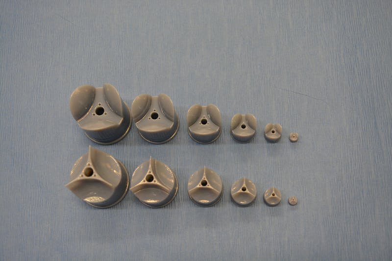
In rotary jet spinning technology, a rotating nozzle extrudes a solution of extracellular matrix (ECM) into nanofibers that wrap themselves around heart valve-shaped mandrels. By using a series of mandrels with different sizes, the manufacturing process becomes fully scalable and is able to provide JetValves for all age groups and heart sizes. Credit: Wyss Institute at Harvard University

From a May 18, 2017 news item on ScienceDaily,

The human heart beats approximately 35 million times every year, effectively pumping blood into the circulation via four different heart valves. Unfortunately, in over four million people each year, these delicate tissues malfunction due to birth defects, age-related deteriorations, and infections, causing cardiac valve disease.

Today, clinicians use either artificial prostheses or fixed animal and cadaver-sourced tissues to replace defective valves. While these prostheses can restore the function of the heart for a while, they are associated with adverse comorbidity and wear down and need to be replaced during invasive and expensive surgeries. Moreover, in children, implanted heart valve prostheses need to be replaced even more often as they cannot grow with the child.

A team lead by Kevin Kit Parker, Ph.D. at Harvard University’s Wyss Institute for Biologically Inspired Engineering recently developed a nanofiber fabrication technique to rapidly manufacture heart valves with regenerative and growth potential. In a paper published in Biomaterials, Andrew Capulli, Ph.D. and colleagues fabricated a valve-shaped nanofiber network that mimics the mechanical and chemical properties of the native valve extracellular matrix (ECM). To achieve this, the team used the Parker lab’s proprietary rotary jet spinning technology — in which a rotating nozzle extrudes an ECM solution into nanofibers that wrap themselves around heart valve-shaped mandrels. “Our setup is like a very fast cotton candy machine that can spin a range of synthetic and natural occurring materials. In this study, we used a combination of synthetic polymers and ECM proteins to fabricate biocompatible JetValves that are hemodynamically competent upon implantation and support cell migration and re-population in vitro. Importantly, we can make human-sized JetValves in minutes — much faster than possible for other regenerative prostheses,” said Parker.

A May 18,2017 Wyss Institute for Biologically Inspired Engineering news release (also on EurekAlert), which originated the news item, expands on the theme of Jetvalves,

To further develop and test the clinical potential of JetValves, Parker’s team collaborated with the translational team of Simon P. Hoerstrup, M.D., Ph.D., at the University of Zurich in Switzerland, which is a partner institution with the Wyss Institute. As a leader in regenerative heart prostheses, Hoerstrup and his team in Zurich have previously developed regenerative, tissue-engineered heart valves to replace mechanical and fixed-tissue heart valves. In Hoerstrup’s approach, human cells directly deposit a regenerative layer of complex ECM on biodegradable scaffolds shaped as heart valves and vessels. The living cells are then eliminated from the scaffolds resulting in an “off-the-shelf” human matrix-based prostheses ready for implantation.

In the paper, the cross-disciplinary team successfully implanted JetValves in sheep using a minimally invasive technique and demonstrated that the valves functioned properly in the circulation and regenerated new tissue. “In our previous studies, the cell-derived ECM-coated scaffolds could recruit cells from the receiving animal’s heart and support cell proliferation, matrix remodeling, tissue regeneration, and even animal growth. While these valves are safe and effective, their manufacturing remains complex and expensive as human cells must be cultured for a long time under heavily regulated conditions. The JetValve’s much faster manufacturing process can be a game-changer in this respect. If we can replicate these results in humans, this technology could have invaluable benefits in minimizing the number of pediatric re-operations,” said Hoerstrup.

In support of these translational efforts, the Wyss Institute for Biologically Inspired Engineering and the University of Zurich announced today a cross-institutional team effort to generate a functional heart valve replacement with the capacity for repair, regeneration, and growth. The team is also working towards a GMP-grade version of their customizable, scalable, and cost-effective manufacturing process that would enable deployment to a large patient population. In addition, the new heart valve would be compatible with minimally invasive procedures to serve both pediatric and adult patients.

The project will be led jointly by Parker and Hoerstrup. Parker is a Core Faculty member of the Wyss Institute and the Tarr Family Professor of Bioengineering and Applied Physics at the Harvard John A. Paulson School of Engineering and Applied Sciences (SEAS). Hoerstrup is Chair and Director of the University of Zurich’s Institute for Regenerative Medicine (IREM), Co-Director of the recently founded Wyss Translational Center Zurich and a Wyss Institute Associate Faculty member.

Since JetValves can be manufactured in all desired shapes and sizes, and take seconds to minutes to produce, the team’s goal is to provide customized, ready-to-use, regenerative heart valves much faster and at much lower cost than currently possible.

“Achieving the goal of minimally invasive, low-cost regenerating heart valves could have tremendous impact on patients’ lives across age-, social- and geographical boundaries. Once again, our collaborative team structure that combines unique and leading expertise in bioengineering, regenerative medicine, surgical innovation and business development across the Wyss Institute and our partner institutions, makes it possible for us to advance technology development in ways not possible in a conventional academic laboratory,” said Wyss Institute Founding Director Donald Ingber, M.D., Ph.D., who is also the Judah Folkman Professor of Vascular Biology at HMS and the Vascular Biology Program at Boston Children’s Hospital, as well as Professor of Bioengineering at SEAS.

This scanning electron microscopy image shows how extracellular matrix (ECM) nanofibers generated with JetValve technology are arranged in parallel networks with physical properties comparable to those found in native heart tissue. Credit: Wyss Institute at Harvard University

Here’s a link to and a citation for the paper,

JetValve: Rapid manufacturing of biohybrid scaffolds for biomimetic heart valve replacement by Andrew K. Capulli, Maximillian Y. Emmert, Francesco S. Pasqualini, b, Debora Kehl, Etem Caliskan, Johan U. Lind, Sean P. Sheehy, Sung Jin Park, Seungkuk Ahn, Benedikt Webe, Josue A. Goss. Biomaterials Volume 133, July 2017, Pages 229–241  https://doi.org/10.1016/j.biomaterials.2017.04.033

This paper is behind a paywall.
3D bioprinting: a conference about the latest trends (May 3 – 5, 2017 at the University of British Columbia, Vancouver)

The University of British Columbia’s (UBC) Peter Wall Institute for Advanced Studies (PWIAS) is hosting along with local biotech firm, Aspect Biosystems, a May 3 -5, 2017 international research roundtable known as ‘Printing the Future of Therapeutics in 3D‘.

A May 1, 2017 UBC news release (received via email) offers some insight into the field of bioprinting from one of the roundtable organizers,

This week, global experts will gather [4] at the University of British
Columbia to discuss the latest trends in 3D bioprinting—a technology
used to create living tissues and organs.

In this Q&A, UBC chemical and biological engineering professor
Vikramaditya Yadav [5], who is also with the Regenerative Medicine
Cluster Initiative in B.C., explains how bioprinting could potentially
transform healthcare and drug development, and highlights Canadian
innovations in this field.

WHY IS 3D BIOPRINTING SIGNIFICANT?

Bioprinted tissues or organs could allow scientists to predict
beforehand how a drug will interact within the body. For every
life-saving therapeutic drug that makes its way into our medicine
cabinets, Health Canada blocks the entry of nine drugs because they are
proven unsafe or ineffective. Eliminating poor-quality drug candidates
to reduce development costs—and therefore the cost to consumers—has
never been more urgent.

In Canada alone, nearly 4,500 individuals are waiting to be matched with
organ donors. If and when bioprinters evolve to the point where they can
manufacture implantable organs, the concept of an organ transplant
waiting list would cease to exist. And bioprinted tissues and organs
from a patient’s own healthy cells could potentially reduce the risk
of transplant rejection and related challenges.

HOW IS THIS TECHNOLOGY CURRENTLY BEING USED?

Skin, cartilage and bone, and blood vessels are some of the tissue types
that have been successfully constructed using bioprinting. Two of the
most active players are the Wake Forest Institute for Regenerative
Medicine in North Carolina, which reports that its bioprinters can make
enough replacement skin to cover a burn with 10 times less healthy
tissue than is usually needed, and California-based Organovo, which
makes its kidney and liver tissue commercially available to
pharmaceutical companies for drug testing.

Beyond medicine, bioprinting has already been commercialized to print
meat and artificial leather. It’s been estimated that the global
bioprinting market will hit $2 billion by 2021.

HOW IS CANADA INVOLVED IN THIS FIELD?

Canada is home to some of the most innovative research clusters and
start-up companies in the field. The UBC spin-off Aspect Biosystems [6]
has pioneered a bioprinting paradigm that rapidly prints on-demand
tissues. It has successfully generated tissues found in human lungs.

Many initiatives at Canadian universities are laying strong foundations
for the translation of bioprinting and tissue engineering into
mainstream medical technologies. These include the Regenerative Medicine
Cluster Initiative in B.C., which is headed by UBC, and the University
of Toronto’s Institute of Biomaterials and Biomedical Engineering.

WHAT ETHICAL ISSUES DOES BIOPRINTING CREATE?

There are concerns about the quality of the printed tissues. It’s
important to note that the U.S. Food and Drug Administration and Health
Canada are dedicating entire divisions to regulation of biomanufactured
products and biomedical devices, and the FDA also has a special division
that focuses on regulation of additive manufacturing – another name
for 3D printing.

These regulatory bodies have an impressive track record that should
assuage concerns about the marketing of substandard tissue. But cost and
pricing are arguably much more complex issues.

Some ethicists have also raised questions about whether society is not
too far away from creating Replicants, à la _Blade Runner_. The idea is
fascinating, scary and ethically grey. In theory, if one could replace
the extracellular matrix of bones and muscles with a stronger substitute
and use cells that are viable for longer, it is not too far-fetched to
create bones or muscles that are stronger and more durable than their
natural counterparts.

WILL DOCTORS BE PRINTING REPLACEMENT BODY PARTS IN 20 YEARS’ TIME?

This is still some way off. Optimistically, patients could see the
technology in certain clinical environments within the next decade.
However, some technical challenges must be addressed in order for this
to occur, beginning with faithful replication of the correct 3D
architecture and vascularity of tissues and organs. The bioprinters
themselves need to be improved in order to increase cell viability after
printing.

These developments are happening as we speak. Regulation, though, will
be the biggest challenge for the field in the coming years.

There are some events open to the public (from the international research roundtable homepage),

OPEN EVENTS

You’re invited to attend the open events associated with Printing the Future of Therapeutics in 3D.

Café Scientifique

Thursday, May 4, 2017
Telus World of Science
5:30 – 8:00pm [all tickets have been claimed as of May 2, 2017 at 3:15 pm PT]

3D Bioprinting: Shaping the Future of Health

Imagine a world where drugs are developed without the use of animals, where doctors know how a patient will react to a drug before prescribing it and where patients can have a replacement organ 3D-printed using their own cells, without dealing with long donor waiting lists or organ rejection. 3D bioprinting could enable this world. Join us for lively discussion and dessert as experts in the field discuss the exciting potential of 3D bioprinting and the ethical issues raised when you can print human tissues on demand. This is also a rare opportunity to see a bioprinter live in action!

Open Session

Friday, May 5, 2017
Peter Wall Institute for Advanced Studies
2:00 – 7:00pm

A Scientific Discussion on the Promise of 3D Bioprinting

The medical industry is struggling to keep our ageing population healthy. Developing effective and safe drugs is too expensive and time-consuming to continue unchanged. We cannot meet the current demand for transplant organs, and people are dying on the donor waiting list every day.  We invite you to join an open session where four of the most influential academic and industry professionals in the field discuss how 3D bioprinting is being used to shape the future of health and what ethical challenges may be involved if you are able to print your own organs.

ROUNDTABLE INFORMATION

The University of British Columbia and the award-winning bioprinting company Aspect Biosystems, are proud to be co-organizing the first “Printing the Future of Therapeutics in 3D” International Research Roundtable. This event will congregate global leaders in tissue engineering research and pharmaceutical industry experts to discuss the rapidly emerging and potentially game-changing technology of 3D-printing living human tissues (bioprinting). The goals are to:

Highlight the state-of-the-art in 3D bioprinting research
Ideate on disruptive innovations that will transform bioprinting from a novel research tool to a broadly adopted systematic practice
Formulate an actionable strategy for industry engagement, clinical translation and societal impact
Present in a public forum, key messages to educate and stimulate discussion on the promises of bioprinting technology

The Roundtable will bring together a unique collection of industry experts and academic leaders to define a guiding vision to efficiently deploy bioprinting technology for the discovery and development of new therapeutics. As the novel technology of 3D bioprinting is more broadly adopted, we envision this Roundtable will become a key annual meeting to help guide the development of the technology both in Canada and globally.

We thank you for your involvement in this ground-breaking event and look forward to you all joining us in Vancouver for this unique research roundtable.

Kind Regards,
The Organizing Committee
Christian Naus, Professor, Cellular & Physiological Sciences, UBC
Vikram Yadav, Assistant Professor, Chemical & Biological Engineering, UBC
Tamer Mohamed, CEO, Aspect Biosystems
Sam Wadsworth, CSO, Aspect Biosystems
Natalie Korenic, Business Coordinator, Aspect Biosystems

I’m glad to see this event is taking place—and with public events too! (Wish I’d seen the Café Scientifique announcement earlier when I first checked for tickets  yesterday. I was hoping there’d been some cancellations today.) Finally, for the interested, you can find Aspect Biosystems here.

Recycling apples to regenerate bone and cartilage tissue

A March 30, 2017 news item on phys.org announces research utilizing apple waste as a matrix for regenerating bones and cartilage,

Researchers from UPM and CSIC [both organizations are in Spain] have employed waste from the agri-food industry to develop biomaterials that act as matrices to regenerate bone and cartilage tissues, which is of great interest for the treatment of diseases related to aging.

The researchers have produced biocompatible materials from apple pomace resulting from juice production. These materials can be used as 3-D matrices for the regeneration of bone and cartilage tissues, useful in regenerative medicine for diseases such as osteoporosis, arthritis or osteoarthritis, all of them rising due to the increasing average age of the population.

A March 30, 2017 Universidad Politécnica de Madrid (UPM) press release, which originated the news item,, expands on the theme,

Apple pomace is an abundant raw material. The world production of apples was more than 70 million tons in 2015, of which the European Union contributed with more than 15%, while half a million tons of which came from Spain. About 75% of apples can be converted into juice and the rest, known as apple pomace, that contains approximately 20–30% dried matter, is used mainly as animal feed or for compost. Since apple pomace is generated in vast quantities and contains a large fraction of water, it poses storage problems and requires immediate treatments to prevent putrefaction. An alternative of great environmental interest is its transformation into value added commodities, thus reducing the volume of waste.

The procedure of the multivalorization of apple pomace carried out by the UPM and CSIC researchers are based on sequential extractions of different bioactive molecules, such as antioxidants or pectin, to finally obtain the waste from which they prepare a biomaterial with suitable porosity and texture to be used in tissue engineering.

The primary extraction of antioxidants and carbohydrates constitutes 2% of the dry weight of apple pomace and pectin extraction is 10%. The extracted chemical cells have a recognized value as nutraceuticals and pectin is a material of great utility in different medical applications, given its high biocompatibility and being part of antitumor drugs or in the treatment of coetaneous wounds.

Furthermore, it has been found that the materials remaining after antioxidant and pectin removal from apple pomace can still be designed with adequate structure, texture and composition to grow diverse types of cells. In this particularly case, the chosen cells were osteoblasts and chondrocytes, both of them related to the regeneration of bone and cartilage tissues because of their application in regenerative medicine in diseases such as osteoporosis, arthritis or osteoarthritis.

Today, there are products in the market with the same applications, however they have a high price reaching over €100 per gram, while waste used in this work hardly reaches €100 per ton. For this reason, there are consistent incentives to convert this waste into final products of great added value.

According to Milagro Ramos, a female researcher of the study, “with this approach we achieve a double goal, firstly using waste as a renewable raw material of high value and chemical diversity, and secondly, to reduce the impact of such waste accumulation on the environment”.

Thanks to the new materials obtained in this work, researchers are developing new technological applications that allow them to structure customized biomaterials through 3D printing techniques.

Here’s a link to and a citation for the paper,

Multivalorization of apple pomace towards materials and chemicals. Waste to wealth by Malcolm Yates, Milagros Ramos Gomez, Maria A. Martin-Luengo, Violeta Zurdo Ibañez, Ana Maria Martinez Serrano. Journal of Cleaner Production Volume 143, 1 February 2017, Pages 847–853  http://doi.org/10.1016/j.jclepro.2016.12.036

This paper is behind a paywall.
A new platform for culturing stem cells: a Multiplexed Artificial Cellular Microenvironment array

Japanese scientists have developed a more precise method for culturing stem cells according to a March 14, 2017 news item on Nanowerk,

A team of researchers in Japan has developed a new platform for culturing human pluripotent stem cells that provides far more control of culture conditions than previous tools by using micro and nanotechnologies.

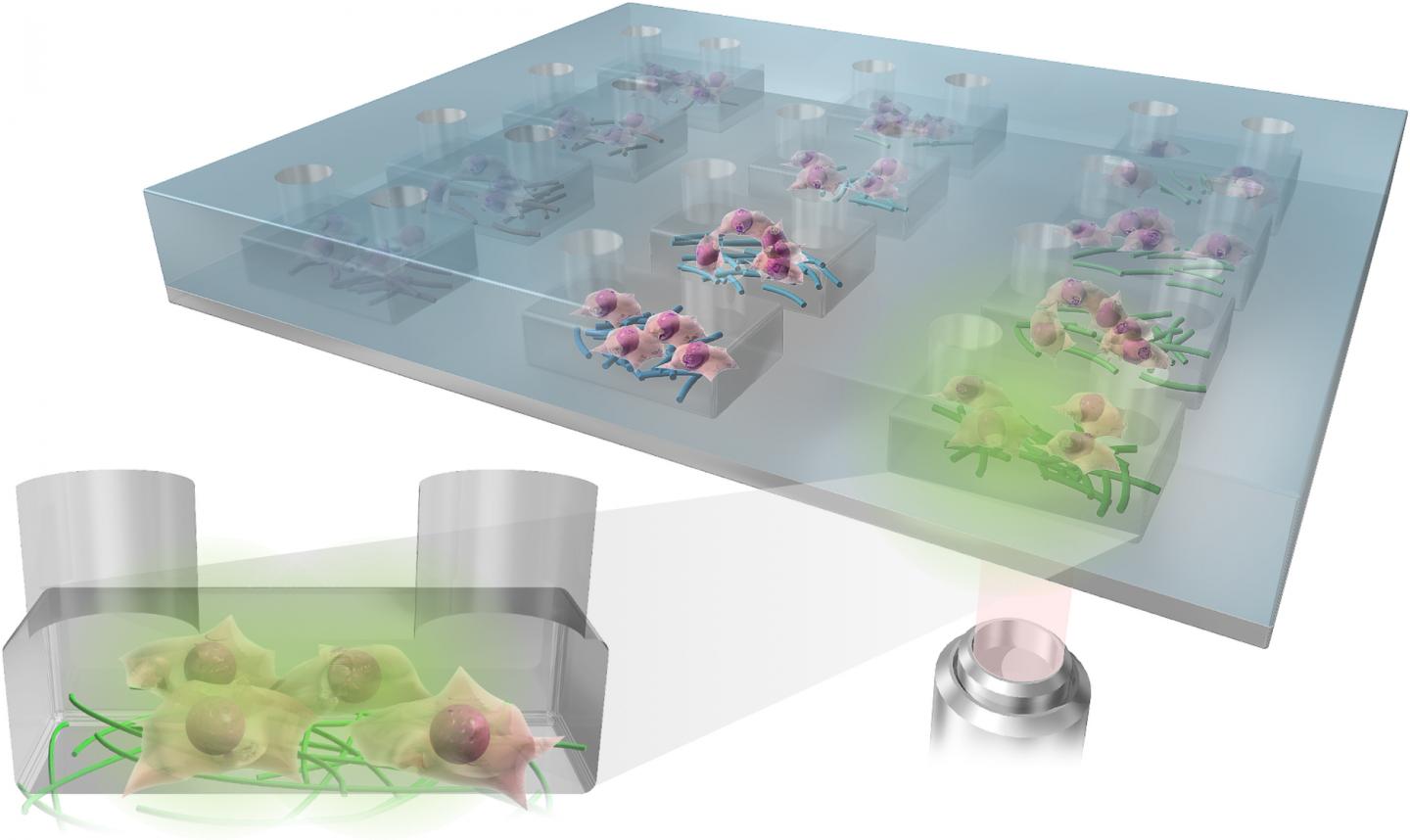
The Multiplexed Artificial Cellular Microenvironment (MACME) array places nanofibres, mimicking cellular matrices, into fluid-filled micro-chambers of precise sizes, which mimic extracellular environments.

Caption: The Multiplexed Artificial Cellular Microenvironment (MACME) array, consisted with a microfluidic structure and nanofibre array for mimicking cellular microenvironments. Credit: Kyoto University iCeMS

A March 17, 2017 Kyoto University press release (also on EurekAlert), which originated the news item, explains the research in more detail,

Human pluripotent stems cells (hPSCs) hold great promise for tissue engineering, regenerative medicine and cell-based therapies because they can become any type of cell. The environment surrounding the cells plays a major role in determining what tissues they become, if they replicate into more cells, or die. However, understanding these interactions has been difficult because researchers have lacked tools that work on the appropriate scale.

Often, stem cells are cultured in a cell culture medium in small petri dishes. While factors such as medium pH levels and nutrients can be controlled, the artificial set up is on the macroscopic scale and does not allow for precise control of the physical environment surrounding the cells.

The MACME array miniaturizes this set up, culturing stem cells in rows of micro-chambers of cell culture medium. It also takes it a step further by placing nanofibers in these chambers to mimic the structures found around cells.

Led by Ken-ichiro Kamei of Kyoto University’s Institute for Integrated Cell-Material Sciences (iCeMS), the team tested a variety of nanofiber materials and densities, micro-chamber heights and initial stem cell densities to determine the best combination that encourages human pluripotent stem cells to replicate.

They stained the cells with several fluorescent markers and used a microscope to see if the cells died, replicated or differentiated into tissues.

Their analysis revealed that gelatin nanofibers and medium-sized chambers that create medium seed cell density provided the best environment for the stem cells to continue to multiply. The quantity and density of neighboring cells strongly influences cell survival.

The array is an “optimal and powerful approach for understanding how environmental cues regulate cellular functions,” the researchers conclude in a recently published paper in the journal Small.

This array appears to be the first time multiple kinds of extracellular environments can be mounted onto a single device, making it much easier to compare how different environments influence cells.

The MACME array could substantially reduce experiment costs compared to conventional tools, in part because it is low volume and requires less cell culture medium. The array does not require any special equipment and is compatible with both commonly used laboratory pipettes and automated pipette systems for performing high-throughput screening.

Here’s a link to and a citation for the paper,

Microfluidic-Nanofiber Hybrid Array for Screening of Cellular Microenvironments by Ken-ichiro Kamei, Yasumasa Mashimo, Momoko Yoshioka, Yumie Tokunaga, Christopher Fockenberg, Shiho Terada, Yoshie Koyama, Minako Nakajima, Teiko Shibata-Seki, Li Liu, Toshihiro Akaike, Eiry Kobatake, Siew-Eng How, Motonari Uesugi, and Yong Chen. Small DOI: 10.1002/smll.201603104 Version of Record online: 8 MAR 2017

© 2017 WILEY-VCH Verlag GmbH & Co. KGaA, Weinheim

This paper is behind a paywall.

3D printed biomimetic blood vessel networks

An artificial blood vessel network that could lead the way to regenerating biologically-based blood vessel networks has been printed in 3D at the University of California at San Diego (UCSD) according to a March 2, 2017 news item on ScienceDaily,

Nanoengineers at the University of California San Diego have 3D printed a lifelike, functional blood vessel network that could pave the way toward artificial organs and regenerative therapies.

The new research, led by nanoengineering professor Shaochen Chen, addresses one of the biggest challenges in tissue engineering: creating lifelike tissues and organs with functioning vasculature — networks of blood vessels that can transport blood, nutrients, waste and other biological materials — and do so safely when implanted inside the body.

A March 2, 2017 UCSD news release (also on EurekAlert), which originated the news item, explains why this is an important development,

Researchers from other labs have used different 3D printing technologies to create artificial blood vessels. But existing technologies are slow, costly and mainly produce simple structures, such as a single blood vessel — a tube, basically. These blood vessels also are not capable of integrating with the body’s own vascular system.

“Almost all tissues and organs need blood vessels to survive and work properly. This is a big bottleneck in making organ transplants, which are in high demand but in short supply,” said Chen, who leads the Nanobiomaterials, Bioprinting, and Tissue Engineering Lab at UC San Diego. “3D bioprinting organs can help bridge this gap, and our lab has taken a big step toward that goal.”

Chen’s lab has 3D printed a vasculature network that can safely integrate with the body’s own network to circulate blood. These blood vessels branch out into many series of smaller vessels, similar to the blood vessel structures found in the body. The work was published in Biomaterials.

Chen’s team developed an innovative bioprinting technology, using their own homemade 3D printers, to rapidly produce intricate 3D microstructures that mimic the sophisticated designs and functions of biological tissues. Chen’s lab has used this technology in the past to create liver tissue and microscopic fish that can swim in the body to detect and remove toxins.

Researchers first create a 3D model of the biological structure on a computer. The computer then transfers 2D snapshots of the model to millions of microscopic-sized mirrors, which are each digitally controlled to project patterns of UV light in the form of these snapshots. The UV patterns are shined onto a solution containing live cells and light-sensitive polymers that solidify upon exposure to UV light. The structure is rapidly printed one layer at a time, in a continuous fashion, creating a 3D solid polymer scaffold encapsulating live cells that will grow and become biological tissue.

“We can directly print detailed microvasculature structures in extremely high resolution. Other 3D printing technologies produce the equivalent of ‘pixelated’ structures in comparison and usually require sacrificial materials and additional steps to create the vessels,” said Wei Zhu, a postdoctoral scholar in Chen’s lab and a lead researcher on the project.

And this entire process takes just a few seconds — a vast improvement over competing bioprinting methods, which normally take hours just to print simple structures. The process also uses materials that are inexpensive and biocompatible.

Chen’s team used medical imaging to create a digital pattern of a blood vessel network found in the body. Using their technology, they printed a structure containing endothelial cells, which are cells that form the inner lining of blood vessels.

The entire structure fits onto a small area measuring 4 millimeters × 5 millimeters, 600 micrometers thick (as thick as a stack containing 12 strands of human hair).

Researchers cultured several structures in vitro for one day, then grafted the resulting tissues into skin wounds of mice. After two weeks, the researchers examined the implants and found that they had successfully grown into and merged with the host blood vessel network, allowing blood to circulate normally.

Chen noted that the implanted blood vessels are not yet capable of other functions, such as transporting nutrients and waste. “We still have a lot of work to do to improve these materials. This is a promising step toward the future of tissue regeneration and repair,” he said.

Moving forward, Chen and his team are working on building patient-specific tissues using human induced pluripotent stem cells, which would prevent transplants from being attacked by a patient’s immune system. And since these cells are derived from a patient’s skin cells, researchers won’t need to extract any cells from inside the body to build new tissue. The team’s ultimate goal is to move their work to clinical trials. “It will take at least several years before we reach that goal,” Chen said.

Here’s a link to and a citation for the paper,

Direct 3D bioprinting of prevascularized tissue constructs with complex microarchitecture by Wei Zhu, Xin Qu, Jie Zhu, Xuanyi Ma, Sherrina Patel, Justin Liu, Pengrui Wang, Cheuk Sun Edwin Lai, Maling Gou, Yang Xu, Kang Zhang, Shaochen Chen. Biomaterials 124 (April 2017) 106-15 http://dx.doi.org/10.1016/j.biomaterials.2017.01.042

This paper is behind a paywall.

There is also an open access copy here on the university website but I cannot confirm that it is identical to the version in the journal.

Better technique for growing organoids taking them from the lab to the clinic

A Nov. 16, 2016 École Polytechnique Fédérale de Lausanne (EPFL) press release (also on EurekAlert) describes a new material for growing organoids,

Organoids are miniature organs that can be grown in the lab from a person’s stem cells. They can be used to model diseases, and in the future could be used to test drugs or even replace damaged tissue in patients. But currently organoids are very difficult to grow in a standardized and controlled way, which is key to designing and using them. EPFL scientists have now solved the problem by developing a patent-pending “hydrogel” that provides a fully controllable and tunable way to grow organoids. …

Organoids need a 3D scaffold

Growing organoids begins with stem cells — immature cells that can grow into any cell type of the human body and that play key roles in tissue function and regeneration. To form an organoid, the stem cells are grown inside three-dimensional gels that contain a mix of biomolecules that promote stem cell renewal and differentiation.

The role of these gels is to mimic the natural environment of the stem cells, which provides them with a protein- and sugar-rich scaffold called the “extracellular matrix”, upon which the stem cells build specific body tissues. The stem cells stick to the extracellular matrix gel, and then “self-organize” into miniature organs like retinas, kidneys, or the gut. These tiny organs retain key aspects of their real-life biology, and can be used to study diseases or test drugs before moving on to human trials.

But the current gels used for organoid growth are derived from mice, and have problems. First, it is impossible to control their makeup from batch to batch, which can cause stem cells to behave inconsistently. Second, their biochemical complexity makes them very difficult to fine-tune for studying the effect of different parameters (e.g. biological molecules, mechanical properties, etc.) on the growth of organoids. Finally, the gels can carry pathogens or immunogens, which means that they are not suitable for growing organoids to be used in the clinic.

A hydrogel solution

The lab of Matthias Lütolf at EPFL’s Institute of Bioengineering has developed a synthetic “hydrogel” that eschews the limitations of conventional, naturally derived gels. The patent-pending gel is made of water and polyethylene glycol, a substance used widely today in various forms, from skin creams and toothpastes to industrial applications and, as in this case, bioengineering.

Nikolce Gjorevski, the first author of the study, and his colleagues used the hydrogel to grow stem cells of the gut into a miniature intestine. The functional hydrogel was not only a goal in and of itself, but also a means to identify the factors that influence the stem cells’ ability to expand and form organoids. By carefully tweaking the hydrogel’s properties, they discovered that separate stages of the organoid formation process require different mechanical environments and biological components.

One such factor is a protein called fibronectin, which helps the stem cells attach to the hydrogel. Lütolf’s lab found that this attachment itself is immensely important for growing organoids, as it triggers a whole host of signals to the stem cell that tell it to grow and build an intestine-like structure. The researchers also discovered an essential role for the mechanical properties, i.e. the physical stiffness, of the gel in regulating intestinal stem cell behavior, shedding light on how cells are able to sense, process and respond to physical stimuli. This insight is particularly valuable – while the influence of biochemical signals on stem cells is well-understood, the effect of physical factors has been more mysterious.

Because the hydrogel is man-made, it is easy to control its chemical composition and key properties, and ensure consistency from batch to batch. And because it is artificial, it does not carry any risk of infection or triggering immune responses. As such, it provides a means of moving organoids from basic research to actual pharmaceutical and clinical applications in the future.

Lütolf’s lab is now researching other types of stem cells in order to extend the capacities of their hydrogel into other tissues.

Here’s a link to and a citation for the paper,

Designer matrices for intestinal stem cell and organoid culture by Nikolce Gjorevski, Norman Sachs, Andrea Manfrin, Sonja Giger, Maiia E. Bragina, Paloma Ordóñez-Morán, Hans Clevers, & Matthias P. Lutolf.  Nature (2016) doi:10.1038/nature20168 Published online 16 November 2016

This paper is behind a paywall.

Brain and machine as one (machine/flesh)

The essay on brains and machines becoming intertwined is making the rounds. First stop on my tour was its Oct. 4, 2016 appearance on the Mail & Guardian, then there was its Oct. 3, 2016 appearance on The Conversation, and finally (moving forward in time) there was its Oct. 4, 2016 appearance on the World Economic Forum website as part of their Final Frontier series.

The essay was written by Richard Jones of Sheffield University (mentioned here many times before but most recently in a Sept. 4, 2014 posting). His book ‘Soft Machines’ provided me with an important and eminently readable introduction to nanotechnology. He is a professor of physics at the University of Sheffield and here’s more from his essay (Oct. 3, 2016 on The Conversation) about brains and machines (Note: Links have been removed),

Imagine a condition that leaves you fully conscious, but unable to move or communicate, as some victims of severe strokes or other neurological damage experience. This is locked-in syndrome, when the outward connections from the brain to the rest of the world are severed. Technology is beginning to promise ways of remaking these connections, but is it our ingenuity or the brain’s that is making it happen?

Ever since an 18th-century biologist called Luigi Galvani made a dead frog twitch we have known that there is a connection between electricity and the operation of the nervous system. We now know that the signals in neurons in the brain are propagated as pulses of electrical potential, whose effects can be detected by electrodes in close proximity. So in principle, we should be able to build an outward neural interface system – that is to say, a device that turns thought into action.

In fact, we already have the first outward neural interface system to be tested in humans. It is called BrainGate and consists of an array of micro-electrodes, implanted into the part of the brain concerned with controlling arm movements. Signals from the micro-electrodes are decoded and used to control the movement of a cursor on a screen, or the motion of a robotic arm.

A crucial feature of these systems is the need for some kind of feedback. A patient must be able to see the effect of their willed patterns of thought on the movement of the cursor. What’s remarkable is the ability of the brain to adapt to these artificial systems, learning to control them better.

You can find out more about BrainGate in my May 17, 2012 posting which also features a video of a woman controlling a mechanical arm so she can drink from a cup coffee by herself for the first time in 15 years.

Jones goes on to describe the cochlear implants (although there’s no mention of the controversy; not everyone believes they’re a good idea) and retinal implants that are currently available. Jones notes this (Note Links have been removed),

The key message of all this is that brain interfaces now are a reality and that the current versions will undoubtedly be improved. In the near future, for many deaf and blind people, for people with severe disabilities – including, perhaps, locked-in syndrome – there are very real prospects that some of their lost capabilities might be at least partially restored.

Until then, our current neural interface systems are very crude. One problem is size; the micro-electrodes in use now, with diameters of tens of microns, may seem tiny, but they are still coarse compared to the sub-micron dimensions of individual nerve fibres. And there is a problem of scale. The BrainGate system, for example, consists of 100 micro-electrodes in a square array; compare that to the many tens of billions of neurons in the brain. The fact these devices work at all is perhaps more a testament to the adaptability of the human brain than to our technological prowess.

Scale models

So the challenge is to build neural interfaces on scales that better match the structures of biology. Here, we move into the world of nanotechnology. There has been much work in the laboratory to make nano-electronic structures small enough to read out the activity of a single neuron. In the 1990s, Peter Fromherz, at the Max Planck Institute for Biochemistry, was a pioneer of using silicon field effect transistors, similar to those used in commercial microprocessors, to interact with cultured neurons. In 2006, Charles Lieber’s group at Harvard succeeded in using transistors made from single carbon nanotubes – whiskers of carbon just one nanometer in diameter – to measure the propagation of single nerve pulses along the nerve fibres.

But these successes have been achieved, not in whole organisms, but in cultured nerve cells which are typically on something like the surface of a silicon wafer. It’s going to be a challenge to extend these methods into three dimensions, to interface with a living brain. Perhaps the most promising direction will be to create a 3D “scaffold” incorporating nano-electronics, and then to persuade growing nerve cells to infiltrate it to create what would in effect be cyborg tissue – living cells and inorganic electronics intimately mixed.

I have featured Charles Lieber and his work here in two recent posts: ‘Bionic’ cardiac patch with nanoelectric scaffolds and living cells on July 11, 2016 and Long-term brain mapping with injectable electronics on Sept. 22, 2016.

For anyone interested in more about the controversy regarding cochlear implants, there’s this page on the Brown University (US) website. You might also want to check out Gregor Wolbring (professor at the University of Calgary) who has written extensively on the concept of ableism (links to his work can be found at the end of this post). I have excerpted from an Aug. 30, 2011 post the portion where Gregor defines ‘ableism’,

From Gregor’s June 17, 2011 posting on the FedCan blog,

The term ableism evolved from the disabled people rights movements in the United States and Britain during the 1960s and 1970s.  It questions and highlights the prejudice and discrimination experienced by persons whose body structure and ability functioning were labelled as ‘impaired’ as sub species-typical. Ableism of this flavor is a set of beliefs, processes and practices, which favors species-typical normative body structure based abilities. It labels ‘sub-normative’ species-typical biological structures as ‘deficient’, as not able to perform as expected.

The disabled people rights discourse and disability studies scholars question the assumption of deficiency intrinsic to ‘below the norm’ labeled body abilities and the favoritism for normative species-typical body abilities. The discourse around deafness and Deaf Culture would be one example where many hearing people expect the ability to hear. This expectation leads them to see deafness as a deficiency to be treated through medical means. In contrast, many Deaf people see hearing as an irrelevant ability and do not perceive themselves as ill and in need of gaining the ability to hear. Within the disabled people rights framework ableism was set up as a term to be used like sexism and racism to highlight unjust and inequitable treatment.

Ableism is, however, much more pervasive.

You can find out more about Gregor and his work here: http://www.crds.org/research/faculty/Gregor_Wolbring2.shtml or here:
https://www.facebook.com/GregorWolbring.

Robots built from living tissue

Biohybrid robots, as they are known, are built from living tissue but not in a Frankenstein kind of way as Victoria Webster PhD candidate at Case Western Reserve University (US) explains in her Aug. 9, 2016 essay on The Conversation (also on phys.org as an Aug. 10, 2016 news item; Note: Links have been removed),

Researchers are increasingly looking for solutions to make robots softer or more compliant – less like rigid machines, more like animals. With traditional actuators – such as motors – this can mean using air muscles or adding springs in parallel with motors. …

But there’s a growing area of research that’s taking a different approach. By combining robotics with tissue engineering, we’re starting to build robots powered by living muscle tissue or cells. These devices can be stimulated electrically or with light to make the cells contract to bend their skeletons, causing the robot to swim or crawl. The resulting biobots can move around and are soft like animals. They’re safer around people and typically less harmful to the environment they work in than a traditional robot might be. And since, like animals, they need nutrients to power their muscles, not batteries, biohybrid robots tend to be lighter too.

Webster explains how these biobots are built,

Researchers fabricate biobots by growing living cells, usually from heart or skeletal muscle of rats or chickens, on scaffolds that are nontoxic to the cells. If the substrate is a polymer, the device created is a biohybrid robot – a hybrid between natural and human-made materials.

If you just place cells on a molded skeleton without any guidance, they wind up in random orientations. That means when researchers apply electricity to make them move, the cells’ contraction forces will be applied in all directions, making the device inefficient at best.

So to better harness the cells’ power, researchers turn to micropatterning. We stamp or print microscale lines on the skeleton made of substances that the cells prefer to attach to. These lines guide the cells so that as they grow, they align along the printed pattern. With the cells all lined up, researchers can direct how their contraction force is applied to the substrate. So rather than just a mess of firing cells, they can all work in unison to move a leg or fin of the device.

Researchers sometimes mimic animals when creating their biobots (Note: Links have been removed),

Others have taken their cues from nature, creating biologically inspired biohybrids. For example, a group led by researchers at California Institute of Technology developed a biohybrid robot inspired by jellyfish. This device, which they call a medusoid, has arms arranged in a circle. Each arm is micropatterned with protein lines so that cells grow in patterns similar to the muscles in a living jellyfish. When the cells contract, the arms bend inwards, propelling the biohybrid robot forward in nutrient-rich liquid.

More recently, researchers have demonstrated how to steer their biohybrid creations. A group at Harvard used genetically modified heart cells to make a biologically inspired manta ray-shaped robot swim. The heart cells were altered to contract in response to specific frequencies of light – one side of the ray had cells that would respond to one frequency, the other side’s cells responded to another.

Amazing, eh? And, this is quite a recent video; it was published on YouTube on July 7, 2016.

Webster goes on to describe work designed to make these robots hardier and more durable so they can leave the laboratory,

… Here at Case Western Reserve University, we’ve recently begun to investigate … by turning to the hardy marine sea slug Aplysia californica. Since A. californica lives in the intertidal region, it can experience big changes in temperature and environmental salinity over the course of a day. When the tide goes out, the sea slugs can get trapped in tide pools. As the sun beats down, water can evaporate and the temperature will rise. Conversely in the event of rain, the saltiness of the surrounding water can decrease. When the tide eventually comes in, the sea slugs are freed from the tidal pools. Sea slugs have evolved very hardy cells to endure this changeable habitat.

We’ve been able to use Aplysia tissue to actuate a biohybrid robot, suggesting that we can manufacture tougher biobots using these resilient tissues. The devices are large enough to carry a small payload – approximately 1.5 inches long and one inch wide.

Webster has written a fascinating piece and, if you have time, I encourage you to read it in its entirety.

Small, soft, and electrically functional: an injectable biomaterial

This development could be looked at as a form of synthetic biology without the genetic engineering. From a July 1, 2016 news item on ScienceDaily,

Ideally, injectable or implantable medical devices should not only be small and electrically functional, they should be soft, like the body tissues with which they interact. Scientists from two UChicago labs set out to see if they could design a material with all three of those properties.

The material they came up with, published online June 27, 2016, in Nature Materials, forms the basis of an ingenious light-activated injectable device that could eventually be used to stimulate nerve cells and manipulate the behavior of muscles and organs.

“Most traditional materials for implants are very rigid and bulky, especially if you want to do electrical stimulation,” said Bozhi Tian, an assistant professor in chemistry whose lab collaborated with that of neuroscientist Francisco Bezanilla on the research.

The new material, in contrast, is soft and tiny — particles just a few micrometers in diameter (far less than the width of a human hair) that disperse easily in a saline solution so they can be injected. The particles also degrade naturally inside the body after a few months, so no surgery would be needed to remove them.

A July 1, 2016 University of Chicago news release (also on EurekAlert) by , which originated the news item, provides more detail,

Each particle is built of two types of silicon that together form a structure full of nano-scale pores, like a tiny sponge. And like a sponge, it is squishy — a hundred to a thousand times less rigid than the familiar crystalline silicon used in transistors and solar cells. “It is comparable to the rigidity of the collagen fibers in our bodies,” said Yuanwen Jiang, Tian’s graduate student. “So we’re creating a material that matches the rigidity of real tissue.”

The material constitutes half of an electrical device that creates itself spontaneously when one of the silicon particles is injected into a cell culture, or, eventually, a human body. The particle attaches to a cell, making an interface with the cell’s plasma membrane. Those two elements together — cell membrane plus particle — form a unit that generates current when light is shined on the silicon particle.

“You don’t need to inject the entire device; you just need to inject one component,” João L. Carvalho-de-Souza , Bezanilla’s postdoc said. “This single particle connection with the cell membrane allows sufficient generation of current that could be used to stimulate the cell and change its activity. After you achieve your therapeutic goal, the material degrades naturally. And if you want to do therapy again, you do another injection.”

The scientists built the particles using a process they call nano-casting. They fabricate a silicon dioxide mold composed of tiny channels — “nano-wires” — about seven nanometers in diameter (less than 10,000 times smaller than the width of a human hair) connected by much smaller “micro-bridges.” Into the mold they inject silane gas, which fills the pores and channels and decomposes into silicon.

And this is where things get particularly cunning. The scientists exploit the fact the smaller an object is, the more the atoms on its surface dominate its reactions to what is around it. The micro-bridges are minute, so most of their atoms are on the surface. These interact with oxygen that is present in the silicon dioxide mold, creating micro-bridges made of oxidized silicon gleaned from materials at hand. The much larger nano-wires have proportionately fewer surface atoms, are much less interactive, and remain mostly pure silicon. [I have a note regarding ‘micro’ and ‘nano’ later in this posting.]

“This is the beauty of nanoscience,” Jiang said. “It allows you to engineer chemical compositions just by manipulating the size of things.”

Web-like nanostructure

Finally, the mold is dissolved. What remains is a web-like structure of silicon nano-wires connected by micro-bridges of oxidized silicon that can absorb water and help increase the structure’s softness. The pure silicon retains its ability to absorb light.

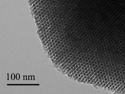
Transmission electron microscopy image shows an ordered nanowire array. The 100-nanometer scale bar is 1,000 times narrower than a hair. Courtesy of Tian Lab

Transmission electron microscopy image shows an ordered nanowire array. The 100-nanometer scale bar is 1,000 times narrower than a hair. Courtesy of
Tian Lab

The scientists have added the particles onto neurons in culture in the lab, shone light on the particles, and seen current flow into the neurons which activates the cells. The next step is to see what happens in living animals. They are particularly interested in stimulating nerves in the peripheral nervous system that connect to organs. These nerves are relatively close to the surface of the body, so near-infra-red wavelength light can reach them through the skin.

Tian imagines using the light-activated devices to engineer human tissue and create artificial organs to replace damaged ones. Currently, scientists can make engineered organs with the correct form but not the ideal function.

To get a lab-built organ to function properly, they will need to be able to manipulate individual cells in the engineered tissue. The injectable device would allow a scientist to do that, tweaking an individual cell using a tightly focused beam of light like a mechanic reaching into an engine and turning a single bolt. The possibility of doing this kind of synthetic biology without genetic engineering [emphasis mine] is enticing.

“No one wants their genetics to be altered,” Tian said. “It can be risky. There’s a need for a non-genetic system that can still manipulate cell behavior. This could be that kind of system.”

Tian’s graduate student Yuanwen Jiang did the material development and characterization on the project. The biological part of the collaboration was done in the lab of Francisco Bezanilla, the Lillian Eichelberger Cannon Professor of Biochemistry and Molecular Biology, by postdoc João L. Carvalho-de-Souza. They were, said Tian, the “heroes” of the work.

I was a little puzzled about the use of the word ‘micro’ in a context suggesting it was smaller than something measured at the nanoscale. Dr. Tian very kindly cleared up my confusion with this response in a July 4, 2016 email,

In fact, the definition of ‘micro’ and ’nano’ have been quite ambiguous in literature. For example, microporous materials (e.g., zeolite) usually refer to materials with pore sizes of less than 2 nm — this is defined based on IUPAC [International Union of Pure and Applied Chemistry] definition (http://goldbook.iupac.org/M03853.html). We used ‘micro-bridges’ because they come from the ‘micropores’ in the original template.

Thank you Dr. Tian for that very clear reply and Steve Koppes for forwarding my request to Dr. Tian!

Here’s a link to and a citation for the paper,

Heterogeneous silicon mesostructures for lipid-supported bioelectric interfaces by Yuanwen Jiang, João L. Carvalho-de-Souza, Raymond C. S. Wong, Zhiqiang Luo, Dieter Isheim, Xiaobing Zuo, Alan W. Nicholls, Il Woong Jung, Jiping Yue, Di-Jia Liu, Yucai Wang, Vincent De Andrade, Xianghui Xiao, Luizetta Navrazhnykh, Dara E. Weiss, Xiaoyang Wu, David N. Seidman, Francisco Bezanilla, & Bozhi Tian. Nature Materials (2016)  doi:10.1038/nmat4673 Published online 27 June 2016

This paper is behind a paywall.

I gather animal testing will be the next step as they continue to develop this exciting technology. Good luck!