Tag Archives: US

Taxonomies (classification schemes) rouse passions

There seems to have been some lively debate among biologists about matters most of us treat as invisible: naming, establishing, and classifying categories. These activities can become quite visible when learning a new language, e.g., French which divides nouns into two genders or German which classifies nouns with any of three genders.

A July 26, 2020 essay by Stephen Garnett (Professor of Conservation and Sustainable Livelihoods, Charles Darwin University, Australia), Les Christidis (Professor, Southern Cross University, Australia), Richard L. Pyle (Associate lecturer, University of Hawaii, US), and Scott Thomson (Research associate, Universidade de São Paulo, Brazil) for The Conversation (also on phys.org but published July 27, 2020) describes a very heated debate over taxonomy,

Taxonomy, or the naming of species, is the foundation of modern biology. It might sound like a fairly straightforward exercise, but in fact it’s complicated and often controversial.

Why? Because there’s no one agreed list of all the world’s species. Competing lists exist for organisms such as mammals and birds, while other less well-known groups have none. And there are more than 30 definitions of what constitutes a species [emphasis mine]. This can make life difficult for biodiversity researchers and those working in areas such as conservation, biosecurity and regulation of the wildlife trade.

In the past few years, a public debate erupted among global taxonomists, including those who authored and contributed to this article, about whether the rules of taxonomy should be changed. Strongly worded ripostes were exchanged. A comparison to Stalin [emphasis mine] was floated.

Here’s how it started,

In May 2017 two of the authors, Stephen Garnett and Les Christidis, published an article in Nature. They argued taxonomy needed rules around what should be called a species, because currently there are none. They wrote:

” … for a discipline aiming to impose order on the natural world, taxonomy (the classification of complex organisms) is remarkably anarchic […] There is reasonable agreement among taxonomists that a species should represent a distinct evolutionary lineage. But there is none about how a lineage should be defined.

‘Species’ are often created or dismissed arbitrarily, according to the individual taxonomist’s adherence to one of at least 30 definitions. Crucially, there is no global oversight of taxonomic decisions — researchers can ‘split or lump’ species with no consideration of the consequences.”

Garnett and Christidis proposed that any changes to the taxonomy of complex organisms be overseen by the highest body in the global governance of biology, the International Union of Biological Sciences (IUBS), which would “restrict […] freedom of taxonomic action.”

… critics rejected the description of taxonomy as “anarchic”. In fact, they argued there are detailed rules around the naming of species administered by groups such as the International Commission on Zoological Nomenclature and the International Code of Nomenclature for algae, fungi, and plants. For 125 years, the codes have been almost universally adopted by scientists.

So in March 2018, 183 researchers – led by Scott Thomson and Richard Pyle – wrote an animated response to the Nature article, published in PLoS Biology [PLoS is Public Library of Science; it is an open access journal].

They wrote that Garnett and Christidis’ IUBS proposal was “flawed in terms of scientific integrity […] but is also untenable in practice”. They argued:

“Through taxonomic research, our understanding of biodiversity and classifications of living organisms will continue to progress. Any system that restricts such progress runs counter to basic scientific principles, which rely on peer review and subsequent acceptance or rejection by the community, rather than third-party regulation.”

In a separate paper, another group of taxonomists accused Garnett and Christidis of trying to suppress freedom of scientific thought, likening them to Stalin’s science advisor Trofim Lysenko.

The various parties did come together,

We hope by 2030, a scientific debate that began with claims of anarchy might lead to a clear governance system – and finally, the world’s first endorsed global list of species.

As for how they got to a “clear governance system”, there’s the rest of the July 26, 2020 essay on The Conversation or there’s the copy on phys.org (published July 27, 2020).

A biohybrid artificial synapse that can communicate with living cells

As I noted in my June 16, 2020 posting, we may have more than one kind of artificial brain in our future. This latest work features a biohybrid. From a June 15, 2020 news item on ScienceDaily,

In 2017, Stanford University researchers presented a new device that mimics the brain’s efficient and low-energy neural learning process [see my March 8, 2017 posting for more]. It was an artificial version of a synapse — the gap across which neurotransmitters travel to communicate between neurons — made from organic materials. In 2019, the researchers assembled nine of their artificial synapses together in an array, showing that they could be simultaneously programmed to mimic the parallel operation of the brain [see my Sept. 17, 2019 posting].

Now, in a paper published June 15 [2020] in Nature Materials, they have tested the first biohybrid version of their artificial synapse and demonstrated that it can communicate with living cells. Future technologies stemming from this device could function by responding directly to chemical signals from the brain. The research was conducted in collaboration with researchers at Istituto Italiano di Tecnologia (Italian Institute of Technology — IIT) in Italy and at Eindhoven University of Technology (Netherlands).

“This paper really highlights the unique strength of the materials that we use in being able to interact with living matter,” said Alberto Salleo, professor of materials science and engineering at Stanford and co-senior author of the paper. “The cells are happy sitting on the soft polymer. But the compatibility goes deeper: These materials work with the same molecules neurons use naturally.”

While other brain-integrated devices require an electrical signal to detect and process the brain’s messages, the communications between this device and living cells occur through electrochemistry — as though the material were just another neuron receiving messages from its neighbor.

A June 15, 2020 Stanford University news release (also on EurekAlert) by Taylor Kubota, which originated the news item, delves further into this recent work,

How neurons learn

The biohybrid artificial synapse consists of two soft polymer electrodes, separated by a trench filled with electrolyte solution – which plays the part of the synaptic cleft that separates communicating neurons in the brain. When living cells are placed on top of one electrode, neurotransmitters that those cells release can react with that electrode to produce ions. Those ions travel across the trench to the second electrode and modulate the conductive state of this electrode. Some of that change is preserved, simulating the learning process occurring in nature.

“In a biological synapse, essentially everything is controlled by chemical interactions at the synaptic junction. Whenever the cells communicate with one another, they’re using chemistry,” said Scott Keene, a graduate student at Stanford and co-lead author of the paper. “Being able to interact with the brain’s natural chemistry gives the device added utility.”

This process mimics the same kind of learning seen in biological synapses, which is highly efficient in terms of energy because computing and memory storage happen in one action. In more traditional computer systems, the data is processed first and then later moved to storage.

To test their device, the researchers used rat neuroendocrine cells that release the neurotransmitter dopamine. Before they ran their experiment, they were unsure how the dopamine would interact with their material – but they saw a permanent change in the state of their device upon the first reaction.

“We knew the reaction is irreversible, so it makes sense that it would cause a permanent change in the device’s conductive state,” said Keene. “But, it was hard to know whether we’d achieve the outcome we predicted on paper until we saw it happen in the lab. That was when we realized the potential this has for emulating the long-term learning process of a synapse.”

A first step

This biohybrid design is in such early stages that the main focus of the current research was simply to make it work.

“It’s a demonstration that this communication melding chemistry and electricity is possible,” said Salleo. “You could say it’s a first step toward a brain-machine interface, but it’s a tiny, tiny very first step.”

Now that the researchers have successfully tested their design, they are figuring out the best paths for future research, which could include work on brain-inspired computers, brain-machine interfaces, medical devices or new research tools for neuroscience. Already, they are working on how to make the device function better in more complex biological settings that contain different kinds of cells and neurotransmitters.

Here’s a link to and a citation for the paper,

A biohybrid synapse with neurotransmitter-mediated plasticity by Scott T. Keene, Claudia Lubrano, Setareh Kazemzadeh, Armantas Melianas, Yaakov Tuchman, Giuseppina Polino, Paola Scognamiglio, Lucio Cinà, Alberto Salleo, Yoeri van de Burgt & Francesca Santoro. Nature Materials (2020) DOI: https://doi.org/10.1038/s41563-020-0703-y Published: 15 June 2020

This paper is behind a paywall.

Living with a mind-controlled prosthetic

This could be described as the second half of an October 10, 2014 post (Mind-controlled prostheses ready for real world activities). Five and a half years later, Sweden’s Chalmers University of Technology has announced mind-controlled prosthetics in daily use that feature the sense of touch. From an April 30, 2020 Chalmers University of Technology press release (also on EurekAlert but published April 29, 2020) by Johanna Wilde,

For the first time, people with arm amputations can experience sensations of touch in a mind-controlled arm prosthesis that they use in everyday life. A study in the New England Journal of Medicine reports on three Swedish patients who have lived, for several years, with this new technology – one of the world’s most integrated interfaces between human and machine.

See the film: “The most natural robotic prosthesis in the world” [Should you not have Swedish language skills, you can click on the subtitle option in the video’s settings field]

The advance is unique: the patients have used a mind-controlled prosthesis in their everyday life for up to seven years. For the last few years, they have also lived with a new function – sensations of touch in the prosthetic hand. This is a new concept for artificial limbs, which are called neuromusculoskeletal prostheses – as they are connected to the user’s nerves, muscles, and skeleton.

The research was led by Max Ortiz Catalan, Associate Professor at Chalmers University of Technology, in collaboration with Sahlgrenska University Hospital, University of Gothenburg, and Integrum AB, all in Gothenburg, Sweden. Researchers at Medical University of Vienna in Austria and the Massachusetts Institute of Technology in the USA were also involved.

“Our study shows that a prosthetic hand, attached to the bone and controlled by electrodes implanted in nerves and muscles, can operate much more precisely than conventional prosthetic hands. We further improved the use of the prosthesis by integrating tactile sensory feedback that the patients use to mediate how hard to grab or squeeze an object. Over time, the ability of the patients to discern smaller changes in the intensity of sensations has improved,” says Max Ortiz Catalan.

“The most important contribution of this study was to demonstrate that this new type of prosthesis is a clinically viable replacement for a lost arm. No matter how sophisticated a neural interface becomes, it can only deliver real benefit to patients if the connection between the patient and the prosthesis is safe and reliable in the long term. Our results are the product of many years of work, and now we can finally present the first bionic arm prosthesis that can be reliably controlled using implanted electrodes, while also conveying sensations to the user in everyday life”, continues Max Ortiz Catalan.

Since receiving their prostheses, the patients have used them daily in all their professional and personal activities.

The new concept of a neuromusculoskeletal prosthesis is unique in that it delivers several different features which have not been presented together in any other prosthetic technology in the world:

[1] It has a direct connection to a person’s nerves, muscles, and skeleton.

[2] It is mind-controlled and delivers sensations that are perceived by the user as arising from the missing hand.

[3] It is self-contained; all electronics needed are contained within the prosthesis, so patients do not need to carry additional equipment or batteries.

[4] It is safe and stable in the long term; the technology has been used without interruption by patients during their everyday activities, without supervision from the researchers, and it is not restricted to confined or controlled environments.

The newest part of the technology, the sensation of touch, is possible through stimulation of the nerves that used to be connected to the biological hand before the amputation. Force sensors located in the thumb of the prosthesis measure contact and pressure applied to an object while grasping. This information is transmitted to the patients’ nerves leading to their brains. Patients can thus feel when they are touching an object, its characteristics, and how hard they are pressing it, which is crucial for imitating a biological hand.

“Currently, the sensors are not the obstacle for restoring sensation,” says Max Ortiz Catalan. “The challenge is creating neural interfaces that can seamlessly transmit large amounts of artificially collected information to the nervous system, in a way that the user can experience sensations naturally and effortlessly.”
The implantation of this new technology took place at Sahlgrenska University Hospital, led by Professor Rickard Brånemark and Doctor Paolo Sassu. Over a million people worldwide suffer from limb loss, and the end goal for the research team, in collaboration with Integrum AB, is to develop a widely available product suitable for as many of these people as possible.

“Right now, patients in Sweden are participating in the clinical validation of this new prosthetic technology for arm amputation,” says Max Ortiz Catalan. “We expect this system to become available outside Sweden within a couple of years, and we are also making considerable progress with a similar technology for leg prostheses, which we plan to implant in a first patient later this year.”

More about: How the technology works:

The implant system for the arm prosthesis is called e-OPRA and is based on the OPRA implant system created by Integrum AB. The implant system anchors the prosthesis to the skeleton in the stump of the amputated limb, through a process called osseointegration (osseo = bone). Electrodes are implanted in muscles and nerves inside the amputation stump, and the e-OPRA system sends signals in both directions between the prosthesis and the brain, just like in a biological arm.

The prosthesis is mind-controlled, via the electrical muscle and nerve signals sent through the arm stump and captured by the electrodes. The signals are passed into the implant, which goes through the skin and connects to the prosthesis. The signals are then interpreted by an embedded control system developed by the researchers. The control system is small enough to fit inside the prosthesis and it processes the signals using sophisticated artificial intelligence algorithms, resulting in control signals for the prosthetic hand’s movements.

The touch sensations arise from force sensors in the prosthetic thumb. The signals from the sensors are converted by the control system in the prosthesis into electrical signals which are sent to stimulate a nerve in the arm stump. The nerve leads to the brain, which then perceives the pressure levels against the hand.

The neuromusculoskeletal implant can connect to any commercially available arm prosthesis, allowing them to operate more effectively.

More about: How the artificial sensation is experienced:

People who lose an arm or leg often experience phantom sensations, as if the missing body part remains although not physically present. When the force sensors in the prosthetic thumb react, the patients in the study feel that the sensation comes from their phantom hand. Precisely where on the phantom hand varies between patients, depending on which nerves in the stump receive the signals. The lowest level of pressure can be compared to touching the skin with the tip of a pencil. As the pressure increases, the feeling becomes stronger and increasingly ‘electric’.

I have read elsewhere that one of the most difficult aspects of dealing with a prosthetic is the loss of touch. This has to be exciting news for a lot of people. Here’s a link to and a citation for the paper,

Self-Contained Neuromusculoskeletal Arm Prostheses by Max Ortiz-Catalan, Enzo Mastinu, Paolo Sassu, Oskar Aszmann, and Rickard Brånemark. N Engl J Med 2020; 382:1732-1738 DOI: 10.1056/NEJMoa1917537 Published: April 30, 2020

This paper is behind a paywall.

Antiviral, antibacterial surface for reducing spread of infectious diseases

In the past several years, scientists have created antibacterial surfaces by fabricating materials with specific types of nanostructures. According to a May 27, 2020 news item on Nanowerk, scientists have now been able to add antiviral properties (Note: A link has been removed),

The novel coronavirus pandemic has caused an increased demand for antimicrobial treatments that can keep surfaces clean, particularly in health care settings. Although some surfaces have been developed that can combat bacteria, what’s been lacking is a surface that can also kill off viruses.

Now, researchers have found a way to impart durable antiviral and antibacterial properties to an aluminum alloy used in hospitals, according to a report in ACS Biomaterials Science & Engineering (“Antiviral and Antibacterial Nanostructured Surfaces with Excellent Mechanical Properties for Hospital Applications”).

A May 27, 2020 American Chemical Society (ACS) news release (also on EurekAlert), which originated the news item, describes the problem and the proposed solution,

Among other mechanisms, viruses and bacteria can spread when a person touches a site where germs have settled, such as a doorframe, handrail or medical device. A healthy person can often fight off these bugs, but hospital patients can be more vulnerable to infection. The number of hospital-acquired infections has been on the decline in the U.S., but they still cause tens of thousands of deaths every year, according to the U.S. Department of Health and Human Services. Chemical disinfectants or coatings containing hydrophobic compounds, silver ions or copper can reduce infectious contaminants on surfaces, but these treatments don’t last. However, nature has developed its own solutions for battling microorganisms, including microscopic structural features that render some insect wings lethal to bacteria. Scientists have replicated this effect by forming surfaces covered with minute pillars and other shapes that distort and kill bacterial cells. But Prasad Yarlagadda and colleagues wanted to inactivate viruses as well as bacteria, so they set out to generate a novel nanoscale topography on long-lasting, industrially relevant materials.

The team experimented with disks of aluminum 6063, which is used in doorframes, window panels, and hospital and medical equipment. Etching the disks with sodium hydroxide for up to 3 hours changed the initially smooth, hydrophobic surface into a ridged, hydrophilic surface. Bacteria or viruses were then applied to the etched disks. Most of the Pseudomonas aeruginosa and Staphylococcus aureus bacteria were inactivated after 3 hours on the surface, while viability of common respiratory viruses dropped within 2 hours; both results were better than with plastic or smooth aluminum surfaces. The disks retained their effectiveness even after tests designed to mimic hospital wear and tear. The researchers note this is the first report to show combined antibacterial and antiviral properties of a durable, nanostructured surface that has the potential to stop the spread of infections arising from physical surfaces in hospitals. This strategy could be extended to surfaces in other public areas, such as cruise ships, planes and airports, they say. The team is now studying the effects of their nano-textured aluminum surfaces on the novel coronavirus.

This approach reminds me of Sharklet, a company fabricating a material designed to mimic a shark’s skin which is naturally antibacterial due to the nanostructures on its skin (see my September 18, 2014 posting).

More about Sharklet later. First, here’s a link to and a citation for the paper about this latest work,

Antiviral and Antibacterial Nanostructured Surfaces with Excellent Mechanical Properties for Hospital Applications by Jafar Hasan, Yanan Xu, Tejasri Yarlagadda, Michael Schuetz, Kirsten Spann, and Prasad KDV Yarlagadda. ACS Biomater. Sci. Eng. 2020, XXXX, XXX, XXX-XXX DOI: https://doi.org/10.1021/acsbiomaterials.0c00348 Publication Date:May 7, 2020 Copyright © 2020 American Chemical Society

This paper is behind a paywall.

Business and science: a Sharklet update

You can find the Sharklet website here. I wasn’t able to find any news about recent business deals other than the company’s acquisition by Peaceful Union in May 2017. From a May 17, 2017 Sharklet news release on Business Wire (and on the company website here),

Sharklet Technologies, Inc., a biotechnology company lauded for the creation and commercialization of Sharklet®, the world’s first micro-texture that inhibits bacterial growth on surfaces, has announced that it has completed a financing event led by Peaceful Union, an equity medical device firm in Hangzhou, China. Terms of the transaction were not disclosed.

The acquisition of the company will enable Sharklet Technologies to accelerate the development of Sharklet for medical devices where chemical-free bacterial inhibition is desired as well as high-touch surfaces prone to bacterial contamination. The company also will accelerate development of a newly enhanced wound dressing technology to encourage healing.

Joe Bagan and Mark Spiecker led the transaction structure. “This is an important day for the company and investors,” said Joe Bagan, former board chair, and Mark Spiecker, former CEO. “Our investors will realize a significant transaction while enabling the company to accelerate growth.”

In concert with the investment, Sharklet Technologies founding member, chief technology officer, and Sharklet inventor Dr. Anthony Brennan, will become chairman of the board assuming duties from chairman Joe Bagan and CEO Mark Spiecker.

Interestingly, Bagan and Spiecker are Chief Executive Officer (CEO) and President, respectively at STAQ Pharma. I wonder if there are plans to sell this company too.

Getting back to Sharklet, I found two items of recent origin about business but I cannot speak to the accuracy or trustworthiness of either item. That said, you will find they provide some detail about Sharklet’s new business directions and new business ties.

While Sharklet’s current business associations have a sketchy quality, it seems that’s not unusual in business, especially where new technologies are concerned. For example, the introduction of electricity into homes and businesses was a tumultuous affair as the 2008 book, ‘Power Struggles; Scientific Authority and the Creation of Practical Electricity Before Edison’ by Michael Brian Schiffer makes clear, from the MIT [Massachusetts Institute of Technology] Press ‘Power Struggles’ webpage,

In 1882, Thomas Edison and his Edison Electric Light Company unveiled the first large-scale electrical system in the world to light a stretch of offices in a city. … After laying out a unified theoretical framework for understanding technological change, Schiffer presents a series of fascinating case studies of pre-Edison electrical technologies, including Volta’s electrochemical battery, the blacksmith’s electric motor, the first mechanical generators, Morse’s telegraph, the Atlantic cable, and the lighting of the Capitol dome. Schiffer discusses claims of “practicality” and “impracticality” (sometimes hotly contested) made for these technologies, and examines the central role of the scientific authority—in particular, the activities of Joseph Henry, mid-nineteenth-century America’s foremost scientist—in determining the fate of particular technologies. These emerging electrical technologies formed the foundation of the modern industrial world. Schiffer shows how and why they became commercial products in the context of an evolving corporate capitalism in which conflicting judgments of practicality sometimes turned into power struggles. [emphases mine]

Even given that the book’s focus is pre-Edison electricity, how do you mention Edison himself without even casually mentioning Nikola Tesla and George Westinghouse in the book’s overview? Getting back to my point, emerging technologies do not emerge easily.

A tangle of silver nanowires for brain-like action

I’ve been meaning to get to this news item from late 2019 as it features work from a team that I’ve been following for a number of years now. First mentioned here in an October 17, 2011 posting, James Gimzewski has been working with researchers at the University of California at Los Angeles (UCLA) and researchers at Japan’s National Institute for Materials Science (NIMS) on neuromorphic computing.

This particular research had a protracted rollout with the paper being published in October 2019 and the last news item about it being published in mid-December 2019.

A December 17, 2029 news item on Nanowerk was the first to alert me to this new work (Note: A link has been removed),

UCLA scientists James Gimzewski and Adam Stieg are part of an international research team that has taken a significant stride toward the goal of creating thinking machines.

Led by researchers at Japan’s National Institute for Materials Science, the team created an experimental device that exhibited characteristics analogous to certain behaviors of the brain — learning, memorization, forgetting, wakefulness and sleep. The paper, published in Scientific Reports (“Emergent dynamics of neuromorphic nanowire networks”), describes a network in a state of continuous flux.

A December 16, 2019 UCLA news release, which originated the news item, offers more detail (Note: A link has been removed),

“This is a system between order and chaos, on the edge of chaos,” said Gimzewski, a UCLA distinguished professor of chemistry and biochemistry, a member of the California NanoSystems Institute at UCLA and a co-author of the study. “The way that the device constantly evolves and shifts mimics the human brain. It can come up with different types of behavior patterns that don’t repeat themselves.”

The research is one early step along a path that could eventually lead to computers that physically and functionally resemble the brain — machines that may be capable of solving problems that contemporary computers struggle with, and that may require much less power than today’s computers do.

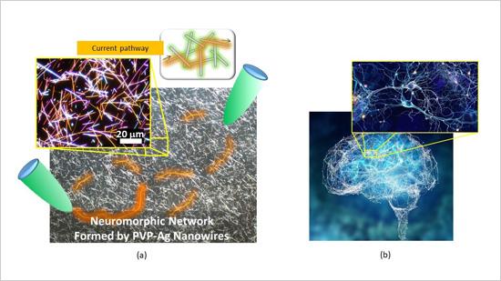
The device the researchers studied is made of a tangle of silver nanowires — with an average diameter of just 360 nanometers. (A nanometer is one-billionth of a meter.) The nanowires were coated in an insulating polymer about 1 nanometer thick. Overall, the device itself measured about 10 square millimeters — so small that it would take 25 of them to cover a dime.

Allowed to randomly self-assemble on a silicon wafer, the nanowires formed highly interconnected structures that are remarkably similar to those that form the neocortex, the part of the brain involved with higher functions such as language, perception and cognition.

One trait that differentiates the nanowire network from conventional electronic circuits is that electrons flowing through them cause the physical configuration of the network to change. In the study, electrical current caused silver atoms to migrate from within the polymer coating and form connections where two nanowires overlap. The system had about 10 million of these junctions, which are analogous to the synapses where brain cells connect and communicate.

The researchers attached two electrodes to the brain-like mesh to profile how the network performed. They observed “emergent behavior,” meaning that the network displayed characteristics as a whole that could not be attributed to the individual parts that make it up. This is another trait that makes the network resemble the brain and sets it apart from conventional computers.

After current flowed through the network, the connections between nanowires persisted for as much as one minute in some cases, which resembled the process of learning and memorization in the brain. Other times, the connections shut down abruptly after the charge ended, mimicking the brain’s process of forgetting.

In other experiments, the research team found that with less power flowing in, the device exhibited behavior that corresponds to what neuroscientists see when they use functional MRI scanning to take images of the brain of a sleeping person. With more power, the nanowire network’s behavior corresponded to that of the wakeful brain.

The paper is the latest in a series of publications examining nanowire networks as a brain-inspired system, an area of research that Gimzewski helped pioneer along with Stieg, a UCLA research scientist and an associate director of CNSI.

“Our approach may be useful for generating new types of hardware that are both energy-efficient and capable of processing complex datasets that challenge the limits of modern computers,” said Stieg, a co-author of the study.

The borderline-chaotic activity of the nanowire network resembles not only signaling within the brain but also other natural systems such as weather patterns. That could mean that, with further development, future versions of the device could help model such complex systems.

In other experiments, Gimzewski and Stieg already have coaxed a silver nanowire device to successfully predict statistical trends in Los Angeles traffic patterns based on previous years’ traffic data.

Because of their similarities to the inner workings of the brain, future devices based on nanowire technology could also demonstrate energy efficiency like the brain’s own processing. The human brain operates on power roughly equivalent to what’s used by a 20-watt incandescent bulb. By contrast, computer servers where work-intensive tasks take place — from training for machine learning to executing internet searches — can use the equivalent of many households’ worth of energy, with the attendant carbon footprint.

“In our studies, we have a broader mission than just reprogramming existing computers,” Gimzewski said. “Our vision is a system that will eventually be able to handle tasks that are closer to the way the human being operates.”

The study’s first author, Adrian Diaz-Alvarez, is from the International Center for Material Nanoarchitectonics at Japan’s National Institute for Materials Science. Co-authors include Tomonobu Nakayama and Rintaro Higuchi, also of NIMS; and Zdenka Kuncic at the University of Sydney in Australia.

Caption: (a) Micrograph of the neuromorphic network fabricated by this research team. The network contains of numerous junctions between nanowires, which operate as synaptic elements. When voltage is applied to the network (between the green probes), current pathways (orange) are formed in the network. (b) A Human brain and one of its neuronal networks. The brain is known to have a complex network structure and to operate by means of electrical signal propagation across the network. Credit: NIMS

A November 11, 2019 National Institute for Materials Science (Japan) press release (also on EurekAlert but dated December 25, 2019) first announced the news,

An international joint research team led by NIMS succeeded in fabricating a neuromorphic network composed of numerous metallic nanowires. Using this network, the team was able to generate electrical characteristics similar to those associated with higher order brain functions unique to humans, such as memorization, learning, forgetting, becoming alert and returning to calm. The team then clarified the mechanisms that induced these electrical characteristics.

The development of artificial intelligence (AI) techniques has been rapidly advancing in recent years and has begun impacting our lives in various ways. Although AI processes information in a manner similar to the human brain, the mechanisms by which human brains operate are still largely unknown. Fundamental brain components, such as neurons and the junctions between them (synapses), have been studied in detail. However, many questions concerning the brain as a collective whole need to be answered. For example, we still do not fully understand how the brain performs such functions as memorization, learning and forgetting, and how the brain becomes alert and returns to calm. In addition, live brains are difficult to manipulate in experimental research. For these reasons, the brain remains a “mysterious organ.” A different approach to brain research?in which materials and systems capable of performing brain-like functions are created and their mechanisms are investigated?may be effective in identifying new applications of brain-like information processing and advancing brain science.

The joint research team recently built a complex brain-like network by integrating numerous silver (Ag) nanowires coated with a polymer (PVP) insulating layer approximately 1 nanometer in thickness. A junction between two nanowires forms a variable resistive element (i.e., a synaptic element) that behaves like a neuronal synapse. This nanowire network, which contains a large number of intricately interacting synaptic elements, forms a “neuromorphic network”. When a voltage was applied to the neuromorphic network, it appeared to “struggle” to find optimal current pathways (i.e., the most electrically efficient pathways). The research team measured the processes of current pathway formation, retention and deactivation while electric current was flowing through the network and found that these processes always fluctuate as they progress, similar to the human brain’s memorization, learning, and forgetting processes. The observed temporal fluctuations also resemble the processes by which the brain becomes alert or returns to calm. Brain-like functions simulated by the neuromorphic network were found to occur as the huge number of synaptic elements in the network collectively work to optimize current transport, in the other words, as a result of self-organized and emerging dynamic processes..

The research team is currently developing a brain-like memory device using the neuromorphic network material. The team intends to design the memory device to operate using fundamentally different principles than those used in current computers. For example, while computers are currently designed to spend as much time and electricity as necessary in pursuit of absolutely optimum solutions, the new memory device is intended to make a quick decision within particular limits even though the solution generated may not be absolutely optimum. The team also hopes that this research will facilitate understanding of the brain’s information processing mechanisms.

This project was carried out by an international joint research team led by Tomonobu Nakayama (Deputy Director, International Center for Materials Nanoarchitectonics (WPI-MANA), NIMS), Adrian Diaz Alvarez (Postdoctoral Researcher, WPI-MANA, NIMS), Zdenka Kuncic (Professor, School of Physics, University of Sydney, Australia) and James K. Gimzewski (Professor, California NanoSystems Institute, University of California Los Angeles, USA).

Here at last is a link to and a citation for the paper,

Emergent dynamics of neuromorphic nanowire networks by Adrian Diaz-Alvarez, Rintaro Higuchi, Paula Sanz-Leon, Ido Marcus, Yoshitaka Shingaya, Adam Z. Stieg, James K. Gimzewski, Zdenka Kuncic & Tomonobu Nakayama. Scientific Reports volume 9, Article number: 14920 (2019) DOI: https://doi.org/10.1038/s41598-019-51330-6 Published: 17 October 2019

This paper is open access.

Nanodevices show (from the inside) how cells change

Embryo cells + nanodevices from University of Bath on Vimeo.

Caption: Five mouse embryos, each containing a nanodevice that is 22-millionths of a metre long. The film begins when the embryos are 2-hours old and continues for 5 hours. Each embryo is about 100-millionths of a metre in diameter. Credit: Professor Tony Perry

Fascinating, yes? As I often watch before reading the caption, these were mysterious grey blobs moving around was my first impression. Given the headline for the May 26, 2020 news item on ScienceDaily, I was expecting the squarish-shaped devices inside,

For the first time, scientists have introduced minuscule tracking devices directly into the interior of mammalian cells, giving an unprecedented peek into the processes that govern the beginning of development.

This work on one-cell embryos is set to shift our understanding of the mechanisms that underpin cellular behaviour in general, and may ultimately provide insights into what goes wrong in ageing and disease.

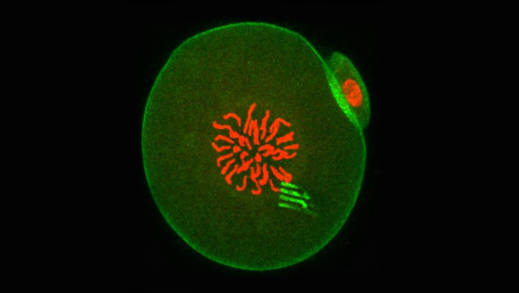
The research, led by Professor Tony Perry from the Department of Biology and Biochemistry at the University of Bath [UK], involved injecting a silicon-based nanodevice together with sperm into the egg cell of a mouse. The result was a healthy, fertilised egg containing a tracking device.

This image looks to have been enhanced with colour,

Fluorescence of an embryo containing a nanodevice. Courtesy: University of Bath

A May 25, 2020 University of Bath press release (also on EurekAlert but published May 26, 2020)

The tiny devices are a little like spiders, complete with eight highly flexible ‘legs’. The legs measure the ‘pulling and pushing’ forces exerted in the cell interior to a very high level of precision, thereby revealing the cellular forces at play and showing how intracellular matter rearranged itself over time.

The nanodevices are incredibly thin – similar to some of the cell’s structural components, and measuring 22 nanometres, making them approximately 100,000 times thinner than a pound coin. This means they have the flexibility to register the movement of the cell’s cytoplasm as the one-cell embryo embarks on its voyage towards becoming a two-cell embryo.

“This is the first glimpse of the physics of any cell on this scale from within,” said Professor Perry. “It’s the first time anyone has seen from the inside how cell material moves around and organises itself.”

WHY PROBE A CELL’S MECHANICAL BEHAVIOUR?

The activity within a cell determines how that cell functions, explains Professor Perry. “The behaviour of intracellular matter is probably as influential to cell behaviour as gene expression,” he said. Until now, however, this complex dance of cellular material has remained largely unstudied. As a result, scientists have been able to identify the elements that make up a cell, but not how the cell interior behaves as a whole.

“From studies in biology and embryology, we know about certain molecules and cellular phenomena, and we have woven this information into a reductionist narrative of how things work, but now this narrative is changing,” said Professor Perry. The narrative was written largely by biologists, who brought with them the questions and tools of biology. What was missing was physics. Physics asks about the forces driving a cell’s behaviour, and provides a top-down approach to finding the answer.

“We can now look at the cell as a whole, not just the nuts and bolts that make it.”

Mouse embryos were chosen for the study because of their relatively large size (they measure 100 microns, or 100-millionths of a metre, in diameter, compared to a regular cell which is only 10 microns [10-millionths of a metre] in diameter). This meant that inside each embryo, there was space for a tracking device.

The researchers made their measurements by examining video recordings taken through a microscope as the embryo developed. “Sometimes the devices were pitched and twisted by forces that were even greater than those inside muscle cells,” said Professor Perry. “At other times, the devices moved very little, showing the cell interior had become calm. There was nothing random about these processes – from the moment you have a one-cell embryo, everything is done in a predictable way. The physics is programmed.”

The results add to an emerging picture of biology that suggests material inside a living cell is not static, but instead changes its properties in a pre-ordained way as the cell performs its function or responds to the environment. The work may one day have implications for our understanding of how cells age or stop working as they should, which is what happens in disease.

The study is published this week in Nature Materials and involved a trans-disciplinary partnership between biologists, materials scientists and physicists based in the UK, Spain and the USA.

Here’s a link to and a citation for the paper,

Tracking intracellular forces and mechanical property changes in mouse one-cell embryo development by Marta Duch, Núria Torras, Maki Asami, Toru Suzuki, María Isabel Arjona, Rodrigo Gómez-Martínez, Matthew D. VerMilyea, Robert Castilla, José Antonio Plaza & Anthony C. F. Perry. Nature Materials (2020) DOI: https://doi.org/10.1038/s41563-020-0685-9 Published 25 May 2020

This paper is behind a paywall.

Quantum processor woven from light

Weaving a quantum processor from light is a jaw-dropping event (as far as I’m concerned). An October 17, 2019 news item on phys.org makes the announcement,

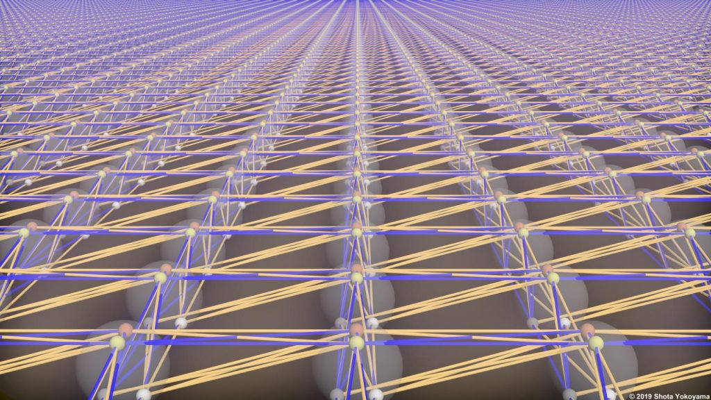
An international team of scientists from Australia, Japan and the United States has produced a prototype of a large-scale quantum processor made of laser light.

Based on a design ten years in the making, the processor has built-in scalability that allows the number of quantum components—made out of light—to scale to extreme numbers. The research was published in Science today [October 18, 2019; Note: I cannot explain the discrepancy between the dates]].

Quantum computers promise fast solutions to hard problems, but to do this they require a large number of quantum components and must be relatively error free. Current quantum processors are still small and prone to errors. This new design provides an alternative solution, using light, to reach the scale required to eventually outperform classical computers on important problems.

Caption: The entanglement structure of a large-scale quantum processor made of light. Credit: Shota Yokoyama 2019

An October 18, 2019 RMIT University (Australia) press release (also on EurekAlert but published October 17, 2019), which originated the news time, expands on the theme,

“While today’s quantum processors are impressive, it isn’t clear if the current designs can be scaled up to extremely large sizes,” notes Dr Nicolas Menicucci, Chief Investigator at the Centre for Quantum Computation and Communication Technology (CQC2T) at RMIT University in Melbourne, Australia.

“Our approach starts with extreme scalability – built in from the very beginning – because the processor, called a cluster state, is made out of light.”

Using light as a quantum processor

A cluster state is a large collection of entangled quantum components that performs quantum computations when measured in a particular way.

“To be useful for real-world problems, a cluster state must be both large enough and have the right entanglement structure. In the two decades since they were proposed, all previous demonstrations of cluster states have failed on one or both of these counts,” says Dr Menicucci. “Ours is the first ever to succeed at both.”

To make the cluster state, specially designed crystals convert ordinary laser light into a type of quantum light called squeezed light, which is then weaved into a cluster state by a network of mirrors, beamsplitters and optical fibres.

The team’s design allows for a relatively small experiment to generate an immense two-dimensional cluster state with scalability built in. Although the levels of squeezing – a measure of quality – are currently too low for solving practical problems, the design is compatible with approaches to achieve state-of-the-art squeezing levels.

The team says their achievement opens up new possibilities for quantum computing with light.

“In this work, for the first time in any system, we have made a large-scale cluster state whose structure enables universal quantum computation.” Says Dr Hidehiro Yonezawa, Chief Investigator, CQC2T at UNSW Canberra. “Our experiment demonstrates that this design is feasible – and scalable.”

###

The experiment was an international effort, with the design developed through collaboration by Dr Menicucci at RMIT, Dr Rafael Alexander from the University of New Mexico and UNSW Canberra researchers Dr Hidehiro Yonezawa and Dr Shota Yokoyama. A team of experimentalists at the University of Tokyo, led by Professor Akira Furusawa, performed the ground-breaking experiment.

Here’s a link to and a citation for the paper,

Generation of time-domain-multiplexed two-dimensional cluster state by Warit Asavanant, Yu Shiozawa, Shota Yokoyama, Baramee Charoensombutamon, Hiroki Emura, Rafael N. Alexander, Shuntaro Takeda, Jun-ichi Yoshikawa, Nicolas C. Menicucci, Hidehiro Yonezawa, Akira Furusawa. Science 18 Oct 2019: Vol. 366, Issue 6463, pp. 373-376 DOI: 10.1126/science.aay2645

This paper is behind a paywall.

Of puke, CRISPR, fruit flies, and monarch butterflies

I’ve never seen an educational institution use a somewhat vulgar slang term such as ‘puke’ before. Especially not in a news release. You’ll find that elsewhere online ‘puke’ has been replaced, in the headline, with the more socially acceptable ‘vomit’.

Since I wanted to catch this historic moment amid concerns that the original version of the news release will disappear, I’m including the entire news release as i saw it on EurekAlert.com (from an October 2, 2019 University of California at Berkeley news release),

News Release 2-Oct-2019

CRISPRed fruit flies mimic monarch butterfly — and could make you puke
Scientists recreate in flies the mutations that let monarch butterfly eat toxic milkweed with impunity

University of California – Berkeley

The fruit flies in Noah Whiteman’s lab may be hazardous to your health.

Whiteman and his University of California, Berkeley, colleagues have turned perfectly palatable fruit flies — palatable, at least, to frogs and birds — into potentially poisonous prey that may cause anything that eats them to puke. In large enough quantities, the flies likely would make a human puke, too, much like the emetic effect of ipecac syrup.

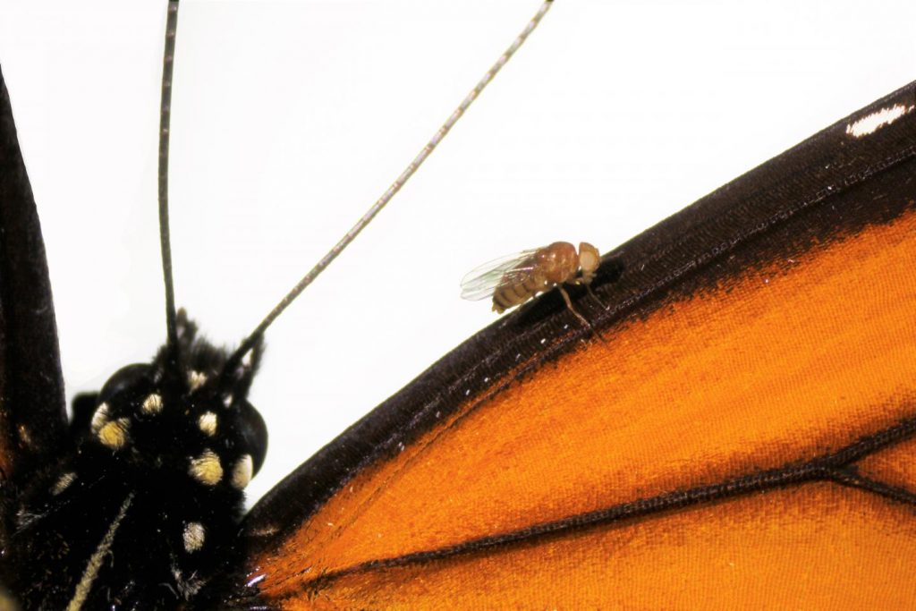
That’s because the team genetically engineered the flies, using CRISPR-Cas9 gene editing, to be able to eat milkweed without dying and to sequester its toxins, just as America’s most beloved butterfly, the monarch, does to deter predators.

This is the first time anyone has recreated in a multicellular organism a set of evolutionary mutations leading to a totally new adaptation to the environment — in this case, a new diet and new way of deterring predators.

Like monarch caterpillars, the CRISPRed fruit fly maggots thrive on milkweed, which contains toxins that kill most other animals, humans included. The maggots store the toxins in their bodies and retain them through metamorphosis, after they turn into adult flies, which means the adult “monarch flies” could also make animals upchuck.

The team achieved this feat by making three CRISPR edits in a single gene: modifications identical to the genetic mutations that allow monarch butterflies to dine on milkweed and sequester its poison. These mutations in the monarch have allowed it to eat common poisonous plants other insects could not and are key to the butterfly’s thriving presence throughout North and Central America.

Flies with the triple genetic mutation proved to be 1,000 times less sensitive to milkweed toxin than the wild fruit fly, Drosophila melanogaster.

Whiteman and his colleagues will describe their experiment in the Oct. 2 [2019] issue of the journal Nature.

Monarch flies

The UC Berkeley researchers created these monarch flies to establish, beyond a shadow of a doubt, which genetic changes in the genome of monarch butterflies were necessary to allow them to eat milkweed with impunity. They found, surprisingly, that only three single-nucleotide substitutions in one gene are sufficient to give fruit flies the same toxin resistance as monarchs.

“All we did was change three sites, and we made these superflies,” said Whiteman, an associate professor of integrative biology. “But to me, the most amazing thing is that we were able to test evolutionary hypotheses in a way that has never been possible outside of cell lines. It would have been difficult to discover this without having the ability to create mutations with CRISPR.”

Whiteman’s team also showed that 20 other insect groups able to eat milkweed and related toxic plants – including moths, beetles, wasps, flies, aphids, a weevil and a true bug, most of which sport the color orange to warn away predators – independently evolved mutations in one, two or three of the same amino acid positions to overcome, to varying degrees, the toxic effects of these plant poisons.

In fact, his team reconstructed the one, two or three mutations that led to each of the four butterfly and moth lineages, each mutation conferring some resistance to the toxin. All three mutations were necessary to make the monarch butterfly the king of milkweed.
Resistance to milkweed toxin comes at a cost, however. Monarch flies are not as quick to recover from upsets, such as being shaken — a test known as “bang” sensitivity.

“This shows there is a cost to mutations, in terms of recovery of the nervous system and probably other things we don’t know about,” Whiteman said. “But the benefit of being able to escape a predator is so high … if it’s death or toxins, toxins will win, even if there is a cost.”

Plant vs. insect

Whiteman is interested in the evolutionary battle between plants and parasites and was intrigued by the evolutionary adaptations that allowed the monarch to beat the milkweed’s toxic defense. He also wanted to know whether other insects that are resistant — though all less resistant than the monarch — use similar tricks to disable the toxin.

“Since plants and animals first invaded land 400 million years ago, this coevolutionary arms race is thought to have given rise to a lot of the plant and animal diversity that we see, because most animals are insects, and most insects are herbivorous: they eat plants,” he said.

Milkweeds and a variety of other plants, including foxglove, the source of digitoxin and digoxin, contain related toxins — called cardiac glycosides — that can kill an elephant and any creature with a beating heart. Foxglove’s effect on the heart is the reason that an extract of the plant, in the genus Digitalis, has been used for centuries to treat heart conditions, and why digoxin and digitoxin are used today to treat congestive heart failure.

These plants’ bitterness alone is enough to deter most animals, but a small minority of insects, including the monarch (Danaus plexippus) and its relative, the queen butterfly (Danaus gilippus), have learned to love milkweed and use it to repel predators.

Whiteman noted that the monarch is a tropical lineage that invaded North America after the last ice age, in part enabled by the three mutations that allowed it to eat a poisonous plant other animals could not, giving it a survival edge and a natural defense against predators.

“The monarch resists the toxin the best of all the insects, and it has the biggest population size of any of them; it’s all over the world,” he said.

The new paper reveals that the mutations had to occur in the right sequence, or else the flies would never have survived the three separate mutational events.

Thwarting the sodium pump

The poisons in these plants, most of them a type of cardenolide, interfere with the sodium/potassium pump (Na+/K+-ATPase) that most of the body’s cells use to move sodium ions out and potassium ions in. The pump creates an ion imbalance that the cell uses to its favor. Nerve cells, for example, transmit signals along their elongated cell bodies, or axons, by opening sodium and potassium gates in a wave that moves down the axon, allowing ions to flow in and out to equilibrate the imbalance. After the wave passes, the sodium pump re-establishes the ionic imbalance.

Digitoxin, from foxglove, and ouabain, the main toxin in milkweed, block the pump and prevent the cell from establishing the sodium/potassium gradient. This throws the ion concentration in the cell out of whack, causing all sorts of problems. In animals with hearts, like birds and humans, heart cells begin to beat so strongly that the heart fails; the result is death by cardiac arrest.

Scientists have known for decades how these toxins interact with the sodium pump: they bind the part of the pump protein that sticks out through the cell membrane, clogging the channel. They’ve even identified two specific amino acid changes or mutations in the protein pump that monarchs and the other insects evolved to prevent the toxin from binding.

But Whiteman and his colleagues weren’t satisfied with this just so explanation: that insects coincidentally developed the same two identical mutations in the sodium pump 14 separate times, end of story. With the advent of CRISPR-Cas9 gene editing in 2012, coinvented by UC Berkeley’s Jennifer Doudna, Whiteman and colleagues Anurag Agrawal of Cornell University and Susanne Dobler of the University of Hamburg in Germany applied to the Templeton Foundation for a grant to recreate these mutations in fruit flies and to see if they could make the flies immune to the toxic effects of cardenolides.

Seven years, many failed attempts and one new grant from the National Institutes of Health later, along with the dedicated CRISPR work of GenetiVision of Houston, Texas, they finally achieved their goal. In the process, they discovered a third critical, compensatory mutation in the sodium pump that had to occur before the last and most potent resistance mutation would stick. Without this compensatory mutation, the maggots died.

Their detective work required inserting single, double and triple mutations into the fruit fly’s own sodium pump gene, in various orders, to assess which ones were necessary. Insects having only one of the two known amino acid changes in the sodium pump gene were best at resisting the plant poisons, but they also had serious side effects — nervous system problems — consistent with the fact that sodium pump mutations in humans are often associated with seizures. However, the third, compensatory mutation somehow reduces the negative effects of the other two mutations.

“One substitution that evolved confers weak resistance, but it is always present and allows for substitutions that are going to confer the most resistance,” said postdoctoral fellow Marianna Karageorgi, a geneticist and evolutionary biologist. “This substitution in the insect unlocks the resistance substitutions, reducing the neurological costs of resistance. Because this trait has evolved so many times, we have also shown that this is not random.”

The fact that one compensatory mutation is required before insects with the most resistant mutation could survive placed a constraint on how insects could evolve toxin resistance, explaining why all 21 lineages converged on the same solution, Whiteman said. In other situations, such as where the protein involved is not so critical to survival, animals might find different solutions.

“This helps answer the question, ‘Why does convergence evolve sometimes, but not other times?'” Whiteman said. “Maybe the constraints vary. That’s a simple answer, but if you think about it, these three mutations turned a Drosophila protein into a monarch one, with respect to cardenolide resistance. That’s kind of remarkable.”

###

The research was funded by the Templeton Foundation and the National Institutes of Health. Co-authors with Whiteman and Agrawal are co-first authors Marianthi Karageorgi of UC Berkeley and Simon Groen, now at New York University; Fidan Sumbul and Felix Rico of Aix-Marseille Université in France; Julianne Pelaez, Kirsten Verster, Jessica Aguilar, Susan Bernstein, Teruyuki Matsunaga and Michael Astourian of UC Berkeley; Amy Hastings of Cornell; and Susanne Dobler of Universität Hamburg in Germany.

Robert Sanders’ Oct. 2, 2019′ news release for the University of California at Berkeley (it’s also been republished as an Oct. 2, 2019 news item on ScienceDaily) has had its headline changed to ‘vomit’ but you’ll find the more vulgar word remains in two locations of the second paragraph of the revised new release.

If you have time, go to the news release on the University of California at Berkeley website just to admire the images that have been embedded in the news release. Here’s one,

Caption: A Drosophila melanogaster “monarch fly” with mutations introduced by CRISPR-Cas9 genome editing (V111, S119 and H122) to the sodium potassium pump, on a wing of a monarch butterfly (Danaus plexippus). Credit & Ccpyright: Julianne Pelaez

Here’s a link to and a citation for the paper,

Genome editing retraces the evolution of toxin resistance in the monarch butterfly by Marianthi Karageorgi, Simon C. Groen, Fidan Sumbul, Julianne N. Pelaez, Kirsten I. Verster, Jessica M. Aguilar, Amy P. Hastings, Susan L. Bernstein, Teruyuki Matsunaga, Michael Astourian, Geno Guerra, Felix Rico, Susanne Dobler, Anurag A. Agrawal & Noah K. Whiteman. Nature (2019) DOI: https://doi.org/10.1038/s41586-019-1610-8 Published 02 October 2019

This paper is behind a paywall.

Words about a word

I’m glad they changed the headline and substituted vomit for puke. I think we need vulgar and/or taboo words to release anger or disgust or other difficult emotions. Incorporating those words into standard language deprives them of that power.

The last word: Genetivision

The company mentioned in the new release, Genetivision, is the place to go for transgenic flies. Here’s a sampling from the their Testimonials webpage,
GenetiVision‘s service has been excellent in the quality and price. The timeliness of its international service has been a big plus. We are very happy with its consistent service and the flies it generates.”
Kwang-Wook Choi, Ph.D.
Department of Biological Sciences
Korea Advanced Institute of Science and Technology


“We couldn’t be happier with GenetiVision. Great prices on both standard P and PhiC31 transgenics, quick turnaround time, and we’re still batting 1000 with transformant success. We used to do our own injections but your service makes it both faster and more cost-effective. Thanks for your service!”
Thomas Neufeld, Ph.D.
Department of Genetics, Cell Biology and Development
University of Minnesota

You can find out more here at the Genetivision website.

Control your electronics devices with your clothing while protecting yourself from bacteria

Purdue University researchers have developed a new fabric innovation that allows the wearer to control electronic devices through the clothing. Courtesy: Purdue University

I like the image but do they really want someone pressing a cufflink? Anyway, being able to turn on your house lights and music system with your clothing would certainly be convenient. From an August 8, 2019 Purdue University (Indiana, US) news release (also on EurekAlert) by Chris Adam,

A new addition to your wardrobe may soon help you turn on the lights and music – while also keeping you fresh, dry, fashionable, clean and safe from the latest virus that’s going around.

Purdue University researchers have developed a new fabric innovation that allows wearers to control electronic devices through clothing.

“It is the first time there is a technique capable to transform any existing cloth item or textile into a self-powered e-textile containing sensors, music players or simple illumination displays using simple embroidery without the need for expensive fabrication processes requiring complex steps or expensive equipment,” said Ramses Martinez, an assistant professor in the School of Industrial Engineering and in the Weldon School of Biomedical Engineering in Purdue’s College of Engineering.

The technology is featured in the July 25 [2019] edition of Advanced Functional Materials.

“For the first time, it is possible to fabricate textiles that can protect you from rain, stains, and bacteria while they harvest the energy of the user to power textile-based electronics,” Martinez said. “These self-powered e-textiles also constitute an important advancement in the development of wearable machine-human interfaces, which now can be washed many times in a conventional washing machine without apparent degradation.

Martinez said the Purdue waterproof, breathable and antibacterial self-powered clothing is based on omniphobic triboelectric nanogeneragtors (RF-TENGs) – which use simple embroidery and fluorinated molecules to embed small electronic components and turn a piece of clothing into a mechanism for powering devices. The Purdue team says the RF-TENG technology is like having a wearable remote control that also keeps odors, rain, stains and bacteria away from the user.

“While fashion has evolved significantly during the last centuries and has easily adopted recently developed high-performance materials, there are very few examples of clothes on the market that interact with the user,” Martinez said. “Having an interface with a machine that we are constantly wearing sounds like the most convenient approach for a seamless communication with machines and the Internet of Things.”

The technology is being patented through the Purdue Research Foundation Office of Technology Commercialization. The researchers are looking for partners to test and commercialize their technology.

Their work aligns with Purdue’s Giant Leaps celebration of the university’s global advancements in artificial intelligence and health as part of Purdue’s 150th anniversary. It is one of the four themes of the yearlong celebration’s Ideas Festival, designed to showcase Purdue as an intellectual center solving real-world issues.

Here’s a link to and a citation for the paper,

Waterproof, Breathable, and Antibacterial Self‐Powered e‐Textiles Based on Omniphobic Triboelectric Nanogenerators by Marina Sala de Medeiros, Daniela Chanci, Carolina Moreno, Debkalpa Goswami, Ramses V. Martinez. Advanced Functional Materials DOI: https://doi.org/10.1002/adfm.201904350 First published online: 25 July 2019

This paper is behind a paywall.

So thin and soft you don’t notice it: new wearable tech

An August 2, 2019 news item on ScienceDaily features some new work on wearable technology that was a bit of a surprise to me,

Wearable human-machine interfaces — devices that can collect and store important health information about the wearer, among other uses — have benefited from advances in electronics, materials and mechanical designs. But current models still can be bulky and uncomfortable, and they can’t always handle multiple functions at one time.

Researchers reported Friday, Aug. 2 [2019], the discovery of a multifunctional ultra-thin wearable electronic device that is imperceptible to the wearer.

I expected this wearable technology to be a piece of clothing that somehow captured health data but it’s not,

While a health care application is mentioned early in the August 2, 2019 University of Houston news release (also on EurekAlert) by Jeannie Kever the primary interest seems to be robots and robotic skin (Note: This news release originated the news item on ScienceDaily),

The device allows the wearer to move naturally and is less noticeable than wearing a Band-Aid, said Cunjiang Yu, Bill D. Cook Associate Professor of Mechanical Engineering at the University of Houston and lead author for the paper, published as the cover story in Science Advances.

“Everything is very thin, just a few microns thick,” said Yu, who also is a principal investigator at the Texas Center for Superconductivity at UH. “You will not be able to feel it.”
It has the potential to work as a prosthetic skin for a robotic hand or other robotic devices, with a robust human-machine interface that allows it to automatically collect information and relay it back to the wearer.

That has applications for health care – “What if when you shook hands with a robotic hand, it was able to instantly deduce physical condition?” Yu asked – as well as for situations such as chemical spills, which are risky for humans but require human decision-making based on physical inspection.

While current devices are gaining in popularity, the researchers said they can be bulky to wear, offer slow response times and suffer a drop in performance over time. More flexible versions are unable to provide multiple functions at once – sensing, switching, stimulation and data storage, for example – and are generally expensive and complicated to manufacture.

The device described in the paper, a metal oxide semiconductor on a polymer base, offers manufacturing advantages and can be processed at temperatures lower than 300 C.

“We report an ultrathin, mechanically imperceptible, and stretchable (human-machine interface) HMI device, which is worn on human skin to capture multiple physical data and also on a robot to offer intelligent feedback, forming a closed-loop HMI,” the researchers wrote. “The multifunctional soft stretchy HMI device is based on a one-step formed, sol-gel-on-polymer-processed indium zinc oxide semiconductor nanomembrane electronics.”

In addition to Yu, the paper’s co-authors include first author Kyoseung Sim, Zhoulyu Rao, Faheem Ershad, Jianming Lei, Anish Thukral and Jie Chen, all of UH; Zhanan Zou and Jianliang Xiao, both of the University of Colorado; and Qing-An Huang of Southeast University in Nanjing, China.

Here’s a link to and a citation for the paper,

Metal oxide semiconductor nanomembrane–based soft unnoticeable multifunctional electronics for wearable human-machine interfaces by Kyoseung Sim, Zhoulyu Rao, Zhanan Zou, Faheem Ershad, Jianming Lei, Anish Thukral, Jie Chen, Qing-An Huang, Jianliang Xiao and Cunjiang Yu. Science Advances 02 Aug 2019: Vol. 5, no. 8, eaav9653 DOI: 10.1126/sciadv.aav9653

This paper appears to be open access.