This piece on new construction materials does have a nanotechnology aspect although it’s not made clear exactly how nanotechnology plays a role.
From a Dec. 28, 2016 news item on phys.org (Note: A link has been removed),
The construction industry is preparing to use textiles from the clothing and footwear industries. Gore-Tex-like membranes, which are usually found in weather-proof jackets and trekking shoes, are now being studied to build breathable, water-resistant walls. Tyvek is one such synthetic textile being used as a “raincoat” for homes.
You can find out more about Tyvek here.on the Dupont website.
A Dec. 21, 2016 press release by Chiara Cecchi for Youris ((European Research Media Center), which originated the news item, proceeds with more about textile-type construction materials,
Camping tents, which have been used for ages to protect against wind, ultra-violet rays and rain, have also inspired the modern construction industry, or “buildtech sector”. This new field of research focuses on the different fibres (animal-based such as wool or silk, plant-based such as linen and cotton and synthetic such as polyester and rayon) in order to develop technical or high-performance materials, thus improving the quality of construction, especially for buildings, dams, bridges, tunnels and roads. This is due to the fibres’ mechanical properties, such as lightness, strength, and also resistance to many factors like creep, deterioration by chemicals and pollutants in the air or rain.
“Textiles play an important role in the modernisation of infrastructure and in sustainable buildings”, explains Andrea Bassi, professor at the Department of Civil and Environmental Engineering (DICA), Politecnico of Milan, “Nylon and fiberglass are mixed with traditional fibres to control thermal and acoustic insulation in walls, façades and roofs. Technological innovation in materials, which includes nanotechnologies [emphasis mine] combined with traditional textiles used in clothes, enables buildings and other constructions to be designed using textiles containing steel polyvinyl chloride (PVC) or ethylene tetrafluoroethylene (ETFE). This gives the materials new antibacterial, antifungal and antimycotic properties in addition to being antistatic, sound-absorbing and water-resistant”.
Rooflys is another example. In this case, coated black woven textiles are placed under the roof to protect roof insulation from mould. These building textiles have also been tested for fire resistance, nail sealability, water and vapour impermeability, wind and UV resistance.
Photo: Production line at the co-operative enterprise CAVAC Biomatériaux, France. Natural fibres processed into a continuous mat (biofib) – Martin Ansell, BRE CICM, University of Bath, UK
In Spain three researchers from the Technical University of Madrid (UPM) have developed a new panel made with textile waste. They claim that it can significantly enhance both the thermal and acoustic conditions of buildings, while reducing greenhouse gas emissions and the energy impact associated with the development of construction materials.
Besides textiles, innovative natural fibre composite materials are a parallel field of the research on insulators that can preserve indoor air quality. These bio-based materials, such as straw and hemp, “can reduce the incidence of mould growth because they breathe. The breathability of materials refers to their ability to absorb and desorb moisture naturally”, says expert Finlay White from Modcell, who contributed to the construction of what they claim are the world’s first commercially available straw houses, “For example, highly insulated buildings with poor ventilation can build-up high levels of moisture in the air. If the moisture meets a cool surface it will condensate and producing mould, unless it is managed. Bio-based materials have the means to absorb moisture so that the risk of condensation is reduced, preventing the potential for mould growth”.
The Bristol-based green technology firm [Modcell] is collaborating with the European Isobio project, which is testing bio-based insulators which perform 20% better than conventional materials. “This would lead to a 5% total energy reduction over the lifecycle of a building”, explains Martin Ansell, from BRE Centre for Innovative Construction Materials (BRE CICM), University of Bath, UK, another partner of the project.
“Costs would also be reduced. We are evaluating the thermal and hygroscopic properties of a range of plant-derived by-products including hemp, jute, rape and straw fibres plus corn cob residues. Advanced sol-gel coatings are being deposited on these fibres to optimise these properties in order to produce highly insulating and breathable construction materials”, Ansell concludes.

You can find Modcell here.
Here’s another image, which I believe is a closeup of the processed fibre shown in the above,
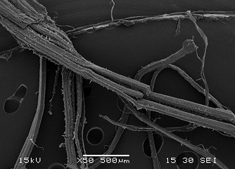
Production line at the co-operative enterprise CAVAC Biomatériaux, France. Natural fibres processed into a continuous mat (biofib) – Martin Ansell, BRE CICM, University of Bath, UK [Note: This caption appears to be a copy of the caption for the previous image]