Tag Archives: regenerative medicine

A bioengineered robot hand with its own nervous system: machine/flesh and a job opening

A November 14, 2017 news item on phys.org announces a grant for a research project which will see engineered robot hands combined with regenerative medicine to imbue neuroprosthetic hands with the sense of touch,

The sense of touch is often taken for granted. For someone without a limb or hand, losing that sense of touch can be devastating. While highly sophisticated prostheses with complex moving fingers and joints are available to mimic almost every hand motion, they remain frustratingly difficult and unnatural for the user. This is largely because they lack the tactile experience that guides every movement. This void in sensation results in limited use or abandonment of these very expensive artificial devices. So why not make a prosthesis that can actually “feel” its environment?

That is exactly what an interdisciplinary team of scientists from Florida Atlantic University and the University of Utah School of Medicine aims to do. They are developing a first-of-its-kind bioengineered robotic hand that will grow and adapt to its environment. This “living” robot will have its own peripheral nervous system directly linking robotic sensors and actuators. FAU’s College of Engineering and Computer Science is leading the multidisciplinary team that has received a four-year, $1.3 million grant from the National Institute of Biomedical Imaging and Bioengineering of the [US] National Institutes of Health for a project titled “Virtual Neuroprosthesis: Restoring Autonomy to People Suffering from Neurotrauma.”

A November14, 2017 Florida Atlantic University (FAU) news release by Gisele Galoustian, which originated the news item, goes into more detail,

With expertise in robotics, bioengineering, behavioral science, nerve regeneration, electrophysiology, microfluidic devices, and orthopedic surgery, the research team is creating a living pathway from the robot’s touch sensation to the user’s brain to help amputees control the robotic hand. A neuroprosthesis platform will enable them to explore how neurons and behavior can work together to regenerate the sensation of touch in an artificial limb.

At the core of this project is a cutting-edge robotic hand and arm developed in the BioRobotics Laboratory in FAU’s College of Engineering and Computer Science. Just like human fingertips, the robotic hand is equipped with numerous sensory receptors that respond to changes in the environment. Controlled by a human, it can sense pressure changes, interpret the information it is receiving and interact with various objects. It adjusts its grip based on an object’s weight or fragility. But the real challenge is figuring out how to send that information back to the brain using living residual neural pathways to replace those that have been damaged or destroyed by trauma.

“When the peripheral nerve is cut or damaged, it uses the rich electrical activity that tactile receptors create to restore itself. We want to examine how the fingertip sensors can help damaged or severed nerves regenerate,” said Erik Engeberg, Ph.D., principal investigator, an associate professor in FAU’s Department of Ocean and Mechanical Engineering, and director of FAU’s BioRobotics Laboratory. “To accomplish this, we are going to directly connect these living nerves in vitro and then electrically stimulate them on a daily basis with sensors from the robotic hand to see how the nerves grow and regenerate while the hand is operated by limb-absent people.”

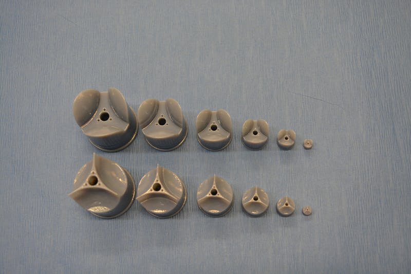
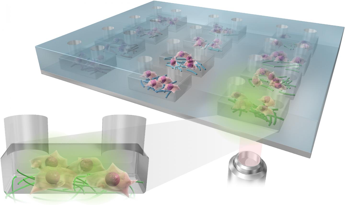
For the study, the neurons will not be kept in conventional petri dishes. Instead, they will be placed in  biocompatible microfluidic chambers that provide a nurturing environment mimicking the basic function of living cells. Sarah E. Du, Ph.D., co-principal investigator, an assistant professor in FAU’s Department of Ocean and Mechanical Engineering, and an expert in the emerging field of microfluidics, has developed these tiny customized artificial chambers with embedded micro-electrodes. The research team will be able to stimulate the neurons with electrical impulses from the robot’s hand to help regrowth after injury. They will morphologically and electrically measure in real-time how much neural tissue has been restored.

Jianning Wei, Ph.D., co-principal investigator, an associate professor of biomedical science in FAU’s Charles E. Schmidt College of Medicine, and an expert in neural damage and regeneration, will prepare the neurons in vitro, observe them grow and see how they fare and regenerate in the aftermath of injury. This “virtual” method will give the research team multiple opportunities to test and retest the nerves without any harm to subjects.

Using an electroencephalogram (EEG) to detect electrical activity in the brain, Emmanuelle Tognoli, Ph.D., co-principal investigator, associate research professor in FAU’s Center for Complex Systems and Brain Sciences in the Charles E. Schmidt College of Science, and an expert in electrophysiology and neural, behavioral, and cognitive sciences, will examine how the tactile information from the robotic sensors is passed onto the brain to distinguish scenarios with successful or unsuccessful functional restoration of the sense of touch. Her objective: to understand how behavior helps nerve regeneration and how this nerve regeneration helps the behavior.

Once the nerve impulses from the robot’s tactile sensors have gone through the microfluidic chamber, they are sent back to the human user manipulating the robotic hand. This is done with a special device that converts the signals coming from the microfluidic chambers into a controllable pressure at a cuff placed on the remaining portion of the amputated person’s arm. Users will know if they are squeezing the object too hard or if they are losing their grip.

Engeberg also is working with Douglas T. Hutchinson, M.D., co-principal investigator and a professor in the Department of Orthopedics at the University of Utah School of Medicine, who specializes in hand and orthopedic surgery. They are developing a set of tasks and behavioral neural indicators of performance that will ultimately reveal how to promote a healthy sensation of touch in amputees and limb-absent people using robotic devices. The research team also is seeking a post-doctoral researcher with multi-disciplinary experience to work on this breakthrough project.

Here’s more about the job opportunity from the FAU BioRobotics Laboratory job posting, (I checked on January 30, 2018 and it seems applications are still being accepted.)

Post-doctoral Opportunity

Dated Posted: Oct. 13, 2017

The BioRobotics Lab at Florida Atlantic University (FAU) invites applications for a NIH NIBIB-funded Postdoctoral position to develop a Virtual Neuroprosthesis aimed at providing a sense of touch in amputees and limb-absent people.

Candidates should have a Ph.D. in one of the following degrees: mechanical engineering, electrical engineering, biomedical engineering, bioengineering or related, with interest and/or experience in transdisciplinary work at the intersection of robotic hands, biology, and biomedical systems. Prior experience in the neural field will be considered an advantage, though not a necessity. Underrepresented minorities and women are warmly encouraged to apply.

The postdoctoral researcher will be co-advised across the department of Mechanical Engineering and the Center for Complex Systems & Brain Sciences through an interdisciplinary team whose expertise spans Robotics, Microfluidics, Behavioral and Clinical Neuroscience and Orthopedic Surgery.

The position will be for one year with a possibility of extension based on performance. Salary will be commensurate with experience and qualifications. Review of applications will begin immediately and continue until the position is filled.

The application should include:

  1. a cover letter with research interests and experiences,
  2. a CV, and
  3. names and contact information for three professional references.

Qualified candidates can contact Erik Engeberg, Ph.D., Associate Professor, in the FAU Department of Ocean and Mechanical Engineering at eengeberg@fau.edu. Please reference AcademicKeys.com in your cover letter when applying for or inquiring about this job announcement.

You can find the apply button on this page. Good luck!

A cheaper way to make artificial organs

In the quest to develop artificial organs, the University of British Columbia (UBC) is the not the first research institution that comes to my mind. It seems I may need to reevaluate now that UBC (Okanagan) has announced some work on bio-inks and artificial organs in a Sept. 12, 2017 news  release (also on EurekAlert) by Patty Wellborn,,

A new bio-ink that may support a more efficient and inexpensive fabrication of human tissues and organs has been created by researchers at UBC’s Okanagan campus.

Keekyoung Kim, an assistant professor at UBC Okanagan’s School of Engineering, says this development can accelerate advances in regenerative medicine.

Using techniques like 3D printing, scientists are creating bio-material products that function alongside living cells. These products are made using a number of biomaterials including gelatin methacrylate (GelMA), a hydrogel that can serve as a building block in bio-printing. This type of bio-material—called bio-ink—are made of living cells, but can be printed and molded into specific organ or tissue shapes.

The UBC team analyzed the physical and biological properties of three different GelMA hydrogels—porcine skin, cold-water fish skin and cold-soluble gelatin. They found that hydrogel made from cold-soluble gelatin (gelatin which dissolves without heat) was by far the best performer and a strong candidate for future 3D organ printing.

“A big drawback of conventional hydrogel is its thermal instability. Even small changes in temperature cause significant changes in its viscosity or thickness,” says Kim. “This makes it problematic for many room temperature bio-fabrication systems, which are compatible with only a narrow range of hydrogel viscosities and which must generate products that are as uniform as possible if they are to function properly.”

Kim’s team created two new hydrogels—one from fish skin, and one from cold-soluble gelatin—and compared their properties to those of porcine skin GelMA. Although fish skin GelMA had some benefits, cold-soluble GelMA was the top overall performer. Not only could it form healthy tissue scaffolds, allowing cells to successfully grow and adhere to it, but it was also thermally stable at room temperature.

The UBC team also demonstrated that cold-soluble GelMA produces consistently uniform droplets at temperatures, thus making it an excellent choice for use in 3D bio-printing.

“We hope this new bio-ink will help researchers create improved artificial organs and lead to the development of better drugs, tissue engineering and regenerative therapies,” Kim says. “The next step is to investigate whether or not cold-soluble GelMA-based tissue scaffolds are can be used long-term both in the laboratory and in real-world transplants.”

Three times cheaper than porcine skin gelatin, cold-soluble gelatin is used primarily in culinary applications.

Here’s a link to and a citation for the paper,

Comparative study of gelatin methacrylate hydrogels from different sources for biofabrication applications by Zongjie Wang, Zhenlin Tian, Fredric Menard, and Keekyoung Kim. Biofabrication, Volume 9, Number 4 Special issue on Bioinks https://doi.org/10.1088/1758-5090/aa83cf Published 21 August 2017

© 2017 IOP Publishing Ltd

This paper is behind a paywall.

‘Origami organs’ for tissue engineering

This is a different approach to tissue engineering and its the consequence of a serendipitous accident.  From an Aug. 7, 2017 Northwestern University news release (also on EurekAlert),

Northwestern Medicine scientists and engineers have invented a range of bioactive “tissue papers” made of materials derived from organs that are thin and flexible enough to even fold into an origami bird. The new biomaterials can potentially be used to support natural hormone production in young cancer patients and aid wound healing.

The tissue papers are made from structural proteins excreted by cells that give organs their form and structure. The proteins are combined with a polymer to make the material pliable.

In the study, individual types of tissue papers were made from ovarian, uterine, kidney, liver, muscle or heart proteins obtained by processing pig and cow organs. Each tissue paper had specific cellular properties of the organ from which it was made.

The article describing the tissue paper and its function will be published Aug. 7 in the journal Advanced Functional Materials.

“This new class of biomaterials has potential for tissue engineering and regenerative medicine as well as drug discovery and therapeutics,” corresponding author Ramille Shah said. “It’s versatile and surgically friendly.”

Shah is an assistant professor of surgery at the Feinberg School of Medicine and an assistant professor of materials science and engineering at McCormick School of Engineering. She also is a member of the Simpson Querrey Institute for BioNanotechnology.

For wound healing, Shah thinks the tissue paper could provide support and the cell signaling needed to help regenerate tissue to prevent scarring and accelerate healing.

The tissue papers are made from natural organs or tissues. The cells are removed, leaving the natural structural proteins – known as the extracellular matrix – that then are dried into a powder and processed into the tissue papers. Each type of paper contains residual biochemicals and protein architecture from its original organ that can stimulate cells to behave in a certain way.

In the lab of reproductive scientist Teresa Woodruff, the tissue paper made from a bovine ovary was used to grow ovarian follicles when they were cultured in vitro. The follicles (eggs and hormone-producing cells) grown on the tissue paper produced hormones necessary for proper function and maturation.

“This could provide another option to restore normal hormone function to young cancer patients who often lose their hormone function as a result of chemotherapy and radiation,” Woodruff, a study coauthor, said.

A strip of the ovarian paper with the follicles could be implanted under the arm to restore hormone production for cancer patients or even women in menopause.

Woodruff is the director of the Oncofertility Consortium and the Thomas J. Watkins Memorial Professor of Obstetrics and Gynecology at Feinberg.

In addition, the tissue paper made from various organs separately supported the growth of adult human stem cells. Scientists placed human bone marrow stem cells on the tissue paper, and all the stem cells attached and multiplied over four weeks.

“That’s a good sign that the paper supports human stem cell growth,” said first author Adam Jakus, who developed the tissue papers. “It’s an indicator that once we start using tissue paper in animal models it will be biocompatible.”

The tissue papers feel and behave much like standard office paper when they are dry, Jakus said. Jakus simply stacks them in a refrigerator or a freezer. He even playfully folded them into an origami bird.

“Even when wet, the tissue papers maintain their mechanical properties and can be rolled, folded, cut and sutured to tissue,” he said.

Jakus was a Hartwell postdoctoral fellow in Shah’s lab for the study and is now chief technology officer and cofounder of the startup company Dimension Inx, LLC, which was also cofounded by Shah. The company will develop, produce and sell 3-D printable materials primarily for medical applications. The Intellectual Property is owned by Northwestern University and will be licensed to Dimension Inx.

An Accidental Spill Sparked Invention

An accidental spill of 3-D printing ink in Shah’s lab by Jakus sparked the invention of the tissue paper. Jakus was attempting to make a 3-D printable ovary ink similar to the other 3-D printable materials he previously developed to repair and regenerate bone, muscle and nerve tissue. When he went to wipe up the spill, the ovary ink had already formed a dry sheet.

“When I tried to pick it up, it felt strong,” Jakus said. “I knew right then I could make large amounts of bioactive materials from other organs. The light bulb went on in my head. I could do this with other organs.”

“It is really amazing that meat and animal by-products like a kidney, liver, heart and uterus can be transformed into paper-like biomaterials that can potentially regenerate and restore function to tissues and organs,” Jakus said. “I’ll never look at a steak or pork tenderloin the same way again.”

For those who like their news in a video,

As someone who once made baklava, that does not look like filo pastry, where an individual sheet is quite thin and rips easily. Enough said.

Here’s a link to and a citation for the paper,

“Tissue Papers” from Organ-Specific Decellularized Extracellular Matrices by Adam E. Jakus, Monica M. Laronda, Alexandra S. Rashedi, Christina M. Robinson, Chris Lee, Sumanas W. Jordan, Kyle E. Orwig, Teresa K. Woodruff, and Ramille N. Shah. Advnaced Functional Materials DOI: 10.1002/adfm.201700992 Version of Record online: 7 AUG 2017

© 2017 WILEY-VCH Verlag GmbH & Co. KGaA, Weinheim

This paper is behind a paywall.

A nano fabrication technique used to create next generation heart valve

I am going to have take the researchers’ word that these somehow lead to healthy heart valve tissue,

In rotary jet spinning technology, a rotating nozzle extrudes a solution of extracellular matrix (ECM) into nanofibers that wrap themselves around heart valve-shaped mandrels. By using a series of mandrels with different sizes, the manufacturing process becomes fully scalable and is able to provide JetValves for all age groups and heart sizes. Credit: Wyss Institute at Harvard University

From a May 18, 2017 news item on ScienceDaily,

The human heart beats approximately 35 million times every year, effectively pumping blood into the circulation via four different heart valves. Unfortunately, in over four million people each year, these delicate tissues malfunction due to birth defects, age-related deteriorations, and infections, causing cardiac valve disease.

Today, clinicians use either artificial prostheses or fixed animal and cadaver-sourced tissues to replace defective valves. While these prostheses can restore the function of the heart for a while, they are associated with adverse comorbidity and wear down and need to be replaced during invasive and expensive surgeries. Moreover, in children, implanted heart valve prostheses need to be replaced even more often as they cannot grow with the child.

A team lead by Kevin Kit Parker, Ph.D. at Harvard University’s Wyss Institute for Biologically Inspired Engineering recently developed a nanofiber fabrication technique to rapidly manufacture heart valves with regenerative and growth potential. In a paper published in Biomaterials, Andrew Capulli, Ph.D. and colleagues fabricated a valve-shaped nanofiber network that mimics the mechanical and chemical properties of the native valve extracellular matrix (ECM). To achieve this, the team used the Parker lab’s proprietary rotary jet spinning technology — in which a rotating nozzle extrudes an ECM solution into nanofibers that wrap themselves around heart valve-shaped mandrels. “Our setup is like a very fast cotton candy machine that can spin a range of synthetic and natural occurring materials. In this study, we used a combination of synthetic polymers and ECM proteins to fabricate biocompatible JetValves that are hemodynamically competent upon implantation and support cell migration and re-population in vitro. Importantly, we can make human-sized JetValves in minutes — much faster than possible for other regenerative prostheses,” said Parker.

A May 18,2017 Wyss Institute for Biologically Inspired Engineering news release (also on EurekAlert), which originated the news item, expands on the theme of Jetvalves,

To further develop and test the clinical potential of JetValves, Parker’s team collaborated with the translational team of Simon P. Hoerstrup, M.D., Ph.D., at the University of Zurich in Switzerland, which is a partner institution with the Wyss Institute. As a leader in regenerative heart prostheses, Hoerstrup and his team in Zurich have previously developed regenerative, tissue-engineered heart valves to replace mechanical and fixed-tissue heart valves. In Hoerstrup’s approach, human cells directly deposit a regenerative layer of complex ECM on biodegradable scaffolds shaped as heart valves and vessels. The living cells are then eliminated from the scaffolds resulting in an “off-the-shelf” human matrix-based prostheses ready for implantation.

In the paper, the cross-disciplinary team successfully implanted JetValves in sheep using a minimally invasive technique and demonstrated that the valves functioned properly in the circulation and regenerated new tissue. “In our previous studies, the cell-derived ECM-coated scaffolds could recruit cells from the receiving animal’s heart and support cell proliferation, matrix remodeling, tissue regeneration, and even animal growth. While these valves are safe and effective, their manufacturing remains complex and expensive as human cells must be cultured for a long time under heavily regulated conditions. The JetValve’s much faster manufacturing process can be a game-changer in this respect. If we can replicate these results in humans, this technology could have invaluable benefits in minimizing the number of pediatric re-operations,” said Hoerstrup.

In support of these translational efforts, the Wyss Institute for Biologically Inspired Engineering and the University of Zurich announced today a cross-institutional team effort to generate a functional heart valve replacement with the capacity for repair, regeneration, and growth. The team is also working towards a GMP-grade version of their customizable, scalable, and cost-effective manufacturing process that would enable deployment to a large patient population. In addition, the new heart valve would be compatible with minimally invasive procedures to serve both pediatric and adult patients.

The project will be led jointly by Parker and Hoerstrup. Parker is a Core Faculty member of the Wyss Institute and the Tarr Family Professor of Bioengineering and Applied Physics at the Harvard John A. Paulson School of Engineering and Applied Sciences (SEAS). Hoerstrup is Chair and Director of the University of Zurich’s Institute for Regenerative Medicine (IREM), Co-Director of the recently founded Wyss Translational Center Zurich and a Wyss Institute Associate Faculty member.

Since JetValves can be manufactured in all desired shapes and sizes, and take seconds to minutes to produce, the team’s goal is to provide customized, ready-to-use, regenerative heart valves much faster and at much lower cost than currently possible.

“Achieving the goal of minimally invasive, low-cost regenerating heart valves could have tremendous impact on patients’ lives across age-, social- and geographical boundaries. Once again, our collaborative team structure that combines unique and leading expertise in bioengineering, regenerative medicine, surgical innovation and business development across the Wyss Institute and our partner institutions, makes it possible for us to advance technology development in ways not possible in a conventional academic laboratory,” said Wyss Institute Founding Director Donald Ingber, M.D., Ph.D., who is also the Judah Folkman Professor of Vascular Biology at HMS and the Vascular Biology Program at Boston Children’s Hospital, as well as Professor of Bioengineering at SEAS.

This scanning electron microscopy image shows how extracellular matrix (ECM) nanofibers generated with JetValve technology are arranged in parallel networks with physical properties comparable to those found in native heart tissue. Credit: Wyss Institute at Harvard University

Here’s a link to and a citation for the paper,

JetValve: Rapid manufacturing of biohybrid scaffolds for biomimetic heart valve replacement by Andrew K. Capulli, Maximillian Y. Emmert, Francesco S. Pasqualini, b, Debora Kehl, Etem Caliskan, Johan U. Lind, Sean P. Sheehy, Sung Jin Park, Seungkuk Ahn, Benedikt Webe, Josue A. Goss. Biomaterials Volume 133, July 2017, Pages 229–241  https://doi.org/10.1016/j.biomaterials.2017.04.033

This paper is behind a paywall.

3D bioprinting: a conference about the latest trends (May 3 – 5, 2017 at the University of British Columbia, Vancouver)

The University of British Columbia’s (UBC) Peter Wall Institute for Advanced Studies (PWIAS) is hosting along with local biotech firm, Aspect Biosystems, a May 3 -5, 2017 international research roundtable known as ‘Printing the Future of Therapeutics in 3D‘.

A May 1, 2017 UBC news release (received via email) offers some insight into the field of bioprinting from one of the roundtable organizers,

This week, global experts will gather [4] at the University of British
Columbia to discuss the latest trends in 3D bioprinting—a technology
used to create living tissues and organs.

In this Q&A, UBC chemical and biological engineering professor
Vikramaditya Yadav [5], who is also with the Regenerative Medicine
Cluster Initiative in B.C., explains how bioprinting could potentially
transform healthcare and drug development, and highlights Canadian
innovations in this field.

WHY IS 3D BIOPRINTING SIGNIFICANT?

Bioprinted tissues or organs could allow scientists to predict
beforehand how a drug will interact within the body. For every
life-saving therapeutic drug that makes its way into our medicine
cabinets, Health Canada blocks the entry of nine drugs because they are
proven unsafe or ineffective. Eliminating poor-quality drug candidates
to reduce development costs—and therefore the cost to consumers—has
never been more urgent.

In Canada alone, nearly 4,500 individuals are waiting to be matched with
organ donors. If and when bioprinters evolve to the point where they can
manufacture implantable organs, the concept of an organ transplant
waiting list would cease to exist. And bioprinted tissues and organs
from a patient’s own healthy cells could potentially reduce the risk
of transplant rejection and related challenges.

HOW IS THIS TECHNOLOGY CURRENTLY BEING USED?

Skin, cartilage and bone, and blood vessels are some of the tissue types
that have been successfully constructed using bioprinting. Two of the
most active players are the Wake Forest Institute for Regenerative
Medicine in North Carolina, which reports that its bioprinters can make
enough replacement skin to cover a burn with 10 times less healthy
tissue than is usually needed, and California-based Organovo, which
makes its kidney and liver tissue commercially available to
pharmaceutical companies for drug testing.

Beyond medicine, bioprinting has already been commercialized to print
meat and artificial leather. It’s been estimated that the global
bioprinting market will hit $2 billion by 2021.

HOW IS CANADA INVOLVED IN THIS FIELD?

Canada is home to some of the most innovative research clusters and
start-up companies in the field. The UBC spin-off Aspect Biosystems [6]
has pioneered a bioprinting paradigm that rapidly prints on-demand
tissues. It has successfully generated tissues found in human lungs.

Many initiatives at Canadian universities are laying strong foundations
for the translation of bioprinting and tissue engineering into
mainstream medical technologies. These include the Regenerative Medicine
Cluster Initiative in B.C., which is headed by UBC, and the University
of Toronto’s Institute of Biomaterials and Biomedical Engineering.

WHAT ETHICAL ISSUES DOES BIOPRINTING CREATE?

There are concerns about the quality of the printed tissues. It’s
important to note that the U.S. Food and Drug Administration and Health
Canada are dedicating entire divisions to regulation of biomanufactured
products and biomedical devices, and the FDA also has a special division
that focuses on regulation of additive manufacturing – another name
for 3D printing.

These regulatory bodies have an impressive track record that should
assuage concerns about the marketing of substandard tissue. But cost and
pricing are arguably much more complex issues.

Some ethicists have also raised questions about whether society is not
too far away from creating Replicants, à la _Blade Runner_. The idea is
fascinating, scary and ethically grey. In theory, if one could replace
the extracellular matrix of bones and muscles with a stronger substitute
and use cells that are viable for longer, it is not too far-fetched to
create bones or muscles that are stronger and more durable than their
natural counterparts.

WILL DOCTORS BE PRINTING REPLACEMENT BODY PARTS IN 20 YEARS’ TIME?

This is still some way off. Optimistically, patients could see the
technology in certain clinical environments within the next decade.
However, some technical challenges must be addressed in order for this
to occur, beginning with faithful replication of the correct 3D
architecture and vascularity of tissues and organs. The bioprinters
themselves need to be improved in order to increase cell viability after
printing.

These developments are happening as we speak. Regulation, though, will
be the biggest challenge for the field in the coming years.

There are some events open to the public (from the international research roundtable homepage),

OPEN EVENTS

You’re invited to attend the open events associated with Printing the Future of Therapeutics in 3D.

Café Scientifique

Thursday, May 4, 2017
Telus World of Science
5:30 – 8:00pm [all tickets have been claimed as of May 2, 2017 at 3:15 pm PT]

3D Bioprinting: Shaping the Future of Health

Imagine a world where drugs are developed without the use of animals, where doctors know how a patient will react to a drug before prescribing it and where patients can have a replacement organ 3D-printed using their own cells, without dealing with long donor waiting lists or organ rejection. 3D bioprinting could enable this world. Join us for lively discussion and dessert as experts in the field discuss the exciting potential of 3D bioprinting and the ethical issues raised when you can print human tissues on demand. This is also a rare opportunity to see a bioprinter live in action!

Open Session

Friday, May 5, 2017
Peter Wall Institute for Advanced Studies
2:00 – 7:00pm

A Scientific Discussion on the Promise of 3D Bioprinting

The medical industry is struggling to keep our ageing population healthy. Developing effective and safe drugs is too expensive and time-consuming to continue unchanged. We cannot meet the current demand for transplant organs, and people are dying on the donor waiting list every day.  We invite you to join an open session where four of the most influential academic and industry professionals in the field discuss how 3D bioprinting is being used to shape the future of health and what ethical challenges may be involved if you are able to print your own organs.

ROUNDTABLE INFORMATION

The University of British Columbia and the award-winning bioprinting company Aspect Biosystems, are proud to be co-organizing the first “Printing the Future of Therapeutics in 3D” International Research Roundtable. This event will congregate global leaders in tissue engineering research and pharmaceutical industry experts to discuss the rapidly emerging and potentially game-changing technology of 3D-printing living human tissues (bioprinting). The goals are to:

Highlight the state-of-the-art in 3D bioprinting research
Ideate on disruptive innovations that will transform bioprinting from a novel research tool to a broadly adopted systematic practice
Formulate an actionable strategy for industry engagement, clinical translation and societal impact
Present in a public forum, key messages to educate and stimulate discussion on the promises of bioprinting technology

The Roundtable will bring together a unique collection of industry experts and academic leaders to define a guiding vision to efficiently deploy bioprinting technology for the discovery and development of new therapeutics. As the novel technology of 3D bioprinting is more broadly adopted, we envision this Roundtable will become a key annual meeting to help guide the development of the technology both in Canada and globally.

We thank you for your involvement in this ground-breaking event and look forward to you all joining us in Vancouver for this unique research roundtable.

Kind Regards,
The Organizing Committee
Christian Naus, Professor, Cellular & Physiological Sciences, UBC
Vikram Yadav, Assistant Professor, Chemical & Biological Engineering, UBC
Tamer Mohamed, CEO, Aspect Biosystems
Sam Wadsworth, CSO, Aspect Biosystems
Natalie Korenic, Business Coordinator, Aspect Biosystems

I’m glad to see this event is taking place—and with public events too! (Wish I’d seen the Café Scientifique announcement earlier when I first checked for tickets  yesterday. I was hoping there’d been some cancellations today.) Finally, for the interested, you can find Aspect Biosystems here.

Recycling apples to regenerate bone and cartilage tissue

A March 30, 2017 news item on phys.org announces research utilizing apple waste as a matrix for regenerating bones and cartilage,

Researchers from UPM and CSIC [both organizations are in Spain] have employed waste from the agri-food industry to develop biomaterials that act as matrices to regenerate bone and cartilage tissues, which is of great interest for the treatment of diseases related to aging.

The researchers have produced biocompatible materials from apple pomace resulting from juice production. These materials can be used as 3-D matrices for the regeneration of bone and cartilage tissues, useful in regenerative medicine for diseases such as osteoporosis, arthritis or osteoarthritis, all of them rising due to the increasing average age of the population.

A March 30, 2017 Universidad Politécnica de Madrid (UPM) press release, which originated the news item,, expands on the theme,

Apple pomace is an abundant raw material. The world production of apples was more than 70 million tons in 2015, of which the European Union contributed with more than 15%, while half a million tons of which came from Spain. About 75% of apples can be converted into juice and the rest, known as apple pomace, that contains approximately 20–30% dried matter, is used mainly as animal feed or for compost. Since apple pomace is generated in vast quantities and contains a large fraction of water, it poses storage problems and requires immediate treatments to prevent putrefaction. An alternative of great environmental interest is its transformation into value added commodities, thus reducing the volume of waste.

The procedure of the multivalorization of apple pomace carried out by the UPM and CSIC researchers are based on sequential extractions of different bioactive molecules, such as antioxidants or pectin, to finally obtain the waste from which they prepare a biomaterial with suitable porosity and texture to be used in tissue engineering.

The primary extraction of antioxidants and carbohydrates constitutes 2% of the dry weight of apple pomace and pectin extraction is 10%. The extracted chemical cells have a recognized value as nutraceuticals and pectin is a material of great utility in different medical applications, given its high biocompatibility and being part of antitumor drugs or in the treatment of coetaneous wounds.

Furthermore, it has been found that the materials remaining after antioxidant and pectin removal from apple pomace can still be designed with adequate structure, texture and composition to grow diverse types of cells. In this particularly case, the chosen cells were osteoblasts and chondrocytes, both of them related to the regeneration of bone and cartilage tissues because of their application in regenerative medicine in diseases such as osteoporosis, arthritis or osteoarthritis.

Today, there are products in the market with the same applications, however they have a high price reaching over €100 per gram, while waste used in this work hardly reaches €100 per ton. For this reason, there are consistent incentives to convert this waste into final products of great added value.

According to Milagro Ramos, a female researcher of the study, “with this approach we achieve a double goal, firstly using waste as a renewable raw material of high value and chemical diversity, and secondly, to reduce the impact of such waste accumulation on the environment”.

Thanks to the new materials obtained in this work, researchers are developing new technological applications that allow them to structure customized biomaterials through 3D printing techniques.

Here’s a link to and a citation for the paper,

Multivalorization of apple pomace towards materials and chemicals. Waste to wealth by Malcolm Yates, Milagros Ramos Gomez, Maria A. Martin-Luengo, Violeta Zurdo Ibañez, Ana Maria Martinez Serrano. Journal of Cleaner Production Volume 143, 1 February 2017, Pages 847–853  http://doi.org/10.1016/j.jclepro.2016.12.036

This paper is behind a paywall.

A new platform for culturing stem cells: a Multiplexed Artificial Cellular Microenvironment array

Japanese scientists have developed a more precise method for culturing stem cells according to a March 14, 2017 news item on Nanowerk,

A team of researchers in Japan has developed a new platform for culturing human pluripotent stem cells that provides far more control of culture conditions than previous tools by using micro and nanotechnologies.

The Multiplexed Artificial Cellular Microenvironment (MACME) array places nanofibres, mimicking cellular matrices, into fluid-filled micro-chambers of precise sizes, which mimic extracellular environments.

Caption: The Multiplexed Artificial Cellular Microenvironment (MACME) array, consisted with a microfluidic structure and nanofibre array for mimicking cellular microenvironments. Credit: Kyoto University iCeMS

A March 17, 2017 Kyoto University press release (also on EurekAlert), which originated the news item, explains the research in more detail,

Human pluripotent stems cells (hPSCs) hold great promise for tissue engineering, regenerative medicine and cell-based therapies because they can become any type of cell. The environment surrounding the cells plays a major role in determining what tissues they become, if they replicate into more cells, or die. However, understanding these interactions has been difficult because researchers have lacked tools that work on the appropriate scale.

Often, stem cells are cultured in a cell culture medium in small petri dishes. While factors such as medium pH levels and nutrients can be controlled, the artificial set up is on the macroscopic scale and does not allow for precise control of the physical environment surrounding the cells.

The MACME array miniaturizes this set up, culturing stem cells in rows of micro-chambers of cell culture medium. It also takes it a step further by placing nanofibers in these chambers to mimic the structures found around cells.

Led by Ken-ichiro Kamei of Kyoto University’s Institute for Integrated Cell-Material Sciences (iCeMS), the team tested a variety of nanofiber materials and densities, micro-chamber heights and initial stem cell densities to determine the best combination that encourages human pluripotent stem cells to replicate.

They stained the cells with several fluorescent markers and used a microscope to see if the cells died, replicated or differentiated into tissues.

Their analysis revealed that gelatin nanofibers and medium-sized chambers that create medium seed cell density provided the best environment for the stem cells to continue to multiply. The quantity and density of neighboring cells strongly influences cell survival.

The array is an “optimal and powerful approach for understanding how environmental cues regulate cellular functions,” the researchers conclude in a recently published paper in the journal Small.

This array appears to be the first time multiple kinds of extracellular environments can be mounted onto a single device, making it much easier to compare how different environments influence cells.

The MACME array could substantially reduce experiment costs compared to conventional tools, in part because it is low volume and requires less cell culture medium. The array does not require any special equipment and is compatible with both commonly used laboratory pipettes and automated pipette systems for performing high-throughput screening.

Here’s a link to and a citation for the paper,

Microfluidic-Nanofiber Hybrid Array for Screening of Cellular Microenvironments by Ken-ichiro Kamei, Yasumasa Mashimo, Momoko Yoshioka, Yumie Tokunaga, Christopher Fockenberg, Shiho Terada, Yoshie Koyama, Minako Nakajima, Teiko Shibata-Seki, Li Liu, Toshihiro Akaike, Eiry Kobatake, Siew-Eng How, Motonari Uesugi, and Yong Chen. Small DOI: 10.1002/smll.201603104 Version of Record online: 8 MAR 2017

© 2017 WILEY-VCH Verlag GmbH & Co. KGaA, Weinheim

This paper is behind a paywall.

Regrowing bone

The ability to grow bone or bone-like material could change life substantially for people with certain kinds of injuries. Scientists at Northwestern University and the University of Chicago have been able to regrow bone in a skull (according to a March 8, 2017 Northwestern University news release (also on EurekAlert),

A team of researchers repaired a hole in a mouse’s skull by regrowing “quality bone,” a breakthrough that could drastically improve the care of people who suffer severe trauma to the skull or face.

The work by a joint team of Northwestern Engineering and University of Chicago researchers was a resounding success, showing that a potent combination of technologies was able to regenerate the skull bone with supporting blood vessels in just the discrete area needed without developing scar tissue — and more rapidly than with previous methods.

“The results are very exciting,” said Guillermo Ameer, professor of biomedical engineering at Northwestern’s McCormick School of Engineering, and professor of surgery at Feinberg School of Medicine.

Supported by the China Scholarship Council, National Institute of Dental and Craniofacial Research, Chicago Community Trust, and National Center for Advancing Translational Sciences, the research was published last week in the journal PLOS One. Russell Reid, associate professor of surgery at the University of Chicago Medical Center, is the article’s corresponding author. Reid, his long-time collaborator Dr. Tong-Chuan He, and colleagues in Hyde Park brought the surgical and biological knowledge and skills. Zari P. Dumanian, affiliated with the medical center’s surgery department, was the paper’s first author.

“This project was a true collaborative team effort in which our Regenerative Engineering Laboratory provided the biomaterials expertise,” Ameer said.

Injuries or defects in the skull or facial bones are very challenging to treat, often requiring the surgeon to graft bone from the patient’s pelvis, ribs, or elsewhere, a painful procedure in itself. Difficulties increase if the injury area is large or if the graft needs to be contoured to the angle of the jaw or the cranial curve.

But if all goes well with this new approach, it may make painful bone grafting obsolete.

In the experiment, the researchers harvested skull cells from the mouse and engineered them to produce a potent protein to promote bone growth. They then used Ameer’s hydrogel, which acted like a temporary scaffolding, to deliver and contain these cells to the affected area. It was the combination of all three technologies that proved so successful, Ameer said.

Using calvaria or skull cells from the subject meant the body didn’t reject those cells.

The protein, BMP9, has been shown to promote bone cell growth more rapidly than other types of BMPs. Importantly, BMP9 also appeared to improve the creation of blood vessels in the area. Being able to safely deliver skull cells that are capable of rapidly regrowing bone in the affected site, in vivo as opposed to using them to grow bone in the laboratory, which would take a very long time, promises a therapy that might be more “surgeon friendly, if you will, and not too complicated to scale up for the patients,” Ameer said.

The scaffolding developed in Ameer’s laboratory, which is a material based on citric acid and called PPCN-g, is a liquid that when warmed to body temperature becomes a gel-like elastic material. “When applied, the liquid, which contains cells capable of producing bone, will conform to the shape of the bone defect to make a perfect fit,” Ameer said. “It then stays in place as a gel, localizing the cells to the site for the duration of the repair.” As the bone regrows, the PPCN-g is reabsorbed by the body.

“What we found is that these cells make natural-looking bone in the presence of the PPCN-g,” Ameer said. “The new bone is very similar to normal bone in that location.”

In fact, the three-part method was successful on a number of fronts: The regenerated bone was better quality, the bone growth was contained to the area defined by the scaffolding, the area healed much more quickly, and the new and old bone were continuous with no scar tissue.

The potential, if the procedure can be adapted to treat people that suffered trauma from car accidents or aggressive cancers that have affected the skull or face, would be huge, and give surgeons a much-sought-after option.

“The reconstruction procedure is a lot easier when you can harvest a few cells, make them produce the BMP9 protein, mix them in the PPCN-g solution, and apply it to the bone defect site to jump-start the new bone growth process where you want it.” Ameer said.

Ameer cautioned that the technology is years away to being used in humans, but added, “We did show proof of concept that we can heal large defects in the skull that would normally not heal on their own using a protein, cells and a new material that come together in a completely new way. Our team is very excited about these findings and the future of reconstructive surgery.”

Here’s a link and a citation for the paper,

Repair of critical sized cranial defects with BMP9-transduced calvarial cells delivered in a thermoresponsive scaffold by Zari P. Dumanian, Viktor Tollemar, Jixing Ye, Minpeng Lu, Yunxiao Zhu, Junyi Liao, Guillermo A. Ameer, Tong-Chuan He, Russell R. Reid. PLOS http://dx.doi.org/10.1371/journal.pone.0172327 Published: March 1, 2017

This is an open access paper.

3D printed biomimetic blood vessel networks

An artificial blood vessel network that could lead the way to regenerating biologically-based blood vessel networks has been printed in 3D at the University of California at San Diego (UCSD) according to a March 2, 2017 news item on ScienceDaily,

Nanoengineers at the University of California San Diego have 3D printed a lifelike, functional blood vessel network that could pave the way toward artificial organs and regenerative therapies.

The new research, led by nanoengineering professor Shaochen Chen, addresses one of the biggest challenges in tissue engineering: creating lifelike tissues and organs with functioning vasculature — networks of blood vessels that can transport blood, nutrients, waste and other biological materials — and do so safely when implanted inside the body.

A March 2, 2017 UCSD news release (also on EurekAlert), which originated the news item, explains why this is an important development,

Researchers from other labs have used different 3D printing technologies to create artificial blood vessels. But existing technologies are slow, costly and mainly produce simple structures, such as a single blood vessel — a tube, basically. These blood vessels also are not capable of integrating with the body’s own vascular system.

“Almost all tissues and organs need blood vessels to survive and work properly. This is a big bottleneck in making organ transplants, which are in high demand but in short supply,” said Chen, who leads the Nanobiomaterials, Bioprinting, and Tissue Engineering Lab at UC San Diego. “3D bioprinting organs can help bridge this gap, and our lab has taken a big step toward that goal.”

Chen’s lab has 3D printed a vasculature network that can safely integrate with the body’s own network to circulate blood. These blood vessels branch out into many series of smaller vessels, similar to the blood vessel structures found in the body. The work was published in Biomaterials.

Chen’s team developed an innovative bioprinting technology, using their own homemade 3D printers, to rapidly produce intricate 3D microstructures that mimic the sophisticated designs and functions of biological tissues. Chen’s lab has used this technology in the past to create liver tissue and microscopic fish that can swim in the body to detect and remove toxins.

Researchers first create a 3D model of the biological structure on a computer. The computer then transfers 2D snapshots of the model to millions of microscopic-sized mirrors, which are each digitally controlled to project patterns of UV light in the form of these snapshots. The UV patterns are shined onto a solution containing live cells and light-sensitive polymers that solidify upon exposure to UV light. The structure is rapidly printed one layer at a time, in a continuous fashion, creating a 3D solid polymer scaffold encapsulating live cells that will grow and become biological tissue.

“We can directly print detailed microvasculature structures in extremely high resolution. Other 3D printing technologies produce the equivalent of ‘pixelated’ structures in comparison and usually require sacrificial materials and additional steps to create the vessels,” said Wei Zhu, a postdoctoral scholar in Chen’s lab and a lead researcher on the project.

And this entire process takes just a few seconds — a vast improvement over competing bioprinting methods, which normally take hours just to print simple structures. The process also uses materials that are inexpensive and biocompatible.

Chen’s team used medical imaging to create a digital pattern of a blood vessel network found in the body. Using their technology, they printed a structure containing endothelial cells, which are cells that form the inner lining of blood vessels.

The entire structure fits onto a small area measuring 4 millimeters × 5 millimeters, 600 micrometers thick (as thick as a stack containing 12 strands of human hair).

Researchers cultured several structures in vitro for one day, then grafted the resulting tissues into skin wounds of mice. After two weeks, the researchers examined the implants and found that they had successfully grown into and merged with the host blood vessel network, allowing blood to circulate normally.

Chen noted that the implanted blood vessels are not yet capable of other functions, such as transporting nutrients and waste. “We still have a lot of work to do to improve these materials. This is a promising step toward the future of tissue regeneration and repair,” he said.

Moving forward, Chen and his team are working on building patient-specific tissues using human induced pluripotent stem cells, which would prevent transplants from being attacked by a patient’s immune system. And since these cells are derived from a patient’s skin cells, researchers won’t need to extract any cells from inside the body to build new tissue. The team’s ultimate goal is to move their work to clinical trials. “It will take at least several years before we reach that goal,” Chen said.

Here’s a link to and a citation for the paper,

Direct 3D bioprinting of prevascularized tissue constructs with complex microarchitecture by Wei Zhu, Xin Qu, Jie Zhu, Xuanyi Ma, Sherrina Patel, Justin Liu, Pengrui Wang, Cheuk Sun Edwin Lai, Maling Gou, Yang Xu, Kang Zhang, Shaochen Chen. Biomaterials 124 (April 2017) 106-15 http://dx.doi.org/10.1016/j.biomaterials.2017.01.042

This paper is behind a paywall.

There is also an open access copy here on the university website but I cannot confirm that it is identical to the version in the journal.