Tag Archives: Spain

EuroScience Open Forum in Toulouse, France from July 9 to July 14, 2018

A March 22, 2018 EuroScience Open Forum (ESOF) 2018 announcement (received via email) trumpets some of the latest news for this event being held July 9 to July 14, 2018 in Toulouse, France. (Located in the south in the region known as the Occitanie, it’s the fourth largest city in France. Toulouse is situated on the River Garonne. See more in its Wikipedia entry.) Here’s the latest from the announcement,

ESOF 2018 Plenary Sessions

Top speakers and hot topics confirmed for the Plenary Sessions at ESOF 2018

Lorna Hughes, Professor at the University of Glasgow, Chair of the Europeana Research Advisory Board, will give a plenary keynote on “Digital humanities”. John Ioannidis, Professor of Medicine and of Health Research and Policy at Stanford University, famous for his PLoS Medicine paper on “Why most Published Research Findings are False”, will talk about “Reproducibility”. A third plenary will involve Marìa Teresa Ruiz, a Chilean astronomer and the 2017 L’Oreal UNESCO award for Women in Science: she will talk about exoplanets.


ESOF under the spotlights

French President’s high patronage: ESOF is at the top of the institutional agendas in 2018.

“Sharing science”. But also putting science at the highest level making it a real political and societal issue in a changing world. ESOF 2018 has officially received the “High Patronage” from the President of the French Republic Emmanuel Macron. ESOF 2018 has also been listed by the French Minister for Europe and Foreign Affairs among the 27 priority events for France.

A constellation of satellites around the ESOF planet!

Second focus on Satellite events:
4th GEO Blue Planet Symposium organised 4-6 July by Mercator Ocean.
ECSJ 2018, 5th European Conference of Science Journalists, co-organised by the French Association of Science Journalists in the News Press (AJSPI) and the Union of European Science Journalists’ Associations (EUSJA) on 8 July.
– Esprit de Découvertes (Discovery spirit) organised by the Académie des Sciences, Inscriptions et Belles Lettres de Toulouse on 8 July.

More Satellite events to come! Don’t forget to stay long enough in order to participate in these focused Satellite Events and … to discover the city.

The programme for ESOF 2018 can be found here.

Science meets poetry

As has become usual, there is a European City of Science event being held in Toulouse in concert (more or less) with and in celebration of the ESOF event. The City of Science event is being held from July 7 – July 16, 2018.

Organizers have not announced much in the way of programming for the City of Science other than a ‘Science meets Poetry’ meeting,

A unique feature of ESOF is the Science meets Poetry day, which is held at every Forum and brings poets and scientists together.

Indeed, there is today a real artistic movement of poets connected with ESOF. Famous participants from earlier meetings include contributors such as the late Seamus Heaney, Roald Hoffmann [sic] Jean-Pierre Luminet and Prince Henrik of Denmark, but many young and aspiring poets are also involved.

The meeting is in two parts:

  • lectures on subjects involving science with poetry
  • a poster session for contributed poems

There are competitions associated with the event and every Science meets Poetry day gives rise to the publication of Proceedings in book form.

In Toulouse, the event will be staged by EuroScience in collaboration with the Académie des Jeux Floraux of Toulouse, the Société des Poètes Français and the European Academy of Sciences Arts and Letters, under patronage of UNESCO. The full programme will be announced later, but includes such themes as a celebration of the number 7 in honour of the seven Troubadours of Toulouse, who held the first Jeux Floraux in the year 1323, Space Travel and the first poets and scientists who wrote about it (including Cyrano de Bergerac and Johannes Kepler), from Metrodorus and Diophantes of Alexandria to Fermat’s Last Theorem, the Poetry of Ecology, Lafayette’s ship the Hermione seen from America and many other thought-provoking subjects.

The meeting will be held in the Hôtel d’Assézat, one of the finest old buildings of the ancient city of Toulouse.

Exceptionally, it will be open to registered participants from ESOF and also to some members of the public within the limits of available space.

Tentative Programme for the Science meets Poetry day on the 12th of July 2018

(some Speakers are still to be confirmed)

  • 09:00 – 09:30 A welcome for the poets : The legendary Troubadours of Toulouse and the poetry of the number 7 (Philippe Dazet-Brun, Académie des Jeux Floraux)
  • 09:30 – 10:00 The science and the poetry of violets from Toulouse (Marie-Thérèse Esquerré-Tugayé  Laboratoire de Recherche en Sciences Végétales, Université Toulouse III-CNRS)
  • 10:00 –10:30  The true Cyrano de Bergerac, gascon poet, and his celebrated travels to the Moon (Jean-Charles Dorge, Société des Poètes Français)
  • 10:30 – 11:00  Coffee Break (with poems as posters)
  • 11:00 – 11:30 Kepler the author and the imaginary travels of the famous astronomer to the Moon. (Uli Rothfuss, die Kogge International Society of German-language authors )
  • 11:30 – 12:00  Spoutnik and Space in Russian Literature (Alla-Valeria Mikhalevitch, Laboratory of the Russian Academy of Sciences  Saint-Petersburg)
  • 12:00 – 12:30  Poems for the planet Mars (James Philip Kotsybar, the ‘Bard of Mars’, California and NASA USA)
  • 12:30 – 14:00  Lunch and meetings of the Juries of poetry competitions
  • 14:00 – 14:30  The voyage of the Hermione and « Lafayette, here we come ! » seen by an American poet (Nick Norwood, University of Columbus Ohio)
  • 14:30 –  15:00 Alexandria, Toulouse and Oxford : the poem rendered by Eutrope and Fermat’s Last Theorem (Chaunes [Jean-Patrick Connerade], European Academy of Sciences, Arts and Letters, UNESCO)
  • 15:00 –15:30  How biology is celebrated in contemporary poetry (Assumpcio Forcada, biologist and poet from Barcelona)
  • 15:30 – 16:00  A book of poems around ecology : a central subject in modern poetry (Sam Illingworth, Metropolitan University of Manchester)
  • 16:00 – 16:30  Coffee break (with poems as posters)
  • 16:30 – 17:00 Toulouse and Europe : poetry at the crossroads of European Languages (Stefka Hrusanova (Bulgarian Academy and Linguaggi-Di-Versi)
  • 17:00 – 17:30 Round Table : seven poets from Toulouse give their views on the theme : Languages, invisible frontiers within both science and poetry
  • 17:30 – 18:00 The winners of the poetry competitions are announced
  • 18:00 – 18:15 Chaunes. Closing remarks

I’m fascinated as in all the years I’ve covered the European City of Science events I’ve never before tripped across a ‘Science meets Poetry’ meeting. Sadly, there’s no contact information for those organizers. However, you can sign up for a newsletter and there are contacts for the larger event, European City of Science or as they are calling it in Toulouse, the Science in the City Festival,

Contact

Camille Rossignol (Toulouse Métropole)

camille.rossignol@toulouse-metropole.fr

+33 (0)5 36 25 27 83

François Lafont (ESOF 2018 / So Toulouse)

francois.lafont@toulouse2018.esof.eu

+33 (0)5 61 14 58 47

Travel grants for media types

One last note and this is for journalists. It’s still possible to apply for a travel grant, which helps ease but not remove the pain of travel expenses. From the ESOF 2018 Media Travel Grants webpage,

ESOF 2018 – ECSJ 2018 Travel Grants

The 5th European Conference of Science Journalists (ECSJ2018) is offering 50 travel + accommodation grants of up to 400€ to international journalists interested in attending ECSJ and ESOF.

We are looking for active professional journalists who cover science or science policy regularly (not necessarily exclusively), with an interest in reflecting on their professional practices and ethics. Applicants can be freelancers or staff, and can work for print, web, or broadcast media.

More information

ESOF 2018 Nature Travel Grants

Springer Nature is a leading research, educational and professional publisher, providing quality content to its communities through a range of innovative platforms, products and services and is home of trusted brands including Nature Research.

Nature Research has supported ESOF since its very first meeting in 2004 and is funding the Nature Travel Grant Scheme for journalists to attend ESOF2018 with the aim of increasing the impact of ESOF. The Nature Travel Grant Scheme offers a lump sum of £400 for journalists based in Europe and £800 for journalists based outside of Europe, to help cover the costs of travel and accommodation to attend ESOF2018.

More information

Good luck!

(My previous posting about this ESOF 2018 was Sept. 4, 2017 [scroll down about 50% of the way] should you be curious.)

Europe’s cathedrals get a ‘lift’ with nanoparticles

That headline is a teensy bit laboured but I couldn’t resist the levels of wordplay available to me. They’re working on a cathedral close to the leaning Tower of Pisa in this video about the latest in stone preservation in Europe.

*ETA August 7, 2019: Video reinserted today.*

I have covered the topic of preserving stone monuments before (most recently in my Oct. 21, 2014 posting). The action in this field seems to be taking place mostly in Europe, specifically Italy, although other countries are also quite involved.

Finally, getting to the European Commission’s latest stone monument preservation project, Nano-Cathedral, a Sept. 26, 2017 news item on Nanowerk announces the latest developments,

Just a few meters from Pisa’s famous Leaning Tower, restorers are defying scorching temperatures to bring back shine to the city’s Cathedral.

Ordinary restoration techniques like laser are being used on much of the stonework that dates back to the 11th century. But a brand new technique is also being used: a new material made of innovative nanoparticles. The aim is to consolidate the inner structure of the stones. It’s being applied mainly on marble.

A March 7, 2017 item on the Euro News website, which originated the Nanowerk news item, provides more detail,

“Marble has very low porosity, which means we have to use nanometric particles in order to go deep inside the stone, to ensure that the treatment is both efficient while still allowing the stone to breathe,” explains Roberto Cela, civil engineer at Opera Della Primaziale Pisana.

The material developed by the European research team includes calcium carbonate, which is a mix of calcium oxide, water and carbon dioxide.

The nano-particles penetrate the stone cementing its decaying structure.

“It is important that these particles have the same chemical nature as the stones that are being treated, so that the physical and mechanical processes that occur over time don’t lead to the break-up of the stones,” says Dario Paolucci, chemist at the University of Pisa.

Vienna’s St Stephen’s is another of the five cathedrals where the new restoration materials are being tested.

The first challenge for researchers is to determine the mechanical characteristics of the cathedral’s stones. Since there are few original samples to work on, they had to figure out a way of “ageing” samples of stones of similar nature to those originally used.

“We tried different things: we tried freeze storage, we tried salts and acids, and we decided to go for thermal ageing,” explains Matea Ban, material scientist at the University of Technology in Vienna. “So what happens is that we heat the stone at certain temperatures. Minerals inside then expand in certain directions, and when they expand they build up stresses to neighbouring minerals and then they crack, and we need those cracks in order to consolidate them.”

Consolidating materials were then applied on a variety of limestones, sandstones and marble – a selection of the different types of stones that were used to build cathedrals around Europe.

What researchers are looking for are very specific properties.

“First of all, the consolidating material has to be well absorbed by the stone,” says petrologist Johannes Weber of the University of Applied Arts in Vienna. “Then, as it evaporates, it has to settle properly within the stone structure. It should not shrink too much. All materials shrink when drying, including consolidating materials. They should adhere to the particles of the stone but shouldn’t completely obstruct its pores.”

Further tests are underway in cathedrals across Europe in the hope of better protecting our invaluable cultural heritage.

There’s a bit more detail about Nano-Cathedral on the Opera della Primaziale Pisana (O₽A) website (from their Nano-Cathedral project page),

With the meeting of June 3 this year the Nano Cathedral project kicked off, supported by the European Union within the nanotechnology field applied to Horizon 2020 cultural heritage with a fund of about 6.5 million euro.

A total of six monumental buildings will be for three years under the eyes and hands of petrographers, geologists, chemists and restorers of the institutes belonging to the Consortium: five cathedrals have been selected to represent the cultural diversity within Europe from the perspective of developing shared values and transnational identity, and a contemporary monumental building entirely clad in Carrara marble, the Opera House of Oslo.

Purpose: the testing of nanomaterials for the conservation of marble and the outer surfaces of our ‘cathedrals’.
The field of investigation to check degradation, testing new consolidating and protective products is the Cathedral of Pisa together with the Cathedrals of Cologne, Vienna, Ghent and Vitoria.
For the selection of case studies we have crosschecked requirements for their historical and architectural value but also for the different types of construction materials – marble, limestone and sandstone – as well as the relocation of six monumental buildings according to European climates.

The Cathedral of Pisa is the most southern, fully positioned in Mediterranean climate, therefore subject to degradation and very different from those which the weather conditions of the Scandinavian peninsula recorded; all the intermediate climate phases are modulated through Ghent, Vitoria, Cologne and Vienna.

At the conclusion of the three-year project, once the analysis in situ and in the laboratory are completed and all the experiments are tested on each different identified portion in each monumental building, an intervention protocol will be defined in detail in order to identify the mineralogical and petrographic characteristics of stone materials and of their degradation, the assessment of the causes and mechanisms of associated alteration, including interactions with factors of environmental pollution. Then we will be able to identify the most appropriate method of restoration and testing of nanotechnology products for the consolidation and protection of different stone materials.

In 2018 we hope to have new materials to protect and safeguard the ‘skin’ of our historic buildings and monuments for a long time.

Back to my headline and the second piece of wordplay, ‘lift’ as in ‘skin lift’ in that last sentence.

I realize this is a bit off topic but it’s worth taking a look at ORA’s home page,

Gabriele D’Annunzio effectively condenses the wonder and admiration that catch whoever visits the Duomo Square of Pisa.

The Opera della Primaziale Pisana (O₽A) is a non-profit organisation which was established in order to oversee the first works for the construction of the monuments in the Piazza del Duomo, subject to its own charter which includes the protection, promotion and enhancement of its heritage, in order to pass the religious and artistic meaning onto future generations.

«L’Ardea roteò nel cielo di Cristo, sul prato dei Miracoli.»
Gabriele d’Annunzio in Forse che sì forse che no (1910)

If you go to the home page, you can buy tickets to visit the monuments surrounding the square and there are other notices including one for a competition (it’s too late to apply but the details are interesting) to construct four stained glass windows for the Pisa cathedral.

Art in the details: A look at the role of art in science—a Sept. 19, 2017 Café Scientifique event in Vancouver, Canada

The Sept. 19, 2017 Café Scientifique event, “Art in the Details A look at the role of art in science,” in Vancouver seems to be part of a larger neuroscience and the arts program at the University of British Columbia. First, the details about the Sept. 13, 2017 event from the eventful Vancouver webpage,

Café Scientifique – Art in the Details: A look at the role of art in science

Art in the Details: A look at the role of art in science With so much beauty in the natural world, why does the misconception that art and science are vastly different persist? Join us for discussion and dessert as we hear from artists, researchers and academic professionals about the role art has played in scientific research – from the formative work of Santiago Ramon Y Cajal to modern imaging, and beyond – and how it might help shape scientific understanding in the future. September 19th, 2017  7:00 – 9:00 pm (doors open at 6:45pm)  TELUS World of Science [also known as Science World], 1455 Quebec St., Vancouver, BC V6A 3Z7 Free Admission [emphasis mine] Experts Dr Carol-Ann Courneya Associate Professor in the Department of Cellular and Physiological Science and Assistant Dean of Student Affairs, Faculty of Medicine, University of British Columbia   Dr Jason Snyder  Assistant Professor, Department of Psychology, University of British Columbia http://snyderlab.com/   Dr Steven Barnes Instructor and Assistant Head—Undergraduate Affairs, Department of Psychology, University of British Columbia http://stevenjbarnes.com/   Moderated By   Bruce Claggett Senior Managing Editor, NEWS 1130   This evening event is presented in collaboration with the Djavad Mowafaghian Centre for Brain Health. Please note: this is a private, adult-oriented event and TELUS World of Science will be closed during this discussion.

The Art in the Details event page on the Science World website provides a bit more information about the speakers (mostly in the form of links to their webpage),,

Experts

Dr Carol-Ann Courneya
Associate Professor in the Department of Cellular and Physiological Science and Assistant Dean of Student Affairs, Faculty of Medicine, University of British Columbia

Dr Jason Snyder 

Assistant Professor, Department of Psychology, University of British Columbi

Dr Steven Barnes

Instructor, Department of Psychology, University of British Columbia

Moderated By  

Bruce Claggett

Senior Managing Editor, NEWS 1130

Should you click though to obtain tickets from either the eventful Vancouver or Science World websites, you’ll find the event is sold out but perhaps the organizers will include a waitlist.

Even if you can’t get a ticket, there’s an exhibition of Santiago Ramon Y Cajal’s work (from the Djavad Mowafaghian Centre for Brain Health’s Beautiful brain’s webpage),

Drawings of Santiago Ramón y Cajal to be shown at UBC

Santiago Ramón y Cajal, injured Purkinje neurons, 1914, ink and pencil on paper. Courtesy of Instituto Cajal (CSIC).

Pictured: Santiago Ramón y Cajal, injured Purkinje neurons, 1914, ink and pencil on paper. Courtesy of Instituto Cajal (CSIC).

The Beautiful Brain is the first North American museum exhibition to present the extraordinary drawings of Santiago Ramón y Cajal (1852–1934), a Spanish pathologist, histologist and neuroscientist renowned for his discovery of neuron cells and their structure, for which he was awarded the Nobel Prize in Physiology and Medicine in 1906. Known as the father of modern neuroscience, Cajal was also an exceptional artist. He combined scientific and artistic skills to produce arresting drawings with extraordinary scientific and aesthetic qualities.

A century after their completion, Cajal’s drawings are still used in contemporary medical publications to illustrate important neuroscience principles, and continue to fascinate artists and visual art audiences. Eighty of Cajal’s drawings will be accompanied by a selection of contemporary neuroscience visualizations by international scientists. The Morris and Helen Belkin Art Gallery exhibition will also include early 20th century works that imaged consciousness, including drawings from Annie Besant’s Thought Forms (1901) and Charles Leadbeater’s The Chakras (1927), as well as abstract works by Lawren Harris that explored his interest in spirituality and mysticism.

After countless hours at the microscope, Cajal was able to perceive that the brain was made up of individual nerve cells or neurons rather than a tangled single web, which was only decisively proven by electron microscopy in the 1950s and is the basis of neuroscience today. His speculative drawings stemmed from an understanding of aesthetics in their compressed detail and lucid composition, as he laboured to clearly represent matter and processes that could not be seen.

In a special collaboration with the Morris and Helen Belkin Art Gallery and the VGH & UBC Hospital Foundation this project will encourage meaningful dialogue amongst artists, curators, scientists and scholars on concepts of neuroplasticity and perception. Public and Academic programs will address the emerging field of art and neuroscience and engage interdisciplinary research of scholars from the sciences and humanities alike.

“This is an incredible opportunity for the neuroscience and visual arts communities at the University and Vancouver,” says Dr. Brian MacVicar, who has been working diligently with Director Scott Watson at the Morris and Helen Belkin Art Gallery and with his colleagues at the University of Minnesota for the past few years to bring this exhibition to campus. “Without Cajal’s impressive body of work, our understanding of the anatomy of the brain would not be so well-formed; Cajal’s legacy has been of critical importance to neuroscience teaching and research over the past century.”

A book published by Abrams accompanies the exhibition, containing full colour reproductions of all 80 of the exhibition drawings, commentary on each of the works and essays on Cajal’s life and scientific contributions, artistic roots and achievements and contemporary neuroscience imaging techniques.

Cajal’s work will be on display at the Morris and Helen Belkin Art Gallery from September 5 to December 3, 2017.

Join the UBC arts and neuroscience communities for a free symposium and dance performance celebrating The Beautiful Brain at UBC on September 7. [link removed]

The Beautiful Brain: The Drawings of Santiago Ramón y Cajal was developed by the Frederick R. Weisman Art Museum, University of Minnesota with the Instituto Cajal. The exhibition at the Morris and Helen Belkin Art Gallery, University British Columbia is presented in partnership with the Djavad Mowafaghian Centre for Brain Health with support from the VGH & UBC Hospital Foundation. We gratefully acknowledge the generous support of the Canada Council for the Arts, the British Columbia Arts Council and Belkin Curator’s Forum members.

The Morris and Helen Belkin Art Gallery’s Beautiful Brain webpage has a listing of upcoming events associated with the exhibition as well as instructions on how to get there (if you click on About),

SEMINAR & READING GROUP: Plasticity at SFU Vancouver and 221A: Wednesdays, October 4, 18, November 1, 15 and 21 at 7 pm

CONVERSATION with Anthony Phillips and Timothy Taylor: Wednesday, October 11, 2017 at 7 pm

LECTURE with Catherine Malabou at the Liu Institute: Thursday, November 23 at 6 pm

CONCERT with UBC Contemporary Players: Friday, December 1 at 2 pm

Cajal was also an exceptional artist and studied as a teenager at the Academy of Arts in Huesca, Spain. He combined scientific and artistic skills to produce arresting drawings with extraordinary scientific and aesthetic qualities. A century after their completion, his drawings are still used in contemporary medical publications to illustrate important neuroscience principles, and continue to fascinate artists and visual art audiences. Eighty of Cajal’s drawings are accompanied by a selection of contemporary neuroscience visualizations by international scientists.

Organizationally, this seems a little higgledy piggledy with the Cafe Scientifique event found on some sites, the Belkin Gallery events found on one site, and no single listing of everything on any one site for the Beautiful Brain. Please let me know if you find something I’ve missed.

Gold nanoparticles used to catalyze biofuel waste and create a useful additive

This work is the result of an international collaboration including Russia (from a May 23, 2017 news item on Nanowerk),

Gold nanoparticles serve as catalysts for obtaining valuable chemical products based on glycerol. Scientists from Tomsk Polytechnic University and their international colleagues are developing gold catalysts to recycle one of the main byproducts of biofuel production. The obtained products are in high demand in medicine, agriculture, cosmetic industry and other sectors.

Scientists from the University of Milano (Italy), the National Autonomous University of Mexico, the Institute of Catalysis and Petrochemistry of Madrid (Spain) and the University of Porto (Portugal) take part in the study of gold nanoparticles.

A May 23, 2027 Tomsk Polytechnic University press release, which originated the news item, expands on the theme,

Today the production of biofuels is an important area in many countries. They can be obtained from a great variety of biomasses. In Latin America it is orange and tangerine peel as well as banana skin. In USA biofuels are produced from corn, in the central part of Russia and Europe – from rape (Brassica napus). When processing these plants into biofuels a large amount of glycerol is formed. Its esters constitute the basis of oils and fats. Glycerol is widely used in cosmetic industry as an individual product. However, much more glycerol is obtained in the production of biofuels – many thousands of tons a year. As a result, unused glycerol merely becomes waste,’ describes the problem Alexey Pestryakov, the Head of the Department of Physical and Analytical Chemistry. ‘Now, a lot of research groups are engaged in this issue as to how to transform excess glycerol into other useful products. Along with our foreign colleagues we offered catalysts based on gold nanoparticles.’

The authors of the research note that catalytic oxidation on gold is one of the most effective techniques to obtain from glycerol such useful products as aldehydes, esters, carboxylic acids and other substances.

‘All these substances are products of fine organic chemistry and are in demand in a wide range of industries, first of all, in the pharmaceutical and cosmetic industries. In agriculture they are applied as part of different feed additives, veterinary drugs, fertilizers, plant treatment products, etc.

Thus, unused glycerol after being processed will further be applied,’ sums up Alexey Pestryakov.

Gold catalysts are super active. They can enter into chemical reactions with other substances at room temperature (other catalysts need to be heated), in some case even under zero. However, gold can be a catalyst only at the nanolevel.

‘If you take a piece of gold, even very tiny, there will be no chemical reaction. In order to make gold become chemically active, the size of its particle should be less than two nanometers. Only then it gets its amazing properties,’ explains the scientist.

As a catalyst gold was discovered not so long ago, in the early 1990s, by Japanese chemists.

To date, TPU scientists and their colleagues are not the only ones who develop such catalysts.

Unlike their counterparts the gold catalysts developed at TPU are more stable (they retain their activity longer).

‘A great challenge in this area is that gold catalysts are very rapidly deactivated, not only during work, but even during storage. Our objective is to ensure their longer shelf life. It is also important to use oxygen as an oxidizer, since toxic and corrosive peroxide compounds are often used for such purposes,’ says Alexey Petryakov.

Here’s a link to and a citation for the paper,

More Insights into Support and Preparation Method Effects in Gold Catalyzed Glycerol Oxidation by Nina Bogdanchikova, Inga Tuzovskaya, Laura Prati, Alberto Villa, Alexey Pestryakov, Mario Farías. Current Organic Synthesis VOLUME: 14 ISSUE: 3 Year: 2017Page: [377 – 382] Pages: 6 DOI: 10.2174/1570179413666161031114833

This paper is behind a paywall. (Scroll down the page to find the article.)

Nanoparticles and strange forces

An April 10, 2017 news item on Nanowerk announces work from the University of New Mexico (UNM), Note: A link has been removed,

A new scientific paper published, in part, by a University of New Mexico physicist is shedding light on a strange force impacting particles at the smallest level of the material world.

The discovery, published in Physical Review Letters (“Lateral Casimir Force on a Rotating Particle near a Planar Surface”), was made by an international team of researchers lead by UNM Assistant Professor Alejandro Manjavacas in the Department of Physics & Astronomy. Collaborators on the project include Francisco Rodríguez-Fortuño (King’s College London, U.K.), F. Javier García de Abajo (The Institute of Photonic Sciences, Spain) and Anatoly Zayats (King’s College London, U.K.).

An April 7,2017 UNM news release by Aaron Hill, which originated the news item, expands on the theme,

The findings relate to an area of theoretical nanophotonics and quantum theory known as the Casimir Effect, a measurable force that exists between objects inside a vacuum caused by the fluctuations of electromagnetic waves. When studied using classical physics, the vacuum would not produce any force on the objects. However, when looked at using quantum field theory, the vacuum is filled with photons, creating a small but potentially significant force on the objects.

“These studies are important because we are developing nanotechnologies where we’re getting into distances and sizes that are so small that these types of forces can dominate everything else,” said Manjavacas. “We know these Casimir forces exist, so, what we’re trying to do is figure out the overall impact they have very small particles.”

Manjavacas’ research expands on the Casimir effect by developing an analytical expression for the lateral Casimir force experienced by nanoparticles rotating near a flat surface.

Imagine a tiny sphere (nanoparticle) rotating over a surface. While the sphere slows down due to photons colliding with it, that rotation also causes the sphere to move in a lateral direction. In our physical world, friction between the sphere and the surface would be needed to achieve lateral movement. However, the nano-world does not follow the same set of rules, eliminating the need for contact between the sphere and the surface for movement to occur.

“The nanoparticle experiences a lateral force as if it were in contact with the surface, even though is actually separated from it,” said Manjavacas. “It’s a strange reaction but one that may prove to have significant impact for engineers.”

While the discovery may seem somewhat obscure, it is also extremely useful for researchers working in the always evolving nanotechnology industry. As part of their work, Manjavacas says they’ve also learned the direction of the force can be controlled by changing the distance between the particle and surface, an understanding that may help nanotech engineers develop better nanoscale objects for healthcare, computing or a variety of other areas.

For Manjavacas, the project and this latest publication are just another step forward in his research into these Casimir forces, which he has been studying throughout his scientific career. After receiving his Ph.D. from Complutense University of Madrid (UCM) in 2013, Manjavacas worked as a postdoctoral research fellow at Rice University before coming to UNM in 2015.

Currently, Manjavacas heads UNM’s Theoretical Nanophotonics research group, collaborating with scientists around the world and locally in New Mexico. In fact, Manjavacas credits Los Alamos National Laboratory Researcher Diego Dalvit, a leading expert on Casimir forces, for helping much of his work progress.

“If I had to name the person who knows the most about Casimir forces, I’d say it was him,” said Manjavacas. “He published a book that’s considered one of the big references on the topic. So, having him nearby and being able to collaborate with other UNM faculty is a big advantage for our research.”

Here’s a link to and a citation for the paper,

Lateral Casimir Force on a Rotating Particle near a Planar Surface by Alejandro Manjavacas, Francisco J. Rodríguez-Fortuño, F. Javier García de Abajo, and Anatoly V. Zayats. Phys. Rev. Lett. (Vol. 118, Iss. 13 — 31 March 2017) 118, 133605 DOI:https://doi.org/10.1103/PhysRevLett.118.133605 Published 31 March 2017

This paper is behind a paywall.

Cleaning wastewater with fruit peel

A March 23, 2017 news item on phys.org announces a water purification process based on fruit peel,’

A collaborative of researchers has developed a process to clean water containing heavy metals and organic pollutants using a new adsorbent material made from the peels of oranges and grapefruits.

A March 23, 2017 University of Granada press release explains more about the research (Note: Links have been removed),

Researchers from the University of Granada (UGR), and from the Center for Electrochemical Research and Technological Development (Centro de Investigación y Desarrollo Tecnológico en Electroquímica, CIDETEQ) and the Center of Engineering and Industrial Development (Centro de Ingeniería y Desarrollo Industrial, CIDESI), both in Mexico, have developed a process that allows to clean waters containing heavy metals and organic compounds considered pollutants, using a new adsorbent material made from the peels of fruits such as oranges and grapefruits.

Said peels are residues which pose a problem for the food industry, given that they take up a great volume and aren’t very useful nowadays. 38.2 million tons of said fruit peels are estimated to be produced worldwide each year in the food industry.

The research, in which the UGR participates, has served for designing a new process by which, thanks to an Instant Controlled Pressure Drop treatment, it is possible to modify the structure of said residues, giving them adsorbent properties such as a greater porosity and surface area.

Researcher Luis Alberto Romero Cano, from the Carbon Materials Research Team (Grupo de Investigación en Materiales de Carbón) at the Faculty of Science, UGR, explains that, by a subsequent chemical treatment, they “have managed to add functional groups to the material, thus making it selective in order to remove metals and organic pollutants present in water”.

A subsequent research carried out by the authors of this paper has showed that it is possible to pack those new materials in fixed bed columns, in a way similar to a filter by which wastewater runs on a constant flux process, like the usual wastewater treatments. This laboratory-scale study has allowed to obtain parameters to design a large-scale use of said materials.

“The results show a great potential for the use of said materials as adsorbents capable of competing with commercial activated carbon for the adsorption and recovery of metals present in wastewater, in a way that it could be possible to carry out sustainable processes in which products with a great commercial value could be obtained from food industry residues”, Romero Cano says.

Here’s a link to and a citation for the paper,

Biosorbents prepared from orange peels using Instant Controlled Pressure Drop for Cu(II) and phenol removal by Luis A. Romero-Cano, Linda V. Gonzalez-Gutierrez, Leonardo A. Baldenegro-Perez. Industrial Crops and Products Volume 84, June 2016, Pages 344–349  http://dx.doi.org/10.1016/j.indcrop.2016.02.027

I’m not sure why they decided to promote this research so long after it was published but I’m glad they did. It’s always good to see work designed to make use of what is currently waste. By the way, this paper is behind a paywall.

Graphene-based neural probes

I have two news bits (dated almost one month apart) about the use of graphene in neural probes, one from the European Union and the other from Korea.

European Union (EU)

This work is being announced by the European Commission’s (a subset of the EU) Graphene Flagship (one of two mega-funding projects announced in 2013; 1B Euros each over ten years for the Graphene Flagship and the Human Brain Project).

According to a March 27, 2017 news item on ScienceDaily, researchers have developed a graphene-based neural probe that has been tested on rats,

Measuring brain activity with precision is essential to developing further understanding of diseases such as epilepsy and disorders that affect brain function and motor control. Neural probes with high spatial resolution are needed for both recording and stimulating specific functional areas of the brain. Now, researchers from the Graphene Flagship have developed a new device for recording brain activity in high resolution while maintaining excellent signal to noise ratio (SNR). Based on graphene field-effect transistors, the flexible devices open up new possibilities for the development of functional implants and interfaces.

The research, published in 2D Materials, was a collaborative effort involving Flagship partners Technical University of Munich (TU Munich; Germany), Institut d’Investigacions Biomèdiques August Pi i Sunyer (IDIBAPS; Spain), Spanish National Research Council (CSIC; Spain), The Biomedical Research Networking Center in Bioengineering, Biomaterials and Nanomedicine (CIBER-BBN; Spain) and the Catalan Institute of Nanoscience and Nanotechnology (ICN2; Spain).

Caption: Graphene transistors integrated in a flexible neural probe enables electrical signals from neurons to be measured with high accuracy and density. Inset: The tip of the probe contains 16 flexible graphene transistors. Credit: ICN2

A March 27, 2017 Graphene Flagship press release on EurekAlert, which originated the news item, describes the work,  in more detail,

The devices were used to record the large signals generated by pre-epileptic activity in rats, as well as the smaller levels of brain activity during sleep and in response to visual light stimulation. These types of activities lead to much smaller electrical signals, and are at the level of typical brain activity. Neural activity is detected through the highly localised electric fields generated when neurons fire, so densely packed, ultra-small measuring devices is important for accurate brain readings.

The neural probes are placed directly on the surface of the brain, so safety is of paramount importance for the development of graphene-based neural implant devices. Importantly, the researchers determined that the graphene-based probes are non-toxic, and did not induce any significant inflammation.

Devices implanted in the brain as neural prosthesis for therapeutic brain stimulation technologies and interfaces for sensory and motor devices, such as artificial limbs, are an important goal for improving quality of life for patients. This work represents a first step towards the use of graphene in research as well as clinical neural devices, showing that graphene-based technologies can deliver the high resolution and high SNR needed for these applications.

First author Benno Blaschke (TU Munich) said “Graphene is one of the few materials that allows recording in a transistor configuration and simultaneously complies with all other requirements for neural probes such as flexibility, biocompability and chemical stability. Although graphene is ideally suited for flexible electronics, it was a great challenge to transfer our fabrication process from rigid substrates to flexible ones. The next step is to optimize the wafer-scale fabrication process and improve device flexibility and stability.”

Jose Antonio Garrido (ICN2), led the research. He said “Mechanical compliance is an important requirement for safe neural probes and interfaces. Currently, the focus is on ultra-soft materials that can adapt conformally to the brain surface. Graphene neural interfaces have shown already great potential, but we have to improve on the yield and homogeneity of the device production in order to advance towards a real technology. Once we have demonstrated the proof of concept in animal studies, the next goal will be to work towards the first human clinical trial with graphene devices during intraoperative mapping of the brain. This means addressing all regulatory issues associated to medical devices such as safety, biocompatibility, etc.”

Caption: The graphene-based neural probes were used to detect rats’ responses to visual stimulation, as well as neural signals during sleep. Both types of signals are small, and typically difficult to measure. Credit: ICN2

Here’s a link to and a citation for the paper,

Mapping brain activity with flexible graphene micro-transistors by Benno M Blaschke, Núria Tort-Colet, Anton Guimerà-Brunet, Julia Weinert, Lionel Rousseau, Axel Heimann, Simon Drieschner, Oliver Kempski, Rosa Villa, Maria V Sanchez-Vives. 2D Materials, Volume 4, Number 2 DOI https://doi.org/10.1088/2053-1583/aa5eff Published 24 February 2017

© 2017 IOP Publishing Ltd

This paper is behind a paywall.

Korea

While this research from Korea was published more recently, the probe itself has not been subjected to in vivo (animal testing). From an April 19, 2017 news item on ScienceDaily,

Electrodes placed in the brain record neural activity, and can help treat neural diseases like Parkinson’s and epilepsy. Interest is also growing in developing better brain-machine interfaces, in which electrodes can help control prosthetic limbs. Progress in these fields is hindered by limitations in electrodes, which are relatively stiff and can damage soft brain tissue.

Designing smaller, gentler electrodes that still pick up brain signals is a challenge because brain signals are so weak. Typically, the smaller the electrode, the harder it is to detect a signal. However, a team from the Daegu Gyeongbuk Institute of Science & Technology [DGIST} in Korea developed new probes that are small, flexible and read brain signals clearly.

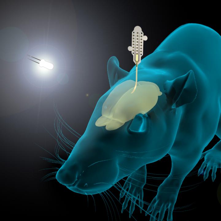
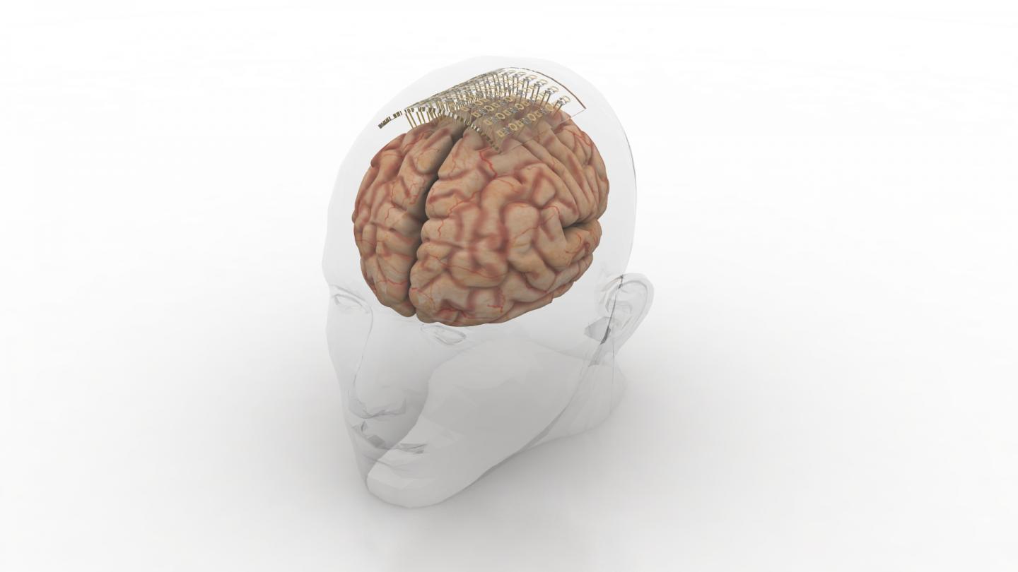
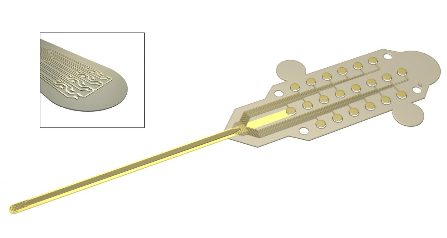
This is a pretty interesting way to illustrate the research,

Caption: Graphene and gold make a better brain probe. Credit: DGIST

An April 19, 2017 DGIST press release (also on EurekAlert), which originated the news item, expands on the theme (Note: A link has been removed),

The probe consists of an electrode, which records the brain signal. The signal travels down an interconnection line to a connector, which transfers the signal to machines measuring and analysing the signals.

The electrode starts with a thin gold base. Attached to the base are tiny zinc oxide nanowires, which are coated in a thin layer of gold, and then a layer of conducting polymer called PEDOT. These combined materials increase the probe’s effective surface area, conducting properties, and strength of the electrode, while still maintaining flexibility and compatibility with soft tissue.

Packing several long, thin nanowires together onto one probe enables the scientists to make a smaller electrode that retains the same effective surface area of a larger, flat electrode. This means the electrode can shrink, but not reduce signal detection. The interconnection line is made of a mix of graphene and gold. Graphene is flexible and gold is an excellent conductor. The researchers tested the probe and found it read rat brain signals very clearly, much better than a standard flat, gold electrode.

“Our graphene and nanowires-based flexible electrode array can be useful for monitoring and recording the functions of the nervous system, or to deliver electrical signals to the brain,” the researchers conclude in their paper recently published in the journal ACS Applied Materials and Interfaces.

The probe requires further clinical tests before widespread commercialization. The researchers are also interested in developing a wireless version to make it more convenient for a variety of applications.

Here’s a link to and a citation for the paper,

Enhancement of Interface Characteristics of Neural Probe Based on Graphene, ZnO Nanowires, and Conducting Polymer PEDOT by Mingyu Ryu, Jae Hoon Yang, Yumi Ahn, Minkyung Sim, Kyung Hwa Lee, Kyungsoo Kim, Taeju Lee, Seung-Jun Yoo, So Yeun Kim, Cheil Moon, Minkyu Je, Ji-Woong Choi, Youngu Lee, and Jae Eun Jang. ACS Appl. Mater. Interfaces, 2017, 9 (12), pp 10577–10586 DOI: 10.1021/acsami.7b02975 Publication Date (Web): March 7, 2017

Copyright © 2017 American Chemical Society

This paper is behind a paywall.

Effective sunscreens from nature

The dream is to find sunscreens that don’t endanger humans or pollute the environment and it seems that Spanish scientists may have taken a step closer to making that dream a reality (from a Jan. 30, 2017 Wiley Publications press release (also on EurekAlert),

The ideal sunscreen should block UVB and UVA radiation while being safe and stable. In the journal Angewandte Chemie, Spanish scientists have introduced a new family of UVA and UVB filters based on natural sunscreen substances found in algae and cyanobacteria. They are highly stable and enhance the effectivity [sic] of commercial sunscreens.

Good news for sunseekers. Commercial [sic] available sunscreen lotions can very effectively protect from dangerous radiation in the ultraviolet [spectrum], but they need to be applied regularly and in high amounts to develop their full potential. One of the most critical issues is the limited stability of the UV filter molecules. Inspired by nature, Diego Sampedro and his colleagues from La Rioja University in Logrono and collaborators from Malaga University and Alcala University, Madrid, Spain, have screened a natural class of UV-protecting [blocking?] molecules for their possible use in skin protection. They adjusted the nature-given motif [sic] to the requirements of chemical synthesis and found that the molecules could indeed boost the sun protection factor of common formulations.

The natural sunscreen molecules are called microsporine-like amino acids (MAAs) and are widespread in the microbial world, most prominently in marine algae and cyanobacteria. MAAs are small molecules derived from amino acids, thermally stable, and they absorb light in the ultraviolet region, protecting the microbial DNA from radiation damage. Thus they are natural sunscreens, which inspired Sampedro and his colleagues to create [a] new class of organic sunscreen compounds.

Theoretical calculations revealed what is chemically needed for a successful design. “We performed a computer calculation of several basic scaffolds [..] to identify the simplest compound that fulfills the requisites for efficient sunscreens”, the authors write. The result of their search was a set of molecules which were readily synthesized, “avoiding the decorating substituents that come from the biosynthetic route.” Thus the small basic molecules can be tuned to give them more favorable properties.

The authors found that the synthesized compounds are characterized by excellent filter capacities in the relevant UV range. In addition they are photostable, much more than, for example, oxybenzene [sic] which is a widely used sunscreen in commercial formulations. They do not react chemically and dissipate radiation as heat (but not to such an extent that the skin temperature would rise as well). And, most importantly, when tested in real formulations, the sun protection factor (SPF) rose by a factor of more than two. Thus they could be promising targets for more stable, more efficient sunscreen lotions. Good news for your next summer vacation.

There’s some unusual phrasing so, I’m guessing that the writer it not accustomed to writing press releases in English. One other comment, it’s oxybenzone that’s often used as an ingredient in commercial sunscreens.

Here’s a link to and a citation for the paper,

Rational Design and Synthesis of Efficient Sunscreens To Boost the Solar Protection Factor by Raúl Losantos, Ignacio Funes-Ardoiz, Dr. José Aguilera, Prof. Enrique Herrera-Ceballos, Dr. Cristina García-Iriepa, Prof. Pedro J. Campos, and Diego Sampedro. Angewandte Chemie International Edition Volume 56, Issue 10, pages 2632–2635, March 1, 2017 DOI: 10.1002/anie.201611627 Version of Record online: 27 JAN 2017

© 2017 Wiley-VCH Verlag GmbH & Co. KGaA, Weinheim

This paper is behind a paywall.

I have previously featured work on another natural sunscreen. In that case it was to be derived from English ivy (July 22, 2010 posting); there was an update on the English ivy work in a May 30, 2016 posting but the researcher has moved in a different direction looking at wound healing and armour as possible applications for the research.

Developing cortical implants for future speech neural prostheses

I’m guessing that graphene will feature in these proposed cortical implants since the project leader is a member of the Graphene Flagship’s Biomedical Technologies Work Package. (For those who don’t know, the Graphene Flagship is one of two major funding initiatives each receiving funding of 1B Euros over 10 years from the European Commission as part of their FET [Future and Emerging Technologies)] Initiative.)  A Jan. 12, 2017 news item on Nanowerk announces the new project (Note: A link has been removed),

BrainCom is a FET Proactive project, funded by the European Commission with 8.35M€ [8.3 million Euros] for the next 5 years, holding its Kick-off meeting on January 12-13 at ICN2 (Catalan Institute of Nanoscience and Nanotechnology) and the UAB [ Universitat Autònoma de Barcelona]. This project, coordinated by ICREA [Catalan Institution for Research and Advanced Studies] Research Prof. Jose A. Garrido from ICN2, will permit significant advances in understanding of cortical speech networks and the development of speech rehabilitation solutions using innovative brain-computer interfaces.

A Jan. 12, 2017 ICN2 press release, which originated the news item expands on the theme (it is a bit repetitive),

More than 5 million people worldwide suffer annually from aphasia, an extremely invalidating condition in which patients lose the ability to comprehend and formulate language after brain damage or in the course of neurodegenerative disorders. Brain-computer interfaces (BCIs), enabled by forefront technologies and materials, are a promising approach to treat patients with aphasia. The principle of BCIs is to collect neural activity at its source and decode it by means of electrodes implanted directly in the brain. However, neurorehabilitation of higher cognitive functions such as language raises serious issues. The current challenge is to design neural implants that cover sufficiently large areas of the brain to allow for reliable decoding of detailed neuronal activity distributed in various brain regions that are key for language processing.

BrainCom is a FET Proactive project funded by the European Commission with 8.35M€ for the next 5 years. This interdisciplinary initiative involves 10 partners including technologists, engineers, biologists, clinicians, and ethics experts. They aim to develop a new generation of neuroprosthetic cortical devices enabling large-scale recordings and stimulation of cortical activity to study high level cognitive functions. Ultimately, the BraimCom project will seed a novel line of knowledge and technologies aimed at developing the future generation of speech neural prostheses. It will cover different levels of the value chain: from technology and engineering to basic and language neuroscience, and from preclinical research in animals to clinical studies in humans.

This recently funded project is coordinated by ICREA Prof. Jose A. Garrido, Group Leader of the Advanced Electronic Materials and Devices Group at the Institut Català de Nanociència i Nanotecnologia (Catalan Institute of Nanoscience and Nanotechnology – ICN2) and deputy leader of the Biomedical Technologies Work Package presented last year in Barcelona by the Graphene Flagship. The BrainCom Kick-Off meeting is held on January 12-13 at ICN2 and the Universitat Autònoma de Barcelona (UAB).

Recent developments show that it is possible to record cortical signals from a small region of the motor cortex and decode them to allow tetraplegic [also known as, quadriplegic] people to activate a robotic arm to perform everyday life actions. Brain-computer interfaces have also been successfully used to help tetraplegic patients unable to speak to communicate their thoughts by selecting letters on a computer screen using non-invasive electroencephalographic (EEG) recordings. The performance of such technologies can be dramatically increased using more detailed cortical neural information.

BrainCom project proposes a radically new electrocorticography technology taking advantage of unique mechanical and electrical properties of novel nanomaterials such as graphene, 2D materials and organic semiconductors.  The consortium members will fabricate ultra-flexible cortical and intracortical implants, which will be placed right on the surface of the brain, enabling high density recording and stimulation sites over a large area. This approach will allow the parallel stimulation and decoding of cortical activity with unprecedented spatial and temporal resolution.

These technologies will help to advance the basic understanding of cortical speech networks and to develop rehabilitation solutions to restore speech using innovative brain-computer paradigms. The technology innovations developed in the project will also find applications in the study of other high cognitive functions of the brain such as learning and memory, as well as other clinical applications such as epilepsy monitoring.

The BrainCom project Consortium members are:

  • Catalan Institute of Nanoscience and Nanotechnology (ICN2) – Spain (Coordinator)
  • Institute of Microelectronics of Barcelona (CNM-IMB-CSIC) – Spain
  • University Grenoble Alpes – France
  • ARMINES/ Ecole des Mines de St. Etienne – France
  • Centre Hospitalier Universitaire de Grenoble – France
  • Multichannel Systems – Germany
  • University of Geneva – Switzerland
  • University of Oxford – United Kingdom
  • Ludwig-Maximilians-Universität München – Germany
  • Wavestone – Luxembourg

There doesn’t seem to be a website for the project but there is a BrainCom webpage on the European Commission’s CORDIS (Community Research and Development Information Service) website.

Nanotech business news from Turkey and from Northern Ireland

I have two nanotech business news bits, one from Turkey and one from Northern Ireland.

Turkey

A Turkish company has sold one of its microscopes to the US National Aeronautics and Space Administration (NASA), according to a Jan. 20, 2017 news item on dailysabah.com,

Turkish nanotechnology company Nanomanyetik has begun selling a powerful microscope to the U.S. space agency NASA, the company’s general director told Anadolu Agency on Thursday [Jan. 19, 2017].

Dr. Ahmet Oral, who also teaches physics at Middle East Technical University, said Nanomanyetik developed a microscope that is able to map surfaces on the nanometric and atomic levels, or extremely small particles.

Nanomanyetik’s foreign customers are drawn to the microscope because of its higher quality yet cheaper price compared to its competitors.

“There are almost 30 firms doing this work,” according to Oral. “Ten of them are active and we are among these active firms. Our aim is to be in the top three,” he said, adding that Nanomanyetik jumps to the head of the line because of its after-sell service.

In addition to sales to NASA, the Ankara-based firm exports the microscope to Brazil, Chile, France, Iran, Israel, Italy, Japan, Poland, South Korea and Spain.

Electronics giant Samsung is also a customer.

“Where does Samsung use this product? There are pixels in the smartphones’ displays. These pixels are getting smaller each year. Now the smallest pixel is 15X10 microns,” he said. Human hair is between 10 and 100 microns in diameter.

“They are figuring inner sides of pixels so that these pixels can operate much better. These patterns are on the nanometer level. They are using these microscopes to see the results of their works,” Oral said.

Nanomanyetik’s microscopes produces good quality, high resolution images and can even display an object’s atoms and individual DNA fibers, according to Oral.

You can find the English language version of the Nanomanyetik (NanoMagnetics Instruments) website here . For those with the language skills there is the Turkish language version, here.

Northern Ireland

A Jan. 22, 2017 news article by Dominic Coyle for The Irish Times (Note: Links have been removed) shares this business news and mention of a world first,

MOF Technologies has raised £1.5 million (€1.73 million) from London-based venture capital group Excelsa Ventures and Queen’s University Belfast’s Qubis research commercialisation group.

MOF Technologies chief executive Paschal McCloskey welcomed the Excelsa investment.

Established in part by Qubis in 2012 in partnership with inventor Prof Stuart James, MOF Technologies began life in a lab at the School of Chemistry and Chemical Engineering at Queen’s.

Its metal organic framework (MOF) technology is seen as having significant potential in areas including gas storage, carbon capture, transport, drug delivery and heat transformation. Though still in its infancy, the market is forecast to grow to £2.2 billion by 2022, the company says.

MOF Technologies last year became the first company worldwide to successfully commercialise MOFs when it agreed a deal with US fruit and vegetable storage provider Decco Worldwide to commercialise MOFs for use in a food application.

TruPick, designed by Decco and using MOF Technologies’ environmentally friendly technology, enables nanomaterials control the effects of ethylene on fruit produce so it maintains freshness in storage or transport.

MOFs are crystalline, sponge-like materials composed of two components – metal ions and organic molecules known as linkers.

“We very quickly recognised the market potential of MOFs in terms of their unmatched ability for gas storage,” said Moritz Bolle from Excelsa Ventures. “This technology will revolutionise traditional applications and open countless new opportunities for industry. We are confident MOF Technologies is the company that will lead this seismic shift in materials science.

You can find MOF Technologies here.