Tag Archives: China

Therapeutic nanoparticles for COVID-19 (disease caused by severe acute respiratory syndrome coronavirus 2 [SARS-CoV-2])—don’t hold your breath!

Last week (specifically, Tuesday, March 3, 2020), someone moved away from me during a class. I’d sneezed.

The irony of the situation is that of the two of us, with my lung issues I’d be the one most at risk of getting very ill and/or dying from COVID-19. ([1] Yes, I confirmed that was the reason she’d moved. [2] The therapeutic nanoparticles news item is coming later) Here are the risk factors to take into account (from the US Centers for Disease Control’s People at Risk for Serious Illness from COVID-19 webpage,

  • Older adults [Note: In one report the age range was stated as ‘people over 70’]
  • People who have serious chronic medical conditions like:
    • Heart disease
    • Diabetes
    • Lung disease

I’m not suggesting that all precautions be abandoned but it would seem that panic might not be called for. Jeremy Samuel Faust, an emergency medicine physician at Brigham and Women’s Hospital in Boston, faculty in its division of health policy and public health, and an instructor at Harvard Medical School, has written a calming March 4, 2020 article (COVID-19 Isn’t As Deadly As We Think; Don’t hoard masks and food. Figure out how to help seniors and the immunosuppressed stay healthy.) for Slate.com (Note: Links have been removed],

There are many compelling reasons to conclude that SARS-CoV-2, the virus that causes COVID-19, is not nearly as deadly as is currently feared. But COVID-19 panic has set in nonetheless. You can’t find hand sanitizer in stores, and N95 face masks are being sold online for exorbitant prices, never mind that neither is the best way to protect against the virus (yes, just wash your hands). The public is behaving as if this epidemic is the next Spanish flu, which is frankly understandable given that initial reports have staked COVID-19 mortality at about 2–3 percent, quite similar to the 1918 pandemic that killed tens of millions of people.

Allow me to be the bearer of good news. These frightening numbers are unlikely to hold. The true case fatality rate, known as CFR, of this virus is likely to be far lower than current reports suggest. Even some lower estimates, such as the 1 percent death rate recently mentioned by the directors of the National Institutes of Health and the Centers for Disease Control and Prevention, likely substantially overstate the case. [emphases mine]

But the most straightforward and compelling evidence that the true case fatality rate of SARS-CoV-2 is well under 1 percent comes not from statistical trends and methodological massage, but from data from the Diamond Princess cruise outbreak and subsequent quarantine off the coast of Japan.

A quarantined boat is an ideal—if unfortunate—natural laboratory to study a virus. Many variables normally impossible to control are controlled. We know that all but one patient boarded the boat without the virus. We know that the other passengers were healthy enough to travel. We know their whereabouts and exposures. While the numbers coming out of China are scary, we don’t know how many of those patients were already ill for other reasons. How many were already hospitalized for another life-threatening illness and then caught the virus? How many were completely healthy, caught the virus, and developed a critical illness? In the real world, we just don’t know.

Here’s the problem with looking at mortality numbers in a general setting: In China, 9 million people die per year, which comes out to 25,000 people every single day, or around 1.5 million people over the past two months alone. A significant fraction of these deaths results from diseases like emphysema/COPD, lower respiratory infections, and cancers of the lung and airway whose symptoms are clinically indistinguishable from the nonspecific symptoms seen in severe COVID-19 cases. And, perhaps unsurprisingly, the death rate from COVID-19 in China spiked precisely among the same age groups in which these chronic diseases first become common. During the peak of the outbreak in China in January and early February, around 25 patients per day were dying with SARS-CoV-2. Most were older patients in whom the chronic diseases listed above are prevalent. Most deaths occurred in Hubei province, an area in which lung cancer and emphysema/COPD are significantly higher than national averages in China, a country where half of all men smoke. How were doctors supposed to sort out which of those 25 out of 25,000 daily deaths were solely due to coronavirus, and which were more complicated? What we need to know is how many excess deaths this virus causes.

This all suggests that COVID-19 is a relatively benign disease for most young people, and a potentially devastating one for the old and chronically ill, albeit not nearly as risky as reported. Given the low mortality rate among younger patients with coronavirus—zero in children 10 or younger among hundreds of cases in China, and 0.2-0.4 percent in most healthy nongeriatric adults (and this is still before accounting for what is likely to be a high number of undetected asymptomatic cases)—we need to divert our focus away from worrying about preventing systemic spread among healthy people—which is likely either inevitable, or out of our control—and commit most if not all of our resources toward protecting those truly at risk of developing critical illness and even death: everyone over 70, and people who are already at higher risk from this kind of virus.

This still largely comes down to hygiene and isolation. But in particular, we need to focus on the right people and the right places. Nursing homes, not schools. Hospitals, not planes. We need to up the hygienic and isolation ante primarily around the subset of people who can’t simply contract SARS-CoV-2 and ride it out the way healthy people should be able to.

Curtis Kim of Vancouver, Canada, has created a website dedicated to tracking the statistics and information about COVID-19 in Canada and around the world. Here’s more about Kim and the website from a March 8, 2020 article by Megan Devlin for the Daily Hive,

Curtis Kim, who studied Computer Systems Technology at the British Columbia Institute of Technology [BCIT], launched the site this week after getting frustrated he was spending so much time on various websites looking for daily coronavirus updates.

The site breaks down the number of cases in Canada, the number of deaths (zero in Canada so far), and the number of people who have recovered. Further down, it provides the same stats for global COVID-19 cases.

There’s also a colour-coded map showing where cases are distributed, and a feed of latest news articles about the virus. Kim also included information about symptoms and how to contact Canadian public health services.

Kim is looking for work and given what I’ve seen of his COVID-19 website, he should have no difficulty. Although I think it might be an idea for him to explain how the ‘lethality’ rate on his website has been obtained since Faust who seems to have more directly relevant experience suggests in his article that the numbers are highly problematic,

My name is Curtis, recently graduated from BCIT. I thought it would be a serious worldwide issue considering the speed of the spread of this virus ever since this COVID-19 occurred. I frequently googled to check up the current status by going through many websites and felt I was wasting time repeatedly searching with same keywords and for sure I wasn’t the only one feeling this way. That’s why I started creating this application. It provides up-to-date information on the COVID-19 broken by province and country around the world, key contact information, and latest news. I like to help people, and want them to understand this situation easily using this application. Hopefully this situation improves soon.

If you have any further inquries about the information on this web application, Please reach me at curtisk808@gmail.com

At about 11:45 am (PT) on March 9, 2020, Kim’s COVID-19 website was updated to include one death in Canada. As you might expect, ti was a resident in a long term care home. Wanyee Li’s March 9, 2020 article for The Star presents the news,

A resident at a long-term care home experiencing a COVID-19 outbreak in North Vancouver has died after contracting the virus, B.C. health officials confirmed Monday [March 9, 2020].

It is the first reported death in Canada linked to the virus.

The outbreak at the Lynn Valley Care Centre has so far been linked to three community transmission cases of the virus.

Provincial Health Officer Dr. Bonnie Henry confirmed five new cases of COVID-19 in B.C. on Monday [March 9, 2020], putting the total in the province at 32.

The five new cases include one health-care worker, two people who are close contacts of an existing case, one person who recently returned from travel to Iran and another who was in Italy recently.

Officials are conducting an investigation into the three community transmission cases at the long-term care home to determine how a health care worker contracted the virus.

I looked up the population figures for the province of British Columbia (BC; Wikipedia entry for Demographics of British Columbia). As of the 2016 census, there were 4,648,055 people in the province. Assuming that population number holds, 67 cases in all of Canada (with 27 cases in BC) of COVID-19 don’t seem like big numbers.

We should definitely take precautions and be careful but there’s no need to panic.

Nanoparticles and a COVID-19 treatment?

Don’t hold you breath. This March 5, 2020 news item on Nanowerk is speculative,

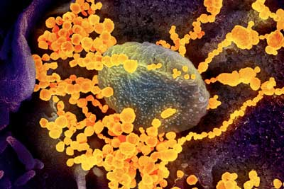
There is no vaccine or specific treatment for COVID-19, the disease caused by the severe acute respiratory syndrome coronavirus 2, or SARS-CoV-2.

Since the outbreak began in late 2019, researchers have been racing to learn more about SARS-CoV-2, which is a strain from a family of viruses known as coronavirus for their crown-like shape.

Northeastern Ûniversity] chemical engineer Thomas Webster, who specializes in developing nano-scale medicine and technology to treat diseases, is part of a contingency of scientists that are contributing ideas and technology to the Centers for Disease Control and Prevention to fight the COVID-19 outbreak.

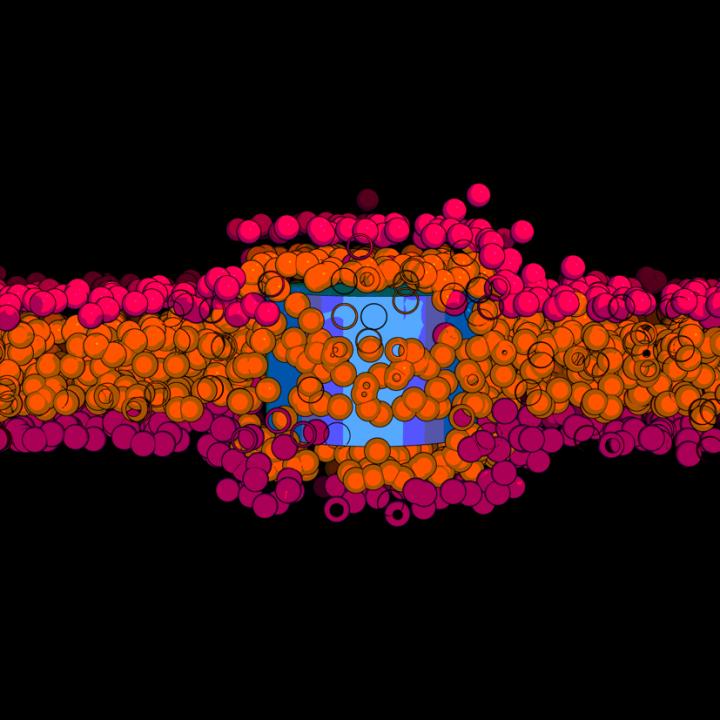
The idea of using nanoparticles, Webster says, is that the virus behind COVID-19 consists of a structure of a similar scale as his nanoparticles. At that scale, matter is ultra-small, about ten thousand times smaller than the width of a single strand of hair.

..

This scanning electron microscope image shows SARS-CoV-2 (round gold objects) emerging from the surface of cells cultured in the lab. SARS-CoV-2, also known as 2019-nCoV, is the virus that causes COVID-19. The virus shown was isolated from a patient in the U.S. (Image: NIAID-RML)

A March 4, 2020 Northeastern University news release by Roberto Molar Candanosa, which originated the news item, delves further into Webster’s thinking process,

Webster is proposing particles of similar sizes that could attach to SARS-CoV-2 viruses, disrupting their structure with a combination of infrared light treatment. That structural change would then halt the ability of the virus to survive and reproduce in the body.

“You have to think in this size range,” says Webster, Art Zafiropoulo Chair of chemical engineering at Northeastern. “In the nanoscale size range, if you want to detect viruses, if you want to deactivate them.”

Finding and neutralizing viruses with nanomedicine is at the core of what Webster and other researchers call theranostics, which focuses on combining therapy and diagnosis. Using that approach, his lab has specialized in nanoparticles to fight the microbes that cause influenza and tuberculosis. 

“It’s not just having one approach to detect whether you have a virus and another approach to use it as a therapy,” he says, “but having the same particle, the same approach, for both your detection and therapy.”

I wish Webster good luck. As for the rest us, let’s wash our hands and keep calm.

Harvard professor and leader in nanoscale electronics charged with making false statements about Chinese funding

I may be mistaken but the implication seems to be that Charles M. Lieber’s lies (he was charged today, January 28, 2020 ) are the ‘tip of the iceberg’ of a very large problem. Ellen Barry’s January 28, 2020 article for the New York Times outlines at least part of what the US government is doing to discover and ultimately discourage the theft of biomedical research from US laboratories.

Dr. Lieber, a leader in the field of nanoscale electronics, was one of three Boston-area scientists accused on Tuesday [January 28, 2020] of working on behalf of China. His case involves work with the Thousand Talents Program, a state-run program that seeks to draw talent educated in other countries.

American officials are investigating hundreds of cases of suspected theft of intellectual property by visiting scientists, nearly all of them Chinese nationals or of Chinese descent. Some are accused of obtaining patents in China based on work that is funded by the United States government, and others of setting up laboratories in China that secretly duplicated American research.

Dr. Lieber, who was arrested on Tuesday [January 28, 2020], stands out among the accused scientists, because he is neither Chinese nor of Chinese descent. …

Lieber is the Chair of Harvard’s Department of Chemistry and Chemical Biology and much more, according to his Wikipedia entry (Note: Links have been removed),

Charles M. Lieber (born 1959) is an American chemist and pioneer in the field of nanoscience and nanotechnology. In 2011, Lieber was recognized by Thomson Reuters as the leading chemist in the world for the decade 2000-2010 based on the impact of his scientific publications.[1] Lieber has published over 400 papers in peer-reviewed scientific journals and has edited and contributed to many books on nanoscience.[2] He is the principal inventor on over fifty issued US patents and applications, and founded the nanotechnology company Nanosys in 2001 and Vista Therapeutics in 2007.[3] He is known for his contributions to the synthesis, assembly and characterization of nanoscale materials and nanodevices, the application of nanoelectronic devices in biology, and as a mentor to numerous leaders in nanoscience.[4] Thompson Reuters predicted Lieber to be a recipient of the 2008 Nobel Prize in Chemistry [to date, January 28, 2020, Lieber has not received a Nobel prize].

Should you search Charles Lieber or Charles M. Lieber on this blog’s search engine, you will find a number of postings about his and his students’ work dating from 2012 to as recently as November 15, 2019.

Here’s another example from Barry’s January 28, 2020 article for the New York Times which illustrates just how shocking this is (Note: Links have been removed),

In 2017 he was named a University Professor, Harvard’s highest faculty rank, one of only 26 professors to hold that status. The same year, he earned the National Institutes of Health Director’s Pioneer Award for inventing syringe-injectable mesh electronics that can integrate with the brain.

Harvard’s president at the time, Drew G. Faust, called him “an extraordinary scientist whose work has transformed nanoscience and nanotechnology and has led to a remarkable range of valuable applications that improve the quality of people’s lives.”

Here’s a bit more about the Chinese program that Lieber is affiliated with,

Launched in 2008, its [China] Thousand Talents Program is an effort to recruit Chinese and foreign academics and entrepreneurs. According to a report in the China Daily, new recruits receive 1 million yuan, or about $146,000, from the central government, and a pledge of 10 million yuan for their ongoing research from the Chinese Academy of Sciences.

The recruitment flows both ways. Researchers of Chinese descent make up nearly half of the work force in American research laboratories, in part because American-born scientists are drawn to the private sector and less interested in academic careers.

I encourage you to read Barry’s entire article. It is jaw-dropping and, where Lieber is concerned, sad. It’s beginning to look like US universities are corrupt. The ‘Jeffrey Epstein (a wealthy and convicted sexual predator and more) connection’ to the Massachusetts Institute of Technology, which led to the resignation of a prominent faculty member (Sept. 19, 2019 article by Anna North for Vox.com), and the Fall 2019 cheating scandal (gaining admission to big name educational institutions by paying someone other than the student to take exams, among many other schemes) suggest a reckoning might be in order.

ETA January 28, 2020 at 1645 hours: I found a January 28, 2020 article by Antonio Regalado for the MIT Technology Review which provides a few more details about Lieber’s situation,

Big money: According to the charging document, Lieber, starting in 2011,  agreed to help set up a research lab at the Wuhan University of Technology and “make strategic visionary and creative research proposals” so that China could do cutting-edge science.

He was well paid for it. Lieber earned a salary when he visited China worth up to $50,000 per month, as well as $150,000 a year in expenses in addition to research funds. According to the complaint, he got paid by way of a Chinese bank account but also was known to send emails asking for cash instead.

Harvard eventually wised up to the existence of a Wuhan lab using its name and logo, but when administrators confronted Lieber, he lied and said he didn’t know about a formal joint program, according to the government complaint.

I imagine the money paid by the Chinese government is in addition to Lieber’s Harvard salary (no doubt a substantial one especially since he’s chair of his department and one of a select number of Harvard’s University Professors) and in addition to any other deals he might have on the side.

So thin and soft you don’t notice it: new wearable tech

An August 2, 2019 news item on ScienceDaily features some new work on wearable technology that was a bit of a surprise to me,

Wearable human-machine interfaces — devices that can collect and store important health information about the wearer, among other uses — have benefited from advances in electronics, materials and mechanical designs. But current models still can be bulky and uncomfortable, and they can’t always handle multiple functions at one time.

Researchers reported Friday, Aug. 2 [2019], the discovery of a multifunctional ultra-thin wearable electronic device that is imperceptible to the wearer.

I expected this wearable technology to be a piece of clothing that somehow captured health data but it’s not,

While a health care application is mentioned early in the August 2, 2019 University of Houston news release (also on EurekAlert) by Jeannie Kever the primary interest seems to be robots and robotic skin (Note: This news release originated the news item on ScienceDaily),

The device allows the wearer to move naturally and is less noticeable than wearing a Band-Aid, said Cunjiang Yu, Bill D. Cook Associate Professor of Mechanical Engineering at the University of Houston and lead author for the paper, published as the cover story in Science Advances.

“Everything is very thin, just a few microns thick,” said Yu, who also is a principal investigator at the Texas Center for Superconductivity at UH. “You will not be able to feel it.”
It has the potential to work as a prosthetic skin for a robotic hand or other robotic devices, with a robust human-machine interface that allows it to automatically collect information and relay it back to the wearer.

That has applications for health care – “What if when you shook hands with a robotic hand, it was able to instantly deduce physical condition?” Yu asked – as well as for situations such as chemical spills, which are risky for humans but require human decision-making based on physical inspection.

While current devices are gaining in popularity, the researchers said they can be bulky to wear, offer slow response times and suffer a drop in performance over time. More flexible versions are unable to provide multiple functions at once – sensing, switching, stimulation and data storage, for example – and are generally expensive and complicated to manufacture.

The device described in the paper, a metal oxide semiconductor on a polymer base, offers manufacturing advantages and can be processed at temperatures lower than 300 C.

“We report an ultrathin, mechanically imperceptible, and stretchable (human-machine interface) HMI device, which is worn on human skin to capture multiple physical data and also on a robot to offer intelligent feedback, forming a closed-loop HMI,” the researchers wrote. “The multifunctional soft stretchy HMI device is based on a one-step formed, sol-gel-on-polymer-processed indium zinc oxide semiconductor nanomembrane electronics.”

In addition to Yu, the paper’s co-authors include first author Kyoseung Sim, Zhoulyu Rao, Faheem Ershad, Jianming Lei, Anish Thukral and Jie Chen, all of UH; Zhanan Zou and Jianliang Xiao, both of the University of Colorado; and Qing-An Huang of Southeast University in Nanjing, China.

Here’s a link to and a citation for the paper,

Metal oxide semiconductor nanomembrane–based soft unnoticeable multifunctional electronics for wearable human-machine interfaces by Kyoseung Sim, Zhoulyu Rao, Zhanan Zou, Faheem Ershad, Jianming Lei, Anish Thukral, Jie Chen, Qing-An Huang, Jianliang Xiao and Cunjiang Yu. Science Advances 02 Aug 2019: Vol. 5, no. 8, eaav9653 DOI: 10.1126/sciadv.aav9653

This paper appears to be open access.

Artificial nose for intelligent olfactory substitution

The signal transmitted into mouse brain can participate in mouse perception and act as the brain stimulator. (Image credit: Prof. ZHAN Yang)

I’m fascinated by the image. Are they suggesting putting implants into people’s brains that can sense dangerous gaseous molecules and convert that into data which can be read on a smartphone? And, are they harvesting bioenergy to supply energy to the implant?

A July 29, 2019 news item on Azonano was not as helpful in answering my questions as I’d hoped (Note: A link has been removed),

An artificial olfactory system based on a self-powered nano-generator has been built by Prof. ZHAN Yang’s team at the Shenzhen Institutes of Advanced Technology (SIAT) of the Chinese Academy of Sciences [CAS], together with colleagues at the University of Electronic Science and Technology of China.

The device, which can detect a variety of odor molecules and identify different odors, has been demonstrated in vivo in animal models. The research titled “An artificial triboelectricity-brain-behavior closed loop for intelligent olfactory substitution” has been reported in Nano Energy.

A July 25, 2019 CAS press release, which originated the news item, provides a little more information,

Odor processing is important to many species. Specific olfactory receptors located on the neurons are involved in odor recognition. These different olfactory receptors form patterned distribution.

Inspired by the biological receptors, the teams collaborated on formulating an artificial olfactory system. Through nano-fabrication on the soft materials and special alignment of material structures, the teams built a self-power device that can code and differentiate different odorant molecules.

This device has been connected to the mouse brain to demonstrate that the olfactory signals can produce appropriate neural stimulation. When the self-powered device generated the electric currents, the mouse displayed behavioral motion changes.

This study, inspired by the biological olfactory system, provides insights on novel design of neural stimulation and brain-machine interface. 

Here’s a link to and a citation for the paper,

An artificial triboelectricity-brain-behavior closed loop for intelligent olfactory substitution by Tianyan Zhong, Mengyang Zhang, Yongming Fu, Yechao Han, Hongye Guan, Haoxuan He, Tianming Zhao, Lili Xing, Xinyu Xue, Yan Zhang, Yang Zhan.Nano Energy Volume 63, September 2019, 103884 DOI: https://doi.org/10.1016/j.nanoen.2019.103884

This paper is behind a paywall.

Smartphone as augmented reality system with software from Brown University

You need to see this,

Amazing, eh? The researchers are scheduled to present this work sometime this week at the ACM Symposium on User Interface Software and Technology (UIST) being held in New Orleans, US, from October 20-23, 2019.

Here’s more about ‘Portal-ble’ in an October 16, 2019 news item on ScienceDaily,

A new software system developed by Brown University [US] researchers turns cell phones into augmented reality portals, enabling users to place virtual building blocks, furniture and other objects into real-world backdrops, and use their hands to manipulate those objects as if they were really there.

The developers hope the new system, called Portal-ble, could be a tool for artists, designers, game developers and others to experiment with augmented reality (AR). The team will present the work later this month at the ACM Symposium on User Interface Software and Technology (UIST 2019) in New Orleans. The source code for Andriod is freely available for download on the researchers’ website, and iPhone code will follow soon.

“AR is going to be a great new mode of interaction,” said Jeff Huang, an assistant professor of computer science at Brown who developed the system with his students. “We wanted to make something that made AR portable so that people could use anywhere without any bulky headsets. We also wanted people to be able to interact with the virtual world in a natural way using their hands.”

An October 16, 2019 Brown University news release (also on EurekAlert), which originated the news item, provides more detail,

Huang said the idea for Portal-ble’s “hands-on” interaction grew out of some frustration with AR apps like Pokemon GO. AR apps use smartphones to place virtual objects (like Pokemon characters) into real-world scenes, but interacting with those objects requires users to swipe on the screen.

“Swiping just wasn’t a satisfying way of interacting,” Huang said. “In the real world, we interact with objects with our hands. We turn doorknobs, pick things up and throw things. So we thought manipulating virtual objects by hand would be much more powerful than swiping. That’s what’s different about Portal-ble.”

The platform makes use of a small infrared sensor mounted on the back of a phone. The sensor tracks the position of people’s hands in relation to virtual objects, enabling users to pick objects up, turn them, stack them or drop them. It also lets people use their hands to virtually “paint” onto real-world backdrops. As a demonstration, Huang and his students used the system to paint a virtual garden into a green space on Brown’s College Hill campus.

Huang says the main technical contribution of the work was developing the right accommodations and feedback tools to enable people to interact intuitively with virtual objects.

“It turns out that picking up a virtual object is really hard if you try to apply real-world physics,” Huang said. “People try to grab in the wrong place, or they put their fingers through the objects. So we had to observe how people tried to interact with these objects and then make our system able accommodate those tendencies.”

To do that, Huang enlisted students in a class he was teaching to come up with tasks they might want to do in the AR world — stacking a set of blocks, for example. The students then asked other people to try performing those tasks using Portal-ble, while recording what people were able to do and what they couldn’t. They could then adjust the system’s physics and user interface to make interactions more successful.

“It’s a little like what happens when people draw lines in Photoshop,” Huang said. “The lines people draw are never perfect, but the program can smooth them out and make them perfectly straight. Those were the kinds of accommodations we were trying to make with these virtual objects.”

The team also added sensory feedback — visual highlights on objects and phone vibrations — to make interactions easier. Huang said he was somewhat surprised that phone vibrations helped users to interact. Users feel the vibrations in the hand they’re using to hold the phone, not in the hand that’s actually grabbing for the virtual object. Still, Huang said, vibration feedback still helped users to more successfully interact with objects.

In follow-up studies, users reported that the accommodations and feedback used by the system made tasks significantly easier, less time-consuming and more satisfying.

Huang and his students plan to continue working with Portal-ble — expanding its object library, refining interactions and developing new activities. They also hope to streamline the system to make it run entirely on a phone. Currently the infrared sensor requires an infrared sensor and external compute stick for extra processing power.

Huang hopes people will download the freely available source code and try it for themselves. 
“We really just want to put this out there and see what people do with it,” he said. “The code is on our website for people to download, edit and build off of. It will be interesting to see what people do with it.

Co-authors on the research paper were Jing Qian, Jiaju Ma, Xiangyu Li, Benjamin Attal, Haoming Lai, James Tompkin and John Hughes. The work was supported by the National Science Foundation (IIS-1552663) and by a gift from Pixar.

You can find the conference paper here on jeffhuang.com,

Portal-ble: Intuitive Free-hand Manipulationin Unbounded Smartphone-based Augmented Reality by Jing Qian, Jiaju Ma, Xiangyu Li∗, Benjamin Attal, Haoming Lai,James Tompkin, John F. Hughes, Jeff Huang. Brown University, Providence RI, USA; Southeast University, Nanjing, China. Presented at ACM Symposium on User Interface Software and Technology (UIST) being held in New Orleans, US

This is the first time I’ve seen an augmented reality system that seems accessible, i.e., affordable. You can find out more on the Portal-ble ‘resource’ page where you’ll also find a link to the source code repository. The researchers, as noted in the news release, have an Android version available now with an iPhone version to be released in the future.

‘Xuan paper’ made fire-resistant with nanowires

Xuan paper is special being both rare and used for calligraphy and art works. Before getting to the ‘fire-resistant’ news, it might be helpful to get some details about Xuan paper as it is typically prepared and used (from a Dec. 29, 2018 news item on xinhuanet.com),

Today’s Chinese artists now have the opportunity to preserve their works much longer than the masters who painted hundreds of years ago.

Chinese researchers have developed a non-flammable version of Xuan paper that has high thermal stability, according to the Chinese Academy of Sciences (CAS).

Xuan paper, a type of handmade paper, was originally produced in ancient China and used for both Chinese calligraphy and paintings. The procedure of making Xuan paper was listed as a world intangible cultural heritage by UNESCO in 2009.

The raw materials need to produce Xuan paper are found in Jingxian County, east China’s Anhui Province and as of late, are in short supply.

The traditional handmade method of Xuan paper involves more than 100 steps and takes nearly two years [emphasis mine]. It has a low output and high cost. Xuan paper made with organic materials often suffers from degradation, yellowing and deteriorating properties during the long-term natural aging process.

Furthermore, the most lethal problem of traditional Xuan paper is its high flammability.

A January 18, 2019 news item on Nanowerk adds a few more details about the traditional paper while describing the ‘new’ Xuan paper (Note: A link has been removed),

Xuan paper is an excellent example of the traditional handmade paper, and features excellent properties of durability, ink wetting, and resistance to insects and mildew. Its excellent durability is attributed to its unique raw materials and handmade manufacturing process under mild conditions.

The bark of pteroceltis tatarinowii, a common species of elm in the area, is used as the main raw material to produce Xuan paper. Limestone particles are deposited on the surface of pteroceltis bark fibers, which can neutralize acids produced by the hydrolysis of plant fibers and from the environment.

Since the raw materials are only produced in Jing County, Anhui Province, China, Xuan paper suffers from a severe shortage. Also, it has the shortcomings such as complicated traditional hand making process and flammability. In a recent paper published in ACS Sustainable Chemistry & Engineering (“Fire-Resistant Inorganic Analogous Xuan Paper with Thousands of Years’ Super-Durability”), a team led by Prof. ZHU Yingjie from Shanghai Institute of Ceramics of Chinese Academy of Sciences developed a new kind of “fire-resistant Xuan paper” based on ultralong hydroxyapatite nanowires.

A January 18, 2019 Chinese Academy of Sciences (CAS) press release, which originated the news item, provides more technical details,

The unique integral structure of the “fire-resistant Xuan paper” with excellent mechanical properties and high flexibility was designed to be similar to the reinforced concrete structure in tall buildings. Ultralong hydroxyapatite nanowires are used as the main building material and are similar to the concrete. Silica glass fibers with micrometer-sized diameters are used as the reinforcing framework material and are similar to supporting steel bars.
In addition, a new kind of inorganic adhesive composed of amorphous nanoparticles was designed, prepared and used as the binder in the “fire-resistant Xuan paper”.

The as-prepared “fire-resistant Xuan paper” well keeps its properties even after the simulated aging for up to 3000 years.

The original whiteness of the “fire-resistant Xuan paper” is 92%, and its whiteness has a slight decrease to 91.6%, with the whiteness retention as high as 99.6% after the simulated aging for 2000 years. Even after the simulated aging for 3000 years, its whiteness only decreases to 86.7% with 94.2% of the whiteness retention. It is much higher than that of the traditional Xuan paper. The whiteness of the traditional unprocessed Xuan paper decreases from initial 70.5% to 47.3% with 67.1% of the whiteness retention after the simulated aging for 2000 years. Its whiteness decreases to 42.2% with 59.9% of the whiteness retention after the simulated aging for 3000 years.

The “fire-resistant Xuan paper” exhibits superior mechanical properties during the simulated aging process.

The retention percentage of tensile strength of the “fire-resistant Xuan paper” is as high as 95.2% aging for 2000 years, and 81.3% aging for 3000 years. In contrast, the average retention percentage of tensile strength of the unprocessed Xuan paper is only 54.9% aging for 2000 years, and 40.4% aging for 3000 years. Furthermore, the “fire-resistant Xuan paper” has an excellent ink wetting performance, which is mainly attributed to the nanoscale porous structure and hydroxyl groups of utralong hydroxyapatite nanowires.

The prevention of mould growth on the paper is a great challenge, because the mould can cause the deterioration of the Xuan paper. In this study, experiments showed that different kinds of mould spores do not breed and spread on the “fire-resistant Xuan paper”, and it is able to maintain a clean surface without the growth of any mould, indicating the excellent anti-mildew performance of the “fire-resistant Xuan paper” even exposure to the external nutrients. On the contrary, the growth and spread of mould are obviously observed on the traditional Xuan paper in the presence of external nutrients, indicating that its anti-mildew performance is not satisfactory.

The most important property is that the “fire-resistant Xuan paper” is fire resistant and highly thermal stable. Thus it can prevent the precious calligraphy and painting works as well as books, documents, and archives from the damage by fire. In addition, the production process of the “fire-resistant Xuan paper” is simple, highly efficient, and it only needs 3~4 days to produce.

Xuan paper is the best material carrier for the calligraphy and painting arts, many of which have been well preserved for hundreds of years.

Here’s a link to and a citation for the paper,

Fire-Resistant Inorganic Analogous Xuan Paper with Thousands of Years’ Super-Durability by Li-Ying Dong and Ying-Jie Zhu. ACS Sustainable Chem. Eng., 2018, 6 (12), pp 17239–17251 DOI: 10.1021/acssuschemeng.8b04630 Publication Date (Web): November 7, 2018

Copyright © 2018 American Chemical Society

This paper is behind a paywall.

One last thing, the researchers have made an image illustrating their work available,

Courtesy: CAS and American Chemical Society

Ouchies no more! Not from bandages, anyway.

An adhesive that US and Chinese scientists have developed shows great promise not just for bandages but wearable robotics too. From a December 14, 2018 news item on Nanowerk,

Researchers from the Harvard John A. Paulson School of Engineering and Applied Sciences (SEAS) and Xi’an Jiaotong University in China have developed a new type of adhesive that can strongly adhere wet materials — such as hydrogel and living tissue — and be easily detached with a specific frequency of light.

The adhesives could be used to attach and painlessly detach wound dressings, transdermal drug delivery devices, and wearable robotics.

A December 18, 2018 SEAS news release by Leah Burrows (also on EurekAlert but published Dec. 14, 2018), which originated the news item, delves further,

“Strong adhesion usually requires covalent bonds, physical interactions, or a combination of both,” said Yang Gao, first author of the paper and researcher at Xi’an Jiaotong University. “Adhesion through covalent bonds is hard to remove and adhesion through physical interactions usually requires solvents, which can be time-consuming and environmentally harmful. Our method of using light to trigger detachment is non-invasive and painless.”

The adhesive uses an aqueous solution of polymer chains spread between two, non-sticky materials — like jam between two slices of bread. On their own, the two materials adhere poorly together but the polymer chains act as a molecular suture, stitching the two materials together by forming a network with the two preexisting polymer networks. This process is known as topological entanglement.

When exposed to ultra-violet light, the network of stitches dissolves, separating the two materials.

The researchers, led by Zhigang Suo, the Allen E. and Marilyn M. Puckett Professor of Mechanics and Materials at SEAS, tested adhesion and detachment on a range of materials, sticking together hydrogels; hydrogels and organic tissue; elastomers; hydrogels and elastomers; and hydrogels and inorganic solids.

“Our strategy works across a range of materials and may enable broad applications,” said Kangling Wu, co-lead author and researcher at Xi’an Jiaotong University in China.
While the researchers focused on using UV light to trigger detachment, their work suggests the possibility that the stitching polymer could detach with near-infrared light, a feature which could be applied to a range of new medical procedures.

“In nature, wet materials don’t like to adhere together,” said Suo. “We have discovered a general approach to overcome this challenge. Our molecular sutures can strongly adhere wet materials together. Furthermore, the strong adhesion can be made permanent, transient, or detachable on demand, in response to a cue. So, as we see it, nature is full of loopholes, waiting to be stitched.”

Here’s a link to and  a citation for the paper,

Photodetachable Adhesion by Yang Gao, Kangling Wu, Zhigang Suo. https://doi.org/10.1002/adma.201806948 First published: 14 December 2018

This paper is behind a paywall.

An artificial synapse tuned by light, a ferromagnetic memristor, and a transparent, flexible artificial synapse

Down the memristor rabbit hole one more time.* I started out with news about two new papers and inadvertently found two more. In a bid to keep this posting to a manageable size, I’m stopping at four.

UK

In a June 19, 2019 Nanowerk Spotlight article, Dr. Neil Kemp discusses memristors and some of his latest work (Note: A link has been removed),

Memristor (or memory resistors) devices are non-volatile electronic memory devices that were first theorized by Leon Chua in the 1970’s. However, it was some thirty years later that the first practical device was fabricated. This was in 2008 when a group led by Stanley Williams at HP Research Labs realized that switching of the resistance between a conducting and less conducting state in metal-oxide thin-film devices was showing Leon Chua’s memristor behaviour.

The high interest in memristor devices also stems from the fact that these devices emulate the memory and learning properties of biological synapses. i.e. the electrical resistance value of the device is dependent on the history of the current flowing through it.

There is a huge effort underway to use memristor devices in neuromorphic computing applications and it is now reasonable to imagine the development of a new generation of artificial intelligent devices with very low power consumption (non-volatile), ultra-fast performance and high-density integration.

These discoveries come at an important juncture in microelectronics, since there is increasing disparity between computational needs of Big Data, Artificial Intelligence (A.I.) and the Internet of Things (IoT), and the capabilities of existing computers. The increases in speed, efficiency and performance of computer technology cannot continue in the same manner as it has done since the 1960s.

To date, most memristor research has focussed on the electronic switching properties of the device. However, for many applications it is useful to have an additional handle (or degree of freedom) on the device to control its resistive state. For example memory and processing in the brain also involves numerous chemical and bio-chemical reactions that control the brain structure and its evolution through development.

To emulate this in a simple solid-state system composed of just switches alone is not possible. In our research, we are interested in using light to mediate this essential control.

We have demonstrated that light can be used to make short and long-term memory and we have shown how light can modulate a special type of learning, called spike timing dependent plasticity (STDP). STDP involves two neuronal spikes incident across a synapse at the same time. Depending on the relative timing of the spikes and their overlap across the synaptic cleft, the connection strength is other strengthened or weakened.

In our earlier work, we were only able to achieve to small switching effects in memristors using light. In our latest work (Advanced Electronic Materials, “Percolation Threshold Enables Optical Resistive-Memory Switching and Light-Tuneable Synaptic Learning in Segregated Nanocomposites”), we take advantage of a percolating-like nanoparticle morphology to vastly increase the magnitude of the switching between electronic resistance states when light is incident on the device.

We have used an inhomogeneous percolating network consisting of metallic nanoparticles distributed in filamentary-like conduction paths. Electronic conduction and the resistance of the device is very sensitive to any disruption of the conduction path(s).

By embedding the nanoparticles in a polymer that can expand or contract with light the conduction pathways are broken or re-connected causing very large changes in the electrical resistance and memristance of the device.

Our devices could lead to the development of new memristor-based artificial intelligence systems that are adaptive and reconfigurable using a combination of optical and electronic signalling. Furthermore, they have the potential for the development of very fast optical cameras for artificial intelligence recognition systems.

Our work provides a nice proof-of-concept but the materials used means the optical switching is slow. The materials are also not well suited to industry fabrication. In our on-going work we are addressing these switching speed issues whilst also focussing on industry compatible materials.

Currently we are working on a new type of optical memristor device that should give us orders of magnitude improvement in the optical switching speeds whilst also retaining a large difference between the resistance on and off states. We hope to be able to achieve nanosecond switching speeds. The materials used are also compatible with industry standard methods of fabrication.

The new devices should also have applications in optical communications, interfacing and photonic computing. We are currently looking for commercial investors to help fund the research on these devices so that we can bring the device specifications to a level of commercial interest.

If you’re interested in memristors, Kemp’s article is well written and quite informative for nonexperts, assuming of course you can tolerate not understanding everything perfectly.

Here are links and citations for two papers. The first is the latest referred to in the article, a May 2019 paper and the second is a paper appearing in July 2019.

Percolation Threshold Enables Optical Resistive‐Memory Switching and Light‐Tuneable Synaptic Learning in Segregated Nanocomposites by Ayoub H. Jaafar, Mary O’Neill, Stephen M. Kelly, Emanuele Verrelli, Neil T. Kemp. Advanced Electronic Materials DOI: https://doi.org/10.1002/aelm.201900197 First published: 28 May 2019

Wavelength dependent light tunable resistive switching graphene oxide nonvolatile memory devices by Ayoub H.Jaafar, N.T.Kemp. DOI: https://doi.org/10.1016/j.carbon.2019.07.007 Carbon Available online 3 July 2019

The first paper (May 2019) is definitely behind a paywall and the second paper (July 2019) appears to be behind a paywall.

Dr. Kemp’s work has been featured here previously in a January 3, 2018 posting in the subsection titled, Shining a light on the memristor.

China

This work from China was announced in a June 20, 2019 news item on Nanowerk,

Memristors, demonstrated by solid-state devices with continuously tunable resistance, have emerged as a new paradigm for self-adaptive networks that require synapse-like functions. Spin-based memristors offer advantages over other types of memristors because of their significant endurance and high energy effciency.

However, it remains a challenge to build dense and functional spintronic memristors with structures and materials that are compatible with existing ferromagnetic devices. Ta/CoFeB/MgO heterostructures are commonly used in interfacial PMA-based [perpendicular magnetic anisotropy] magnetic tunnel junctions, which exhibit large tunnel magnetoresistance and are implemented in commercial MRAM [magnetic random access memory] products.

“To achieve the memristive function, DW is driven back and forth in a continuous manner in the CoFeB layer by applying in-plane positive or negative current pulses along the Ta layer, utilizing SOT that the current exerts on the CoFeB magnetization,” said Shuai Zhang, a coauthor in the paper. “Slowly propagating domain wall generates a creep in the detection area of the device, which yields a broad range of intermediate resistive states in the AHE [anomalous Hall effect] measurements. Consequently, AHE resistance is modulated in an analog manner, being controlled by the pulsed current characteristics including amplitude, duration, and repetition number.”

“For a follow-up study, we are working on more neuromorphic operations, such as spike-timing-dependent plasticity and paired pulsed facilitation,” concludes You. …

Here’s are links to and citations for the paper (Note: It’s a little confusing but I believe that one of the links will take you to the online version, as for the ‘open access’ link, keep reading),

A Spin–Orbit‐Torque Memristive Device by Shuai Zhang, Shijiang Luo, Nuo Xu, Qiming Zou, Min Song, Jijun Yun, Qiang Luo, Zhe Guo, Ruofan Li, Weicheng Tian, Xin Li, Hengan Zhou, Huiming Chen, Yue Zhang, Xiaofei Yang, Wanjun Jiang, Ka Shen, Jeongmin Hong, Zhe Yuan, Li Xi, Ke Xia, Sayeef Salahuddin, Bernard Dieny, Long You. Advanced Electronic Materials Volume 5, Issue 4 April 2019 (print version) 1800782 DOI: https://doi.org/10.1002/aelm.201800782 First published [online]: 30 January 2019 Note: there is another DOI, https://doi.org/10.1002/aelm.201970022 where you can have open access to Memristors: A Spin–Orbit‐Torque Memristive Device (Adv. Electron. Mater. 4/2019)

The paper published online in January 2019 is behind a paywall and the paper (almost the same title) published in April 2019 has a new DOI and is open access. Final note: I tried accessing the ‘free’ paper and opened up a free file for the artwork featuring the work from China on the back cover of the April 2019 of Advanced Electronic Materials.

Korea

Usually when I see the words transparency and flexibility, I expect to see graphene is one of the materials. That’s not the case for this paper (link to and citation for),

Transparent and flexible photonic artificial synapse with piezo-phototronic modulator: Versatile memory capability and higher order learning algorithm by Mohit Kumar, Joondong Kim, Ching-Ping Wong. Nano Energy Volume 63, September 2019, 103843 DOI: https://doi.org/10.1016/j.nanoen.2019.06.039 Available online 22 June 2019

Here’s the abstract for the paper where you’ll see that the material is made up of zinc oxide silver nanowires,

An artificial photonic synapse having tunable manifold synaptic response can be an essential step forward for the advancement of novel neuromorphic computing. In this work, we reported the development of highly transparent and flexible two-terminal ZnO/Ag-nanowires/PET photonic artificial synapse [emphasis mine]. The device shows purely photo-triggered all essential synaptic functions such as transition from short-to long-term plasticity, paired-pulse facilitation, and spike-timing-dependent plasticity, including in the versatile memory capability. Importantly, strain-induced piezo-phototronic effect within ZnO provides an additional degree of regulation to modulate all of the synaptic functions in multi-levels. The observed effect is quantitatively explained as a dynamic of photo-induced electron-hole trapping/detraining via the defect states such as oxygen vacancies. We revealed that the synaptic functions can be consolidated and converted by applied strain, which is not previously applied any of the reported synaptic devices. This study will open a new avenue to the scientific community to control and design highly transparent wearable neuromorphic computing.

This paper is behind a paywall.

Jiggly jell-o as a new hydrogen fuel catalyst

Jello [uploaded from https://www.organicauthority.com/eco-chic-table/new-jell-o-mold-jiggle-chic-holidays]

I’m quite intrigued by this ‘jell-o’ story. It’s hard to believe a childhood dessert might prove to have an application as a catalyst for producing hydrogen fuel. From a December 14, 2018 news item on Nanowerk,

A cheap and effective new catalyst developed by researchers at the University of California, Berkeley, can generate hydrogen fuel from water just as efficiently as platinum, currently the best — but also most expensive — water-splitting catalyst out there.

The catalyst, which is composed of nanometer-thin sheets of metal carbide, is manufactured using a self-assembly process that relies on a surprising ingredient: gelatin, the material that gives Jell-O its jiggle.

Two-dimensional metal carbides spark a reaction that splits water into oxygen and valuable hydrogen gas. Berkeley researchers have discovered an easy new recipe for cooking up these nanometer-thin sheets that is nearly as simple as making Jell-O from a box. (Xining Zang graphic, copyright Wiley)

A December 13, 2018 University of California at Berkeley (UC Berkeley) news release by Kara Manke (also on EurekAlert but published on Dec. 14, 2018), which originated the news item, provides more technical detail,

“Platinum is expensive, so it would be desirable to find other alternative materials to replace it,” said senior author Liwei Lin, professor of mechanical engineering at UC Berkeley. “We are actually using something similar to the Jell-O that you can eat as the foundation, and mixing it with some of the abundant earth elements to create an inexpensive new material for important catalytic reactions.”

The work appears in the Dec. 13 [2018] print edition of the journal Advanced Materials.

A zap of electricity can break apart the strong bonds that tie water molecules together, creating oxygen and hydrogen gas, the latter of which is an extremely valuable source of energy for powering hydrogen fuel cells. Hydrogen gas can also be used to help store energy from renewable yet intermittent energy sources like solar and wind power, which produce excess electricity when the sun shines or when the wind blows, but which go dormant on rainy or calm days.

A black and white image of metal carbide under high magnification.

When magnified, the two-dimensional metal carbides resemble sheets of cell[o]phane. (Xining Zang photo, copyright Wiley)

But simply sticking an electrode in a glass of water is an extremely inefficient method of generating hydrogen gas. For the past 20 years, scientists have been searching for catalysts that can speed up this reaction, making it practical for large-scale use.

“The traditional way of using water gas to generate hydrogen still dominates in industry. However, this method produces carbon dioxide as byproduct,” said first author Xining Zang, who conducted the research as a graduate student in mechanical engineering at UC Berkeley. “Electrocatalytic hydrogen generation is growing in the past decade, following the global demand to lower emissions. Developing a highly efficient and low-cost catalyst for electrohydrolysis will bring profound technical, economical and societal benefit.”

To create the catalyst, the researchers followed a recipe nearly as simple as making Jell-O from a box. They mixed gelatin and a metal ion — either molybdenum, tungsten or cobalt — with water, and then let the mixture dry.

“We believe that as gelatin dries, it self-assembles layer by layer,” Lin said. “The metal ion is carried by the gelatin, so when the gelatin self-assembles, your metal ion is also arranged into these flat layers, and these flat sheets are what give Jell-O its characteristic mirror-like surface.”

Heating the mixture to 600 degrees Celsius triggers the metal ion to react with the carbon atoms in the gelatin, forming large, nanometer-thin sheets of metal carbide. The unreacted gelatin burns away.

The researchers tested the efficiency of the catalysts by placing them in water and running an electric current through them. When stacked up against each other, molybdenum carbide split water the most efficiently, followed by tungsten carbide and then cobalt carbide, which didn’t form thin layers as well as the other two. Mixing molybdenum ions with a small amount of cobalt boosted the performance even more.

“It is possible that other forms of carbide may provide even better performance,” Lin said.

On the left, an illustration of blue spheres, representing gelatin molecules, arranged in a lattice shape. On the right, an illustration of thin sheets of metal carbide.

Molecules in gelatin naturally self-assemble in flat sheets, carrying the metal ions with them (left). Heating the mixture to 600 degrees Celsius burns off the gelatin, leaving nanometer-thin sheets of metal carbide. (Xining Zang illustration, copyright Wiley)

The two-dimensional shape of the catalyst is one of the reasons why it is so successful. That is because the water has to be in contact with the surface of the catalyst in order to do its job, and the large surface area of the sheets mean that the metal carbides are extremely efficient for their weight.

Because the recipe is so simple, it could easily be scaled up to produce large quantities of the catalyst, the researchers say.

“We found that the performance is very close to the best catalyst made of platinum and carbon, which is the gold standard in this area,” Lin said. “This means that we can replace the very expensive platinum with our material, which is made in a very scalable manufacturing process.”

Co-authors on the study are Lujie Yang, Buxuan Li and Minsong Wei of UC Berkeley, J. Nathan Hohman and Chenhui Zhu of Lawrence Berkeley National Lab; Wenshu Chen and Jiajun Gu of Shanghai Jiao Tong University; Xiaolong Zou and Jiaming Liang of the Shenzhen Institute; and Mohan Sanghasadasa of the U.S. Army RDECOM AMRDEC.

Here’s a link to and a citation for the paper,

Self‐Assembly of Large‐Area 2D Polycrystalline Transition Metal Carbides for Hydrogen Electrocatalysis by Xining Zang, Wenshu Chen, Xiaolong Zou, J. Nathan Hohman, Lujie Yang
Buxuan Li, Minsong Wei, Chenhui Zhu, Jiaming Liang, Mohan Sanghadasa, Jiajun Gu, Liwei Lin. Advanced Materials Volume30, Issue 50 December 13, 2018 1805188 DOI: https://doi.org/10.1002/adma.201805188 First published [online]: 09 October 2018

This paper is behind a paywall.

Membrane stretching as a new transport mechanism for nanomaterials

This work comes from Catalonia, Spain by way of a collaboration between Chinese, German, and, of course, Spanish scientists. From a December 12, 2018 Universitat Rovira i Virgili press release (also on EurekAlert),

Increasing awareness of bioeffects and toxicity of nanomaterials interacting with cells puts in focus the mechanisms by which nanomaterials can cross lipid membranes. Apart from well-discussed energy-dependent endocytosis for large objects and passive diffusion through membranes by solute molecules, there can exist other transport mechanisms based on physical principles. Based on this hypothesis, the team of theoretical physics at Universitat Rovira i Virgili in Tarragona, led by Dr. Vladimir Baulin, designed a research project to investigate the interaction between nanotube and lipid membranes. In computer simulations, the researchers studied what they call a “model bilayer”, composed only by one type of lipids. Based on their calculations, the team of Dr. Baulin observed that ultra -short nanotube (10nm length) can insert perpendicularly to the lipid bilayer core.

They observed that these nanotubes stay trapped in the cell membrane, as commonly accepted by the scientific community. But a surprise appears when they stretched their model cell membrane, then inserted nanotubes which were trapped in the bilayer, suddenly started to escape from the bilayer on both sides. This means that it is possible to control the transport of nanomaterial across a cell membrane by tuning the membrane tension.

This is where Dr. Baulin contacted Dr. Jean-Baptiste Fleury at the Saarland University (Germany) to confirm this mechanism and to study experimentally this tension-mediated transport phenomena. Dr. Fleury and his team, designed a microfluidic experiment with a well-controlled phospholipid bilayer, an experimental model for cell membranes and added ultra-small carbon nanotubes (10nm in length) in solution. The nanotubes had an adsorbed lipid monolayer that guarantees their stable dispersion and prevent their clustering. Using a combination of optical fluorescent microscopy and electrophysiological measurements, the team of Dr. Fleury could follow individual nanotube crossing a bilayer and unravel their pathway on a molecular level. And as predicted by the simulations, they observed that nanotubes inserted into the bilayer by dissolving their lipid coating into the artificial membrane. When a tension of 4mN/m was applied to the bilayer, nanotubes spontaneously escaped the bilayer just in few milliseconds, while at lower tensions nanotubes remain trapped inside the membrane.

This discovery of translocation of tiny nanotubes through barriers protecting cells, i.e. lipid bilayer, may raise concerns about safety of nanomaterials for public health and suggest new mechanical mechanisms to control the drug delivery.

Caption: Nanotubes trapped inside the membrane. Credit: © URV

Here’s a link to and a citation for the paper,

Tension-Induced Translocation of an Ultrashort Carbon Nanotube through a Phospholipid Bilayer by Yachong Guo, Marco Werner, Ralf Seemann, Vladimir A. Baulin, and Jean-Baptiste Fleury. ACS Nano, Article ASAP DOI: 10.1021/acsnano.8b04657 Publication Date (Web): November 19, 2018

Copyright © 2018 American Chemical Society

This paper is behind a paywall.