Tag Archives: gold nanoparticles

Algae outbreaks (dead zones) in wetlands and waterways

It’s been over seven years since I first started writing about Duke University’s  Center for the Environmental Implications of Nanotechnology and mesocosms (miniature ecosystems) and the impact that nanoparticles may have on plants and water (see August 11, 2011 posting). Since then, their focus has shifted from silver nanoparticles and their impact on plants, fish, bacteria, etc. to a more general examination of metallic nanoparticles and water. A June 25, 2018 news item on ScienceDaily announces some of their latest work,

The last 10 years have seen a surge in the use of tiny substances called nanomaterials in agrochemicals like pesticides and fungicides. The idea is to provide more disease protection and better yields for crops, while decreasing the amount of toxins sprayed on agricultural fields.

But when combined with nutrient runoff from fertilized cropland and manure-filled pastures, these “nanopesticides” could also mean more toxic algae outbreaks for nearby streams, lakes and wetlands, a new study finds.

A June 25, 2018 Duke University news release (also on EurekAlert) by Robin A. Smith, which originated the news item, provides more detail,

Too small to see with all but the most powerful microscopes, engineered nanomaterials are substances manufactured to be less than 100 nanometers in diameter, many times smaller than a hair’s breadth.

Their nano-scale gives them different chemical and physical properties from their bulk counterparts, including more surface area for reactions and interactions.

Those interactions could intensify harmful algal blooms in wetlands, according to experiments led by Marie Simonin, a postdoctoral associate with biology professor Emily Bernhardt at Duke University.

Carbon nanotubes and teeny tiny particles of silver, titanium dioxide and other metals are already added to hundreds of commercial products to make everything from faster, lighter electronics, self-cleaning fabrics, and smarter food packaging that can monitor food for spoilage. They are also used on farms for slow- or controlled-release plant fertilizers and pesticides and more targeted delivery, and because they are effective at lower doses than conventional products.

These and other applications have generated tremendous interest and investment in nanomaterials. However the potential risks to human health or the environment aren’t fully understood, Simonin said.

Most of the 260,000 to 309,000 metric tons of nanomaterials produced worldwide each year are eventually disposed in landfills, according to a previous study. But of the remainder, up to 80,400 metric tons per year are released into soils, and up to 29,200 metric tons end up in natural bodies of water.

“And these emerging contaminants don’t end up in water bodies alone,” Simonin said. “They probably co-occur with nutrient runoff. There are likely multiple stressors interacting.”

Algae outbreaks already plague polluted waters worldwide, said Steven Anderson, a research analyst in the Bernhardt Lab at Duke and one of the authors of the research.

Nitrogen and phosphorous pollution makes its way into wetlands and waterways in the form of agricultural runoff and untreated wastewater. The excessive nutrients cause algae to grow out of control, creating a thick mat of green scum or slime on the surface of the water that blocks sunlight from reaching other plants.

These nutrient-fueled “blooms” eventually reduce oxygen levels to the point where fish and other organisms can’t survive, creating dead zones in the water. Some algal blooms also release toxins that can make pets and people who swallow them sick.

To find out how the combined effects of nutrient runoff and nanoparticle contamination would affect this process, called eutrophication, the researchers set up 18 separate 250-liter tanks with sandy sloped bottoms to mimic small wetlands.

Each open-air tank was filled with water, soil and a variety of wetland plants and animals such as waterweed and mosquitofish.

Over the course of the nine-month experiment, some tanks got a weekly dose of algae-promoting nitrates and phosphates like those found in fertilizers, some tanks got nanoparticles — either copper or gold — and some tanks got both.

Along the way the researchers monitored water chemistry, plant and algae growth and metabolism, and nanoparticle accumulation in plant tissues.

“The results were surprising,” Simonin said. The nanoparticles had tiny effects individually, but when added together with nutrients, even low concentrations of gold and copper nanoparticles used in fungicides and other products turned the once-clear water a murky pea soup color, its surface covered with bright green smelly mats of floating algae.

Over the course of the experiment, big algal blooms were more than three times more frequent and more persistent in tanks where nanoparticles and nutrients were added together than where nutrients were added alone. The algae overgrowths also reduced dissolved oxygen in the water.

It’s not clear yet how nanoparticle exposure shifts the delicate balance between plants and algae as they compete for nutrients and other resources. But the results suggest that nanoparticles and other “metal-based synthetic chemicals may be playing an under-appreciated role in the global trends of increasing eutrophication,” the researchers said.

Here’s a link to and a citation for the paper,

Engineered nanoparticles interact with nutrients to intensify eutrophication in a wetland ecosystem experiment by Marie Simonin, Benjamin P. Colman, Steven M. Anderson, Ryan S. King, Matthew T. Ruis, Astrid Avellan, Christina M. Bergemann, Brittany G. Perrotta, Nicholas K. Geitner, Mengchi Ho, Belen de la Barrera, Jason M. Unrine, Gregory V. Lowry, Curtis J. Richardson, Mark R. Wiesner, Emily S. Bernhardt. Ecological Applications, 2018; DOI: 10.1002/eap.1742 First published: 25 June 2018

This paper is behind a paywall.

Chinese scientists strike gold in plant tissues

I have heard of phytomining in soil remediation efforts (reclaiming nanoscale metals in plants near mining operations; you can find a more detailed definition here at Wiktionary) but, in this case, scientists have discovered plant tissues with nanoscale gold in an area which has no known deposits of gold. From a June 14, 2018 news item on Nanowwerk (Note: A link has been removed),

Plants containing the element gold are already widely known. The flowering perennial plant alfafa, for example, has been cultivated by scientists to contain pure gold in its plant tissue. Now researchers from the Sun Yat-sen University in China have identified and investigated the characteristics of gold nanoparticles in two plant species growing in their natural environments.

The study, led by Xiaoen Luo, is published in Environmental Chemistry Letters (“Discovery of nano-sized gold particles in natural plant tissues”) and has implications for the way gold nanoparticles are produced and absorbed from the environment.

A June 14, 2018 Springer Publications press release, which originated the news item, delves further and proposes a solution to the mystery,

Xiaoen Luo and her colleagues investigated the perennial shrub B. nivea and the annual or biennial weed Erigeron Canadensis. The researchers collected and prepared samples of both plants so that they could be examined using the specialist analytical tool called field-emission transmission electron microscope (TEM).

Gold-bearing nanoparticles – tiny gold particles fused with another element such as oxygen or copper – were found in both types of plant. In E. Canadensis these particles were around 20-50 nm in diameter and had an irregular form. The gold-bearing particles in B. nivea were circular, elliptical or bone-rod shaped with smooth edges and were 5-15 nm.

“The abundance of gold in the crust is very low and there was no metal deposit in the sampling area so we speculate that the source of these gold nanoparticles is a nearby electroplating plant that uses gold in its operations, “ explains Jianjin Cao who is a co-author of the study.

Most of the characteristics of the nanoparticles matched those of artificial particles rather than naturally occurring nanoparticles, which would support this theory. The researchers believe that the gold-bearing particles were absorbed through the pores of the plants directly, indicating that gold could be accumulated from the soil, water or air.

“Discovering gold-bearing nanoparticles in natural plant tissues is of great significance and allows new possibilities to clean up areas contaminated with nanoparticles, and also to enrich gold nanoparticles using plants,” says Xiaoen Luo.

The researchers plan to further study the migration mechanism, storage locations and growth patterns of gold nanoparticles in plants and also verify the absorbing capacity of different plants for gold nanoparticles in polluted areas.

For anyone who’d like to find out more about electroplating, there’s this January 25, 2018 article by Anne Marie Helmenstine for ThoughtCo.

Here’s a link to and a citation for the paper,

Discovery of nano-sized gold particles in natural plant tissues by Xiaoen Luo (Luo, X.) and Jianjin Cao (Cao, J.). Environ Chem Lett (2018) pp 1–8 https://doi.org/10.1007/s10311-018-0749-0 First published online 14 June 2018

This paper appears to be open access.

Create gold nanoparticles and nanowires with water droplets.

For some reason it took a lot longer than usual to find this research paper despite having the journal (Nature Communications), the title (Spontaneous formation …), and the authors’ names. Thankfully, success was wrested from the jaws of defeat (I don’t care if that is trite; it’s how I felt) and links, etc. follow at the end as usual.

An April 19, 2018 Stanford University news release (also on EurekAlert) spins fascinating tale,

An experiment that, by design, was not supposed to turn up anything of note instead produced a “bewildering” surprise, according to the Stanford scientists who made the discovery: a new way of creating gold nanoparticles and nanowires using water droplets.

The technique, detailed April 19 [2018] in the journal Nature Communications, is the latest discovery in the new field of on-droplet chemistry and could lead to more environmentally friendly ways to produce nanoparticles of gold and other metals, said study leader Richard Zare, a chemist in the School of Humanities and Sciences and a co-founder of Stanford Bio-X.

“Being able to do reactions in water means you don’t have to worry about contamination. It’s green chemistry,” said Zare, who is the Marguerite Blake Wilbur Professor in Natural Science at Stanford.

Noble metal

Gold is known as a noble metal because it is relatively unreactive. Unlike base metals such as nickel and copper, gold is resistant to corrosion and oxidation, which is one reason it is such a popular metal for jewelry.

Around the mid-1980s, however, scientists discovered that gold’s chemical aloofness only manifests at large, or macroscopic, scales. At the nanometer scale, gold particles are very chemically reactive and make excellent catalysts. Today, gold nanostructures have found a role in a wide variety of applications, including bio-imaging, drug delivery, toxic gas detection and biosensors.

Until now, however, the only reliable way to make gold nanoparticles was to combine the gold precursor chloroauric acid with a reducing agent such as sodium borohydride.

The reaction transfers electrons from the reducing agent to the chloroauric acid, liberating gold atoms in the process. Depending on how the gold atoms then clump together, they can form nano-size beads, wires, rods, prisms and more.

A spritz of gold

Recently, Zare and his colleagues wondered whether this gold-producing reaction would proceed any differently with tiny, micron-size droplets of chloroauric acid and sodium borohydide. How large is a microdroplet? “It is like squeezing a perfume bottle and out spritzes a mist of microdroplets,” Zare said.

From previous experiments, the scientists knew that some chemical reactions proceed much faster in microdroplets than in larger solution volumes.

Indeed, the team observed that gold nanoparticle grew over 100,000 times faster in microdroplets. However, the most striking observation came while running a control experiment in which they replaced the reducing agent – which ordinarily releases the gold particles – with microdroplets of water.

“Much to our bewilderment, we found that gold nanostructures could be made without any added reducing agents,” said study first author Jae Kyoo Lee, a research associate.

Viewed under an electron microscope, the gold nanoparticles and nanowires appear fused together like berry clusters on a branch.

The surprise finding means that pure water microdroplets can serve as microreactors for the production of gold nanostructures. “This is yet more evidence that reactions in water droplets can be fundamentally different from those in bulk water,” said study coauthor Devleena Samanta, a former graduate student in Zare’s lab and co-author on the paper.

If the process can be scaled up, it could eliminate the need for potentially toxic reducing agents that have harmful health side effects or that can pollute waterways, Zare said.

It’s still unclear why water microdroplets are able to replace a reducing agent in this reaction. One possibility is that transforming the water into microdroplets greatly increases its surface area, creating the opportunity for a strong electric field to form at the air-water interface, which may promote the formation of gold nanoparticles and nanowires.

“The surface area atop a one-liter beaker of water is less than one square meter. But if you turn the water in that beaker into microdroplets, you will get about 3,000 square meters of surface area – about the size of half a football field,” Zare said.

The team is exploring ways to utilize the nanostructures for various catalytic and biomedical applications and to refine their technique to create gold films.

“We observed a network of nanowires that may allow the formation of a thin layer of nanowires,” Samanta said.

Here’s a link and a citation for the paper,

Spontaneous formation of gold nanostructures in aqueous microdroplets by Jae Kyoo Lee, Devleena Samanta, Hong Gil Nam, & Richard N. Zare. Nature Communicationsvolume 9, Article number: 1562 (2018) doi:10.1038/s41467-018-04023-z Published online: 19 April 2018

Not unsurprisingly given Zare’s history as recounted in the news release, this paper is open access.

3D printed all liquid electronics

Even after watching the video, I still don’t quite believe it. A March 28, 2018 news item on ScienceDaily announces the work,

Scientists from the Department of Energy’s Lawrence Berkeley National Laboratory (Berkeley Lab [or LBNL]) have developed a way to print 3-D structures composed entirely of liquids. Using a modified 3-D printer, they injected threads of water into silicone oil — sculpting tubes made of one liquid within another liquid.

They envision their all-liquid material could be used to construct liquid electronics that power flexible, stretchable devices. The scientists also foresee chemically tuning the tubes and flowing molecules through them, leading to new ways to separate molecules or precisely deliver nanoscale building blocks to under-construction compounds.

A March 28, 2018 Berkeley Lab March 26, 2018 news release (also on EurekAlert), which originated the news item, describe the work in more detail,

The researchers have printed threads of water between 10 microns and 1 millimeter in diameter, and in a variety of spiraling and branching shapes up to several meters in length. What’s more, the material can conform to its surroundings and repeatedly change shape.

“It’s a new class of material that can reconfigure itself, and it has the potential to be customized into liquid reaction vessels for many uses, from chemical synthesis to ion transport to catalysis,” said Tom Russell, a visiting faculty scientist in Berkeley Lab’s Materials Sciences Division. He developed the material with Joe Forth, a postdoctoral researcher in the Materials Sciences Division, as well as other scientists from Berkeley Lab and several other institutions. They report their research March 24 [2018] in the journal Advanced Materials.

The material owes its origins to two advances: learning how to create liquid tubes inside another liquid, and then automating the process.

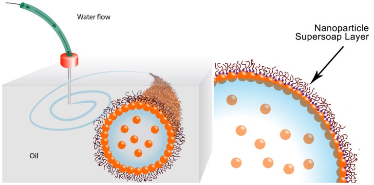
For the first step, the scientists developed a way to sheathe tubes of water in a special nanoparticle-derived surfactant that locks the water in place. The surfactant, essentially soap, prevents the tubes from breaking up into droplets. Their surfactant is so good at its job, the scientists call it a nanoparticle supersoap.

The supersoap was achieved by dispersing gold nanoparticles into water and polymer ligands into oil. The gold nanoparticles and polymer ligands want to attach to each other, but they also want to remain in their respective water and oil mediums. The ligands were developed with help from Brett Helms at the Molecular Foundry, a DOE Office of Science User Facility located at Berkeley Lab.

In practice, soon after the water is injected into the oil, dozens of ligands in the oil attach to individual nanoparticles in the water, forming a nanoparticle supersoap. These supersoaps jam together and vitrify, like glass, which stabilizes the interface between oil and water and locks the liquid structures in position.

This stability means we can stretch water into a tube, and it remains a tube. Or we can shape water into an ellipsoid, and it remains an ellipsoid,” said Russell. “We’ve used these nanoparticle supersoaps to print tubes of water that last for several months.”

Next came automation. Forth modified an off-the-shelf 3-D printer by removing the components designed to print plastic and replacing them with a syringe pump and needle that extrudes liquid. He then programmed the printer to insert the needle into the oil substrate and inject water in a predetermined pattern.

“We can squeeze liquid from a needle, and place threads of water anywhere we want in three dimensions,” said Forth. “We can also ping the material with an external force, which momentarily breaks the supersoap’s stability and changes the shape of the water threads. The structures are endlessly reconfigurable.”

This image illustrates how the water is printed,

These schematics show the printing of water in oil using a nanoparticle supersoap. Gold nanoparticles in the water combine with polymer ligands in the oil to form an elastic film (nanoparticle supersoap) at the interface, locking the structure in place. (Credit: Berkeley Lab)

Here’s a link to and a citation for the paper,

Reconfigurable Printed Liquids by Joe Forth, Xubo Liu, Jaffar Hasnain, Anju Toor, Karol Miszta, Shaowei Shi, Phillip L. Geissler, Todd Emrick, Brett A. Helms, Thomas P. Russell. Advanced Materials https://doi.org/10.1002/adma.201707603 First published: 24 March 2018

This paper is behind a paywall.

Tractor beams for artificial cells

This particular piece has videos of cells moving around. I won’t be including all of them but they are weirdly fascinating. First, a May 14, 2018 news item on Nanowerk announces the latest in tractor beam news from the Imperial College London (ICL; UK),

Researchers have used lasers to connect, arrange and merge artificial cells, paving the way for networks of artificial cells that act like tissues.

The team say that by altering artificial cell membranes they can now get the cells to stick together like ‘stickle bricks’ – allowing them to be arranged into whole new structures.

Biological cells can perform complex functions, but are difficult to controllably engineer.

Artificial cells, however, can in principle be made to order. Now, researchers from Imperial College London and Loughborough University have demonstrated a new level of complexity with artificial cells by arranging them into basic tissue structures with different types of connectivity.

These structures could be used to perform functions like initiating chemical reactions or moving chemicals around networks of artificial and biological cells. This could be useful in carrying out chemical reactions in ultra-small volumes, in studying the mechanisms through which cells communicate with one another, and in the development of a new generation of smart biomaterials.

A May 14, 2018 ICL press release by Hayley Dunning , which originated the news item, provides more detail,

Cells are the basic units of biology, which are capable of working together as a collective when arranged into tissues. In order to do this, cells must be connected and be capable of exchanging materials with one another.

The team were able to link up artificial cells into a range of new architectures, the results of which are published today in Nature Communications.

The artificial cells have a membrane-like layer as their shell, which the researchers engineered to ‘stick’ to each other. In order to get the cells to come close enough, the team first had to manipulate the cells with ‘optical tweezers’ that act like mini ‘tractor beams’ dragging and dropping cells into any position. Once connected in this way the cells can be moved as one unit.

Lead researcher Dr Yuval Elani, an EPSRC Research Fellow from the Department of Chemistry at Imperial, said: “Artificial cell membranes usually bounce off each other like rubber balls. By altering the biophysics of the membranes in our cells, we got them instead to stick to each other like stickle bricks.

“With this, we were able to form networks of cells connected by ‘biojunctions’. By reinserting biological components such as proteins in the membrane, we could get the cells to communicate and exchange material with one another. This mimics what is seen in nature, so it’s a great step forward in creating biological-like artificial cell tissues.”

Building up complexity

The team were also able to engineer a ‘tether’ between two cells. Here the membranes are not stuck together, but a tendril of membrane material links them so that they can be moved together.

Once they had perfected the cell-sticking process, the team were able to build up more complex arrangements. These include lines of cells, 2D shapes like squares, and 3D shapes like pyramids. Once the cells are stuck together, they can be rearranged, and also pulled by the laser beam as an ensemble

Finally, the team were also able to connect two cells, and then make them merge into one larger cell. This was achieved by coating the membranes with gold nanoparticles.

When the laser beam at the heart of the ‘optical tweezer’ technology was concentrated at the junction between the two cells, the nanoparticles resonated, breaking the membranes at that point. The membrane then reforms as a whole.

Merging cells in this way allowed whatever chemicals they were carrying to mix within the new, larger cell, kicking off chemical reactions. This could be useful, for example, for delivering materials such as drugs into cells, and in changing the composition of cells in real time, getting them to adopt new functions.

Professor Oscar Ces, also from the Department of Chemistry at Imperial, said: “Connecting artificial cells together is a valuable technology in the wider toolkit we are assembling for creating these biological systems using bottom-up approaches.

“We can now start to scale up basic cell technologies into larger tissue-scale networks, with precise control over the kind of architecture we create.”

Here’s one of the videos that has been embedded with ICL press release,

You can see the whole series if you go to the May 14, 2018 ICL press release.

Here’s a link to and a citation for the paper,

Sculpting and fusing biomimetic vesicle networks using optical tweezers by Guido Bolognesi, Mark S. Friddin, Ali Salehi-Reyhani, Nathan E. Barlow, Nicholas J. Brooks, Oscar Ces, & Yuval Elani. Nature Communicationsvolume 9, Article number: 1882 (2018) doi:10.1038/s41467-018-04282-w Published: 14 May 2018

This paper is open access.

Cloaking devices made from DNA and gold nanoparticles using top-down lithography

This new technique seems promising but there’ve been a lot of ‘cloaking’ devices announced in the years I’ve been blogging and, in all likelihood, I was late to the party so I’m exercising a little caution before getting too excited. For the latest development in cloaking devices, there’s a January 18, 2018 news item on Nanowerk,

Northwestern University researchers have developed a first-of-its-kind technique for creating entirely new classes of optical materials and devices that could lead to light bending and cloaking devices — news to make the ears of Star Trek’s Spock perk up.

Using DNA [deoxyribonucleic acid] as a key tool, the interdisciplinary team took gold nanoparticles of different sizes and shapes and arranged them in two and three dimensions to form optically active superlattices. Structures with specific configurations could be programmed through choice of particle type and both DNA-pattern and sequence to exhibit almost any color across the visible spectrum, the scientists report.

A January 18, 2018 Northwestern University news release (also on EurekAlert) by Megan Fellman, which originated the news item, delves into more detail (Note: Links have been removed),

“Architecture is everything when designing new materials, and we now have a new way to precisely control particle architectures over large areas,” said Chad A. Mirkin, the George B. Rathmann Professor of Chemistry in the Weinberg College of Arts and Sciences at Northwestern. “Chemists and physicists will be able to build an almost infinite number of new structures with all sorts of interesting properties. These structures cannot be made by any known technique.”

The technique combines an old fabrication method — top-down lithography, the same method used to make computer chips — with a new one — programmable self-assembly driven by DNA. The Northwestern team is the first to combine the two to achieve individual particle control in three dimensions.

The study was published online by the journal Science today (Jan. 18). Mirkin and Vinayak P. Dravid and Koray Aydin, both professors in Northwestern’s McCormick School of Engineering, are co-corresponding authors.

Scientists will be able to use the powerful and flexible technique to build metamaterials — materials not found in nature — for a range of applications including sensors for medical and environmental uses.

The researchers used a combination of numerical simulations and optical spectroscopy techniques to identify particular nanoparticle superlattices that absorb specific wavelengths of visible light. The DNA-modified nanoparticles — gold in this case — are positioned on a pre-patterned template made of complementary DNA. Stacks of structures can be made by introducing a second and then a third DNA-modified particle with DNA that is complementary to the subsequent layers.

In addition to being unusual architectures, these materials are stimuli-responsive: the DNA strands that hold them together change in length when exposed to new environments, such as solutions of ethanol that vary in concentration. The change in DNA length, the researchers found, resulted in a change of color from black to red to green, providing extreme tunability of optical properties.

“Tuning the optical properties of metamaterials is a significant challenge, and our study achieves one of the highest tunability ranges achieved to date in optical metamaterials,” said Aydin, assistant professor of electrical engineering and computer science at McCormick.

“Our novel metamaterial platform — enabled by precise and extreme control of gold nanoparticle shape, size and spacing — holds significant promise for next-generation optical metamaterials and metasurfaces,” Aydin said.

The study describes a new way to organize nanoparticles in two and three dimensions. The researchers used lithography methods to drill tiny holes — only one nanoparticle wide — in a polymer resist, creating “landing pads” for nanoparticle components modified with strands of DNA. The landing pads are essential, Mirkin said, since they keep the structures that are grown vertical.

The nanoscopic landing pads are modified with one sequence of DNA, and the gold nanoparticles are modified with complementary DNA. By alternating nanoparticles with complementary DNA, the researchers built nanoparticle stacks with tremendous positional control and over a large area. The particles can be different sizes and shapes (spheres, cubes and disks, for example).

“This approach can be used to build periodic lattices from optically active particles, such as gold, silver and any other material that can be modified with DNA, with extraordinary nanoscale precision,” said Mirkin, director of Northwestern’s International Institute for Nanotechnology.

Mirkin also is a professor of medicine at Northwestern University Feinberg School of Medicine and professor of chemical and biological engineering, biomedical engineering and materials science and engineering in the McCormick School.

The success of the reported DNA programmable assembly required expertise with hybrid (soft-hard) materials and exquisite nanopatterning and lithographic capabilities to achieve the requisite spatial resolution, definition and fidelity across large substrate areas. The project team turned to Dravid, a longtime collaborator of Mirkin’s who specializes in nanopatterning, advanced microscopy and characterization of soft, hard and hybrid nanostructures.

Dravid contributed his expertise and assisted in designing the nanopatterning and lithography strategy and the associated characterization of the new exotic structures. He is the Abraham Harris Professor of Materials Science and Engineering in McCormick and the founding director of the NUANCE center, which houses the advanced patterning, lithography and characterization used in the DNA-programmed structures.

Here’s a link to and a citation for the paper,

Building superlattices from individual nanoparticles via template-confined DNA-mediated assembly by Qing-Yuan Lin, Jarad A. Mason, Zhongyang, Wenjie Zhou, Matthew N. O’Brien, Keith A. Brown, Matthew R. Jones, Serkan Butun, Byeongdu Lee, Vinayak P. Dravid, Koray Aydin, Chad A. Mirkin. Science 18 Jan 2018: eaaq0591 DOI: 10.1126/science.aaq0591

This paper is behind a paywall.

As noted earlier, it could be a while before cloaking devices are made available. In the meantime, you may find this image inspiring,

Caption: Northwestern University researchers have developed a new method to precisely arrange nanoparticles of different sizes and shapes in two and three dimensions, resulting in optically active superlattices. Credit: Northwestern University

Gold’s origin in the universe due to cosmic collision

An hypothesis for gold’s origins was first mentioned here in a May 26, 2016 posting,

The link between this research and my side project on gold nanoparticles is a bit tenuous but this work on the origins for gold and other precious metals being found in the stars is so fascinating and I’m determined to find a connection.

An artist's impression of two neutron stars colliding. (Credit: Dana Berry / Skyworks Digital, Inc.) Courtesy: Kavli Foundation

An artist’s impression of two neutron stars colliding. (Credit: Dana Berry / Skyworks Digital, Inc.) Courtesy: Kavli Foundation

From a May 19, 2016 news item on phys.org,

The origin of many of the most precious elements on the periodic table, such as gold, silver and platinum, has perplexed scientists for more than six decades. Now a recent study has an answer, evocatively conveyed in the faint starlight from a distant dwarf galaxy.

In a roundtable discussion, published today [May 19, 2016?], The Kavli Foundation spoke to two of the researchers behind the discovery about why the source of these heavy elements, collectively called “r-process” elements, has been so hard to crack.

From the Spring 2016 Kavli Foundation webpage hosting the  “Galactic ‘Gold Mine’ Explains the Origin of Nature’s Heaviest Elements” Roundtable ,

Astronomers studying a galaxy called Reticulum II have just discovered that its stars contain whopping amounts of these metals—collectively known as “r-process” elements (See “What is the R-Process?”). Of the 10 dwarf galaxies that have been similarly studied so far, only Reticulum II bears such strong chemical signatures. The finding suggests some unusual event took place billions of years ago that created ample amounts of heavy elements and then strew them throughout the galaxy’s reservoir of gas and dust. This r-process-enriched material then went on to form Reticulum II’s standout stars.

Based on the new study, from a team of researchers at the Kavli Institute at the Massachusetts Institute of Technology, the unusual event in Reticulum II was likely the collision of two, ultra-dense objects called neutron stars. Scientists have hypothesized for decades that these collisions could serve as a primary source for r-process elements, yet the idea had lacked solid observational evidence. Now armed with this information, scientists can further hope to retrace the histories of galaxies based on the contents of their stars, in effect conducting “stellar archeology.”

Researchers have confirmed the hypothesis according to an Oct. 16, 2017 news item on phys.org,

Gold’s origin in the Universe has finally been confirmed, after a gravitational wave source was seen and heard for the first time ever by an international collaboration of researchers, with astronomers at the University of Warwick playing a leading role.

Members of Warwick’s Astronomy and Astrophysics Group, Professor Andrew Levan, Dr Joe Lyman, Dr Sam Oates and Dr Danny Steeghs, led observations which captured the light of two colliding neutron stars, shortly after being detected through gravitational waves – perhaps the most eagerly anticipated phenomenon in modern astronomy.

Marina Koren’s Oct. 16, 2017 article for The Atlantic presents a richly evocative view (Note: Links have been removed),

Some 130 million years ago, in another galaxy, two neutron stars spiraled closer and closer together until they smashed into each other in spectacular fashion. The violent collision produced gravitational waves, cosmic ripples powerful enough to stretch and squeeze the fabric of the universe. There was a brief flash of light a million trillion times as bright as the sun, and then a hot cloud of radioactive debris. The afterglow hung for several days, shifting from bright blue to dull red as the ejected material cooled in the emptiness of space.

Astronomers detected the aftermath of the merger on Earth on August 17. For the first time, they could see the source of universe-warping forces Albert Einstein predicted a century ago. Unlike with black-hole collisions, they had visible proof, and it looked like a bright jewel in the night sky.

But the merger of two neutron stars is more than fireworks. It’s a factory.

Using infrared telescopes, astronomers studied the spectra—the chemical composition of cosmic objects—of the collision and found that the plume ejected by the merger contained a host of newly formed heavy chemical elements, including gold, silver, platinum, and others. Scientists estimate the amount of cosmic bling totals about 10,000 Earth-masses of heavy elements.

I’m not sure exactly what this image signifies but it did accompany Koren’s article so presumably it’s a representation of colliding neutron stars,
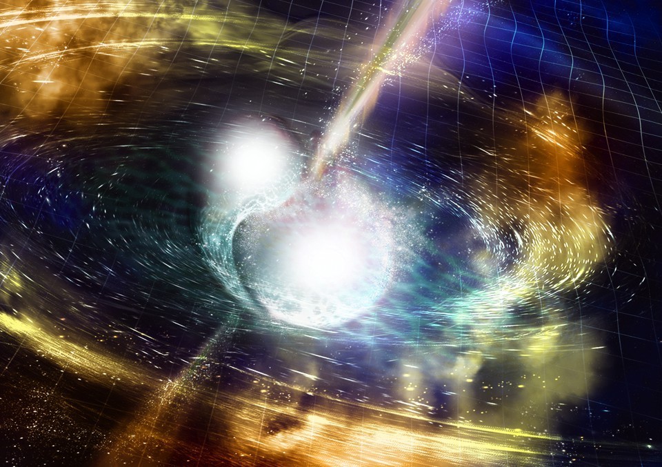
NSF / LIGO / Sonoma State University /A. Simonnet. Downloaded from: https://www.theatlantic.com/science/archive/2017/10/the-making-of-cosmic-bling/543030/

An Oct. 16, 2017 University of Warwick press release (also on EurekAlert), which originated the news item on phys.org, provides more detail,

Huge amounts of gold, platinum, uranium and other heavy elements were created in the collision of these compact stellar remnants, and were pumped out into the universe – unlocking the mystery of how gold on wedding rings and jewellery is originally formed.

The collision produced as much gold as the mass of the Earth. [emphasis mine]

This discovery has also confirmed conclusively that short gamma-ray bursts are directly caused by the merging of two neutron stars.

The neutron stars were very dense – as heavy as our Sun yet only 10 kilometres across – and they collided with each other 130 million years ago, when dinosaurs roamed the Earth, in a relatively old galaxy that was no longer forming many stars.

They drew towards each other over millions of light years, and revolved around each other increasingly quickly as they got closer – eventually spinning around each other five hundred times per second.

Their merging sent ripples through the fabric of space and time – and these ripples are the elusive gravitational waves spotted by the astronomers.

The gravitational waves were detected by the Advanced Laser Interferometer Gravitational-Wave Observatory (Adv-LIGO) on 17 August this year [2017], with a short duration gamma-ray burst detected by the Fermi satellite just two seconds later.

This led to a flurry of observations as night fell in Chile, with a first report of a new source from the Swope 1m telescope.

Longstanding collaborators Professor Levan and Professor Nial Tanvir (from the University of Leicester) used the facilities of the European Southern Observatory to pinpoint the source in infrared light.

Professor Levan’s team was the first one to get observations of this new source with the Hubble Space Telescope. It comes from a galaxy called NGC 4993, 130 million light years away.

Andrew Levan, Professor in the Astronomy & Astrophysics group at the University of Warwick, commented: “Once we saw the data, we realised we had caught a new kind of astrophysical object. This ushers in the era of multi-messenger astronomy, it is like being able to see and hear for the first time.”

Dr Joe Lyman, who was observing at the European Southern Observatory at the time was the first to alert the community that the source was unlike any seen before.

He commented: “The exquisite observations obtained in a few days showed we were observing a kilonova, an object whose light is powered by extreme nuclear reactions. This tells us that the heavy elements, like the gold or platinum in jewellery are the cinders, forged in the billion degree remnants of a merging neutron star.”

Dr Samantha Oates added: “This discovery has answered three questions that astronomers have been puzzling for decades: what happens when neutron stars merge? What causes the short duration gamma-ray bursts? Where are the heavy elements, like gold, made? In the space of about a week all three of these mysteries were solved.”

Dr Danny Steeghs said: “This is a new chapter in astrophysics. We hope that in the next few years we will detect many more events like this. Indeed, in Warwick we have just finished building a telescope designed to do just this job, and we expect it to pinpoint these sources in this new era of multi-messenger astronomy”.

Congratulations to all of the researchers involved in this work!

Many, many research teams were  involved. Here’s a sampling of their news releases which focus on their areas of research,

University of the Witwatersrand (South Africa)

https://www.eurekalert.org/pub_releases/2017-10/uotw-wti101717.php

Weizmann Institute of Science (Israel)

https://www.eurekalert.org/pub_releases/2017-10/wios-cns101717.php

Carnegie Institution for Science (US)

https://www.eurekalert.org/pub_releases/2017-10/cifs-dns101217.php

Northwestern University (US)

https://www.eurekalert.org/pub_releases/2017-10/nu-adc101617.php

National Radio Astronomy Observatory (US)

https://www.eurekalert.org/pub_releases/2017-10/nrao-ru101317.php

Max-Planck-Gesellschaft (Germany)

https://www.eurekalert.org/pub_releases/2017-10/m-gwf101817.php

Penn State (Pennsylvania State University; US)

https://www.eurekalert.org/pub_releases/2017-10/ps-stl101617.php

University of California – Davis

https://www.eurekalert.org/pub_releases/2017-10/uoc–cns101717.php

The American Association for the Advancement of Science’s (AAAS) magazine, Science, has published seven papers on this research. Here’s an Oct. 16, 2017 AAAS news release with an overview of the papers,

https://www.eurekalert.org/pub_releases/2017-10/aaft-btf101617.php

I’m sure there are more news releases out there and that there will be many more papers published in many journals, so if this interests, I encourage you to keep looking.

Two final pieces I’d like to draw your attention to: one answers basic questions and another focuses on how artists knew what to draw when neutron stars collide.

Keith A Spencer’s Oct. 18, 2017 piece on salon.com answers a lot of basic questions for those of us who don’t have a background in astronomy. Here are a couple of examples,

What is a neutron star?

Okay, you know how atoms have protons, neutrons, and electrons in them? And you know how protons are positively charged, and electrons are negatively charged, and neutrons are neutral?

Yeah, I remember that from watching Bill Nye as a kid.

Totally. Anyway, have you ever wondered why the negatively-charged electrons and the positively-charged protons don’t just merge into each other and form a neutral neutron? I mean, they’re sitting there in the atom’s nucleus pretty close to each other. Like, if you had two magnets that close, they’d stick together immediately.

I guess now that you mention it, yeah, it is weird.

Well, it’s because there’s another force deep in the atom that’s preventing them from merging.

It’s really really strong.

The only way to overcome this force is to have a huge amount of matter in a really hot, dense space — basically shove them into each other until they give up and stick together and become a neutron. This happens in very large stars that have been around for a while — the core collapses, and in the aftermath, the electrons in the star are so close to the protons, and under so much pressure, that they suddenly merge. There’s a big explosion and the outer material of the star is sloughed off.

Okay, so you’re saying under a lot of pressure and in certain conditions, some stars collapse and become big balls of neutrons?

Pretty much, yeah.

So why do the neutrons just stick around in a huge ball? Aren’t they neutral? What’s keeping them together? 

Gravity, mostly. But also the strong nuclear force, that aforementioned weird strong force. This isn’t something you’d encounter on a macroscopic scale — the strong force only really works at the type of distances typified by particles in atomic nuclei. And it’s different, fundamentally, than the electromagnetic force, which is what makes magnets attract and repel and what makes your hair stick up when you rub a balloon on it.

So these neutrons in a big ball are bound by gravity, but also sticking together by virtue of the strong nuclear force. 

So basically, the new ball of neutrons is really small, at least, compared to how heavy it is. That’s because the neutrons are all clumped together as if this neutron star is one giant atomic nucleus — which it kinda is. It’s like a giant atom made only of neutrons. If our sun were a neutron star, it would be less than 20 miles wide. It would also not be something you would ever want to get near.

Got it. That means two giant balls of neutrons that weighed like, more than our sun and were only ten-ish miles wide, suddenly smashed into each other, and in the aftermath created a black hole, and we are just now detecting it on Earth?

Exactly. Pretty weird, no?

Spencer does a good job of gradually taking you through increasingly complex explanations.

For those with artistic interests, Neel V. Patel tries to answer a question about how artists knew what draw when neutron stars collided in his Oct. 18, 2017 piece for Slate.com,

All of these things make this discovery easy to marvel at and somewhat impossible to picture. Luckily, artists have taken up the task of imagining it for us, which you’ve likely seen if you’ve already stumbled on coverage of the discovery. Two bright, furious spheres of light and gas spiraling quickly into one another, resulting in a massive swell of lit-up matter along with light and gravitational waves rippling off speedily in all directions, towards parts unknown. These illustrations aren’t just alluring interpretations of a rare phenomenon; they are, to some extent, the translation of raw data and numbers into a tangible visual that gives scientists and nonscientists alike some way of grasping what just happened. But are these visualizations realistic? Is this what it actually looked like? No one has any idea. Which is what makes the scientific illustrators’ work all the more fascinating.

“My goal is to represent what the scientists found,” says Aurore Simmonet, a scientific illustrator based at Sonoma State University in Rohnert Park, California. Even though she said she doesn’t have a rigorous science background (she certainly didn’t know what a kilonova was before being tasked to illustrate one), she also doesn’t believe that type of experience is an absolute necessity. More critical, she says, is for the artist to have an interest in the subject matter and in learning new things, as well as a capacity to speak directly to scientists about their work.

Illustrators like Simmonet usually start off work on an illustration by asking the scientist what’s the biggest takeaway a viewer should grasp when looking at a visual. Unfortunately, this latest discovery yielded a multitude of papers emphasizing different conclusions and highlights. With so many scientific angles, there’s a stark challenge in trying to cram every important thing into a single drawing.

Clearly, however, the illustrations needed to center around the kilonova. Simmonet loves colors, so she began by discussing with the researchers what kind of color scheme would work best. The smash of two neutron stars lends itself well to deep, vibrant hues. Simmonet and Robin Dienel at the Carnegie Institution for Science elected to use a wide array of colors and drew bright cracking to show pressure forming at the merging. Others, like Luis Calcada at the European Southern Observatory, limited the color scheme in favor of emphasizing the bright moment of collision and the signal waves created by the kilonova.

Animators have even more freedom to show the event, since they have much more than a single frame to play with. The Conceptual Image Lab at NASA’s [US National Aeronautics and Space Administration] Goddard Space Flight Center created a short video about the new findings, and lead animator Brian Monroe says the video he and his colleagues designed shows off the evolution of the entire process: the rising action, climax, and resolution of the kilonova event.

The illustrators try to adhere to what the likely physics of the event entailed, soliciting feedback from the scientists to make sure they’re getting it right. The swirling of gas, the direction of ejected matter upon impact, the reflection of light, the proportions of the objects—all of these things are deliberately framed such that they make scientific sense. …

Do take a look at Patel’s piece, if for no other reason than to see all of the images he has embedded there. You may recognize Aurore Simmonet’s name from the credit line in the second image I have embedded here.
Training drugs

This summarizes some of what’s happening in nanomedicine and provides a plug (boost) for the  University of Cambridge’s nanotechnology programmes (from a June 26, 2017 news item on Nanowerk),

Nanotechnology is creating new opportunities for fighting disease – from delivering drugs in smart packaging to nanobots powered by the world’s tiniest engines.

Chemotherapy benefits a great many patients but the side effects can be brutal.
When a patient is injected with an anti-cancer drug, the idea is that the molecules will seek out and destroy rogue tumour cells. However, relatively large amounts need to be administered to reach the target in high enough concentrations to be effective. As a result of this high drug concentration, healthy cells may be killed as well as cancer cells, leaving many patients weak, nauseated and vulnerable to infection.

One way that researchers are attempting to improve the safety and efficacy of drugs is to use a relatively new area of research known as nanothrapeutics to target drug delivery just to the cells that need it.

Professor Sir Mark Welland is Head of the Electrical Engineering Division at Cambridge. In recent years, his research has focused on nanotherapeutics, working in collaboration with clinicians and industry to develop better, safer drugs. He and his colleagues don’t design new drugs; instead, they design and build smart packaging for existing drugs.

The University of Cambridge has produced a video interview (referencing a 1966 movie ‘Fantastic Voyage‘ in its title)  with Sir Mark Welland,

A June 23, 2017 University of Cambridge press release, which originated the news item, delves further into the topic of nanotherapeutics (nanomedicine) and nanomachines,

Nanotherapeutics come in many different configurations, but the easiest way to think about them is as small, benign particles filled with a drug. They can be injected in the same way as a normal drug, and are carried through the bloodstream to the target organ, tissue or cell. At this point, a change in the local environment, such as pH, or the use of light or ultrasound, causes the nanoparticles to release their cargo.

Nano-sized tools are increasingly being looked at for diagnosis, drug delivery and therapy. “There are a huge number of possibilities right now, and probably more to come, which is why there’s been so much interest,” says Welland. Using clever chemistry and engineering at the nanoscale, drugs can be ‘taught’ to behave like a Trojan horse, or to hold their fire until just the right moment, or to recognise the target they’re looking for.

“We always try to use techniques that can be scaled up – we avoid using expensive chemistries or expensive equipment, and we’ve been reasonably successful in that,” he adds. “By keeping costs down and using scalable techniques, we’ve got a far better chance of making a successful treatment for patients.”

In 2014, he and collaborators demonstrated that gold nanoparticles could be used to ‘smuggle’ chemotherapy drugs into cancer cells in glioblastoma multiforme, the most common and aggressive type of brain cancer in adults, which is notoriously difficult to treat. The team engineered nanostructures containing gold and cisplatin, a conventional chemotherapy drug. A coating on the particles made them attracted to tumour cells from glioblastoma patients, so that the nanostructures bound and were absorbed into the cancer cells.

Once inside, these nanostructures were exposed to radiotherapy. This caused the gold to release electrons that damaged the cancer cell’s DNA and its overall structure, enhancing the impact of the chemotherapy drug. The process was so effective that 20 days later, the cell culture showed no evidence of any revival, suggesting that the tumour cells had been destroyed.

While the technique is still several years away from use in humans, tests have begun in mice. Welland’s group is working with MedImmune, the biologics R&D arm of pharmaceutical company AstraZeneca, to study the stability of drugs and to design ways to deliver them more effectively using nanotechnology.

“One of the great advantages of working with MedImmune is they understand precisely what the requirements are for a drug to be approved. We would shut down lines of research where we thought it was never going to get to the point of approval by the regulators,” says Welland. “It’s important to be pragmatic about it so that only the approaches with the best chance of working in patients are taken forward.”

The researchers are also targeting diseases like tuberculosis (TB). With funding from the Rosetrees Trust, Welland and postdoctoral researcher Dr Íris da luz Batalha are working with Professor Andres Floto in the Department of Medicine to improve the efficacy of TB drugs.

Their solution has been to design and develop nontoxic, biodegradable polymers that can be ‘fused’ with TB drug molecules. As polymer molecules have a long, chain-like shape, drugs can be attached along the length of the polymer backbone, meaning that very large amounts of the drug can be loaded onto each polymer molecule. The polymers are stable in the bloodstream and release the drugs they carry when they reach the target cell. Inside the cell, the pH drops, which causes the polymer to release the drug.

In fact, the polymers worked so well for TB drugs that another of Welland’s postdoctoral researchers, Dr Myriam Ouberaï, has formed a start-up company, Spirea, which is raising funding to develop the polymers for use with oncology drugs. Ouberaï is hoping to establish a collaboration with a pharma company in the next two years.

“Designing these particles, loading them with drugs and making them clever so that they release their cargo in a controlled and precise way: it’s quite a technical challenge,” adds Welland. “The main reason I’m interested in the challenge is I want to see something working in the clinic – I want to see something working in patients.”

Could nanotechnology move beyond therapeutics to a time when nanomachines keep us healthy by patrolling, monitoring and repairing the body?

Nanomachines have long been a dream of scientists and public alike. But working out how to make them move has meant they’ve remained in the realm of science fiction.

But last year, Professor Jeremy Baumberg and colleagues in Cambridge and the University of Bath developed the world’s tiniest engine – just a few billionths of a metre [nanometre] in size. It’s biocompatible, cost-effective to manufacture, fast to respond and energy efficient.

The forces exerted by these ‘ANTs’ (for ‘actuating nano-transducers’) are nearly a hundred times larger than those for any known device, motor or muscle. To make them, tiny charged particles of gold, bound together with a temperature-responsive polymer gel, are heated with a laser. As the polymer coatings expel water from the gel and collapse, a large amount of elastic energy is stored in a fraction of a second. On cooling, the particles spring apart and release energy.

The researchers hope to use this ability of ANTs to produce very large forces relative to their weight to develop three-dimensional machines that swim, have pumps that take on fluid to sense the environment and are small enough to move around our bloodstream.

Working with Cambridge Enterprise, the University’s commercialisation arm, the team in Cambridge’s Nanophotonics Centre hopes to commercialise the technology for microfluidics bio-applications. The work is funded by the Engineering and Physical Sciences Research Council and the European Research Council.

“There’s a revolution happening in personalised healthcare, and for that we need sensors not just on the outside but on the inside,” explains Baumberg, who leads an interdisciplinary Strategic Research Network and Doctoral Training Centre focused on nanoscience and nanotechnology.

“Nanoscience is driving this. We are now building technology that allows us to even imagine these futures.”

I have featured Welland and his work here before and noted his penchant for wanting to insert nanodevices into humans as per this excerpt from an April 30, 2010 posting,
Getting back to the Cambridge University video, do go and watch it on the Nanowerk site. It is fun and very informative and approximately 17 mins. I noticed that they reused part of their Nokia morph animation (last mentioned on this blog here) and offered some thoughts from Professor Mark Welland, the team leader on that project. Interestingly, Welland was talking about yet another possibility. (Sometimes I think nano goes too far!) He was suggesting that we could have chips/devices in our brains that would allow us to think about phoning someone and an immediate connection would be made to that person. Bluntly—no. Just think what would happen if the marketers got access and I don’t even want to think what a person who suffers psychotic breaks (i.e., hearing voices) would do with even more input. Welland starts to talk at the 11 minute mark (I think). For an alternative take on the video and more details, visit Dexter Johnson’s blog, Nanoclast, for this posting. Hint, he likes the idea of a phone in the brain much better than I do.

I’m not sure what could have occasioned this latest press release and related video featuring Welland and nanotherapeutics other than guessing that it was a slow news period.

Surgery on nanoparticles?

Chemists performed “surgery” on a 23-gold-atom nanoparticle according to a June 12, 2017 news item on Nanowerk (Note: A link has been removed),

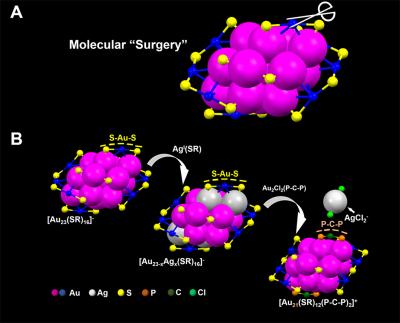
A team of chemists led by Carnegie Mellon University’s [CMU] Rongchao Jin has for the first time conducted site-specific surgery on a nanoparticle. The procedure, which allows for the precise tailoring of nanoparticles, stands to advance the field of nanochemistry.

The surgical technique developed by Qi Li, the study’s lead author and a 3rd year graduate student in the Jin group, will allow researchers to enhance nanoparticles’ functional properties, such as catalytic activity and photoluminescence, increasing their usefulness in a wide variety of fields including health care, electronics and manufacturing. The findings were published in Science Advances (“Molecular “surgery” on a 23-gold-atom nanoparticle”).

Here’s an image the researchers have provided,

Caption: Carnegie Mellon chemists used a two-step metal exchange method to remove two S-Au-S staples from the surface of a nanoparticle. Credit: Carnegie Mellon University

A June 12, 2017 CMU press release (also on EurekAlert), which originated the news item, provides more details about the research,

“Nanochemistry is a relatively new field, it’s only about 20 years old. We’ve been racing to catch up to fields like organic chemistry that are more than 100 years old,” said Jin, a chemistry professor in the Mellon College of Science. “Organic chemists have been able to tailor the functional groups of molecules for quite some time, like tailoring penicillin for better medical functions, for example. We dreamed that we could do something similar in nanoscience. Developing atomically precise nanoparticles has allowed us to make this dream come true.”

In order to make this “nano-surgery” a reality, researchers needed to begin with atomically precise nanoparticles that could be reliably produced time after time. Jin’s lab has been at the forefront of this research. Working with gold nanoparticles, he and his team have developed methods to precisely control the number of atoms in each nanoparticle, resulting in uniformly-sized nanoparticles with every batch. With reliably precise particles, Jin and colleagues were able to identify the particles’ structures, and begin to tease out how that structure impacted the particles’ properties and functionality.

With these well-defined nanoparticles in hand, Jin’s next step was to find a way to surgically tailor the particles in order to learn more about­ – and hopefully enhance – their functionality.

In their recent study, Jin and colleagues performed nano-surgery on a gold nanoparticle made up of 23 gold atoms surrounded by a protective surface of ligands in staple-like motifs. Using a two-step metal exchange method, they removed two S-Au-S staples from the particle’s surface. In doing this they revealed the structural factors that determine the particle’s optical properties and established the role that the surface plays in photoluminescence. Significantly, the surgery increased the particle’s photoluminescence by about 10-fold. Photoluminescence plays a critical role in biological imaging, cancer diagnosis and LED technology, among other applications.

Jin and coworkers are now trying to generalize this site-specific surgery method to other nanoparticles.

Here’s a link to and a citation for the paper,

Molecular “surgery” on a 23-gold-atom nanoparticle by Qi Li, Tian-Yi Luo, Michael G. Taylor, Shuxin Wang, Xiaofan Zhu, Yongbo Song, Giannis Mpourmpakis, Nathaniel L. Rosi, and Rongchao Jin. Science Advances 19 May 2017: Vol. 3, no. 5, e1603193 DOI: 10.1126/sciadv.1603193

This paper is open access.

Gold nanoparticles used to catalyze biofuel waste and create a useful additive

This work is the result of an international collaboration including Russia (from a May 23, 2017 news item on Nanowerk),

Gold nanoparticles serve as catalysts for obtaining valuable chemical products based on glycerol. Scientists from Tomsk Polytechnic University and their international colleagues are developing gold catalysts to recycle one of the main byproducts of biofuel production. The obtained products are in high demand in medicine, agriculture, cosmetic industry and other sectors.

Scientists from the University of Milano (Italy), the National Autonomous University of Mexico, the Institute of Catalysis and Petrochemistry of Madrid (Spain) and the University of Porto (Portugal) take part in the study of gold nanoparticles.

A May 23, 2027 Tomsk Polytechnic University press release, which originated the news item, expands on the theme,

Today the production of biofuels is an important area in many countries. They can be obtained from a great variety of biomasses. In Latin America it is orange and tangerine peel as well as banana skin. In USA biofuels are produced from corn, in the central part of Russia and Europe – from rape (Brassica napus). When processing these plants into biofuels a large amount of glycerol is formed. Its esters constitute the basis of oils and fats. Glycerol is widely used in cosmetic industry as an individual product. However, much more glycerol is obtained in the production of biofuels – many thousands of tons a year. As a result, unused glycerol merely becomes waste,’ describes the problem Alexey Pestryakov, the Head of the Department of Physical and Analytical Chemistry. ‘Now, a lot of research groups are engaged in this issue as to how to transform excess glycerol into other useful products. Along with our foreign colleagues we offered catalysts based on gold nanoparticles.’

The authors of the research note that catalytic oxidation on gold is one of the most effective techniques to obtain from glycerol such useful products as aldehydes, esters, carboxylic acids and other substances.

‘All these substances are products of fine organic chemistry and are in demand in a wide range of industries, first of all, in the pharmaceutical and cosmetic industries. In agriculture they are applied as part of different feed additives, veterinary drugs, fertilizers, plant treatment products, etc.

Thus, unused glycerol after being processed will further be applied,’ sums up Alexey Pestryakov.

Gold catalysts are super active. They can enter into chemical reactions with other substances at room temperature (other catalysts need to be heated), in some case even under zero. However, gold can be a catalyst only at the nanolevel.

‘If you take a piece of gold, even very tiny, there will be no chemical reaction. In order to make gold become chemically active, the size of its particle should be less than two nanometers. Only then it gets its amazing properties,’ explains the scientist.

As a catalyst gold was discovered not so long ago, in the early 1990s, by Japanese chemists.

To date, TPU scientists and their colleagues are not the only ones who develop such catalysts.

Unlike their counterparts the gold catalysts developed at TPU are more stable (they retain their activity longer).

‘A great challenge in this area is that gold catalysts are very rapidly deactivated, not only during work, but even during storage. Our objective is to ensure their longer shelf life. It is also important to use oxygen as an oxidizer, since toxic and corrosive peroxide compounds are often used for such purposes,’ says Alexey Petryakov.

Here’s a link to and a citation for the paper,

More Insights into Support and Preparation Method Effects in Gold Catalyzed Glycerol Oxidation by Nina Bogdanchikova, Inga Tuzovskaya, Laura Prati, Alberto Villa, Alexey Pestryakov, Mario Farías. Current Organic Synthesis VOLUME: 14 ISSUE: 3 Year: 2017Page: [377 – 382] Pages: 6 DOI: 10.2174/1570179413666161031114833

This paper is behind a paywall. (Scroll down the page to find the article.)